Review
. 2022 Jul 18;15(7):1180-1189.
doi: 10.18240/ijo.2022.07.20. eCollection 2022.

Molecules related to diabetic retinopathy in the vitreous and involved pathways

Affiliations
Review

Molecules related to diabetic retinopathy in the vitreous and involved pathways

Hua-Qin Xia et al. Int J Ophthalmol. .

Abstract

Diabetic retinopathy (DR) is one of the most common complications of diabetes and major cause of blindness among people over 50 years old. Current studies showed that the vascular endothelial growth factor (VEGF) played a central role in the pathogenesis of DR, and application of anti-VEGF has been widely acknowledged in treatment of DR targeting retinal neovascularization. However, anti-VEGF therapy has several limitations such as drug resistance. It is essential to develop new drugs for future clinical practice. The vitreous takes up 80% of the whole globe volume and is in direct contact with the retina, making it possible to explore the pathogenesis of DR by studying related factors in the vitreous. This article reviewed recent studies on DR-related factors in the vitreous, elaborating the VEGF upstream hypoxia-inducible factor (HIF) pathway and downstream pathways phosphatidylinositol diphosphate (PIP2), phosphoinositide-3-kinase (PI3K), and mitogen-activated protein kinase (MAPK) pathways. Moreover, factors other than VEGF contributing to the pathogenesis of DR in the vitreous were also summarized, which included factors in four major systems, kallikrein-kinin system such as bradykinin, plasma kallikrein, and coagulation factor XII, oxidative stress system such as lipid peroxide, and superoxide dismutase, inflammation-related factors such as interleukin-1β/6/13/37, and interferon-γ, matrix metalloproteinase (MMP) system such as MMP-9/14. Additionally, we also introduced other DR-related factors such as adiponectin, certain specific amino acids, non-coding RNA and renin (pro) receptor in separate studies.

Keywords: diabetic retinopathy; molecular pathway; vascular endothelial growth factor; vitreous.


Figures

Figure 1
Figure 1. Scheme of VEGF pathway
In the process of DR, VEGF acts as a core molecule, and is mainly induced by hypoxia. Hypoxia increases the expression of HIF and other growth factors (such as bFGF and PDGF), thereby increasing the transcription of the VEGF gene, translating more VEGF and secreting it out of the cell. Subsequently, VEGF is combined with VEGFR of the target cell (mainly via VEGFR2 in DR), and it performs physiological effect mainly through three ways: PI3K-Akt signaling pathway, PLC/PIP2 signaling pathway and MAPK signaling pathway. These pathways subsequently activate nuclear transcription factors and thereby changing gene expression, which ultimately leads to cell proliferation and vascular proliferation. In addition, VEGFR can also activate Paxillin through the FAK pathway, eventually leading to rearrangement of the cytoskeleton and cell migration. VEGF: Vascular endothelial growth factor; VEGFR: Vascular endothelial growth factor receptor; DR: Diabetic retinopathy; HIF: Hypoxia-inducible factor; bFGF: Basic fibroblast growth factor; PDGF: Platelet derived growth factor; MAPK: Mitogen-activated protein kinase.
Figure 2
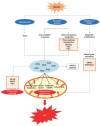
Figure 2. Scheme of molecules and pathways involved in DR and their relationship
KKS pathway, inflammation, MMP, and oxidative stress are all involved in the process of DR, and they are closely related to each other. The inflammation response can activate the KKS pathway, MMP and oxidative stress, and bradykinin produced by KKS, MMP and ROS induced by oxidative stress in turn promote the inflammatory response. Besides, oxidative stress can activate MMP via ROS, and MMP can promote oxidative stress by damaging mitochondria. As for association with VEGF, KKS function independently in the vitreous, while inflammation elevate the expression of VEGF and in turn VEGF intensify the inflammation response. Oxidative stress downregulates certain mi-Rs, which ultimately increase the expression of VEGF, and VEGF can increase the expression of MMP-9. Apart from these four pathways, there are some other molecules are involved, some of them are protective molecules and others are destructive. DR: Diabetic retinopathy; KKS: Kallikrein-Kinin System; MMP: Matrix metalloproteinase; VEGF: Vascular endothelial growth factor; ROS: Reactive oxygen species.
Figure 3
Figure 3. Scheme of KKS pathway and functions
Retinal vascular hemorrhage activates a series of molecules such as CA-1, heparin, etc. These molecules then activate the KKS pathway. The KKS pathway then produces a large amount of bradykinin, which combines with bradykinin receptor 1 and bradykinin receptor 2 on the retina to play a pathogenic role. Among them, B1R mainly induces inflammatory response, while B2R mainly causes dilation of retinal blood vessels.
Figure 4
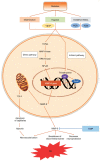
Figure 4. Scheme of oxidative stress pathway
Under the condition of diabetes, hyperglycemia induce the activation of Nox2 in the cell, and at the same time, the excessive glucose will be metabolized by auto-oxidation and four other glucose metabolism pathways, thereby generating a large amount of ROS. These ROS subsequently disturb mitochondrial biogenesis and mitochondrial DNA repair, abnormally methylate mitochondrial DNA, and reduce the expression of antioxidants. The series of reactions ultimately lead to mitochondrial dysfunction and induce cell apoptosis. In addition, ROS can promote the expression of NF-κB, MAPK and other molecules, thereby intensifying the inflammatory response. ROS can also activate the MMP-9 pathway, which also contributes to DR. NF-κB: Nuclear factor kappa-B; MMP: Matrix metalloproteinase; ROS: Reactive oxygen species; DR: Diabetic retinopathy.
Figure 5
Figure 5. Inflammation factors involved in DR
Inflammation is closely related to the other three systems in this article. MMP-9, oxidative stress products ROS and KKS system products BK can all induce the production of inflammatory factors. We divide the classic inflammatory factors into three categories: pro-inflammatory molecules, anti-inflammatory molecular and pleiotropic cytokines, at the same time we introduce some newly discovered inflammatory factors related to DR that exist in the vitreous. DR: Diabetic retinopathy; KKS: Kallikrein-Kinin system; BK: Bradykinins; MMP: Matrix metalloproteinase; ROS: Reactive oxygen species.
Figure 6
Figure 6. Scheme of MMP-9 pathway
Inflammation, hypoxia and oxidative stress caused by diabetes can activate MMP-9 expression through direct and indirect pathways. Subsequently, MMP-9 caused mitochondrial dysfunction and the mitochondria released Cyt C to induce the apoptosis of vascular endothelial cells, further aggravating tissue hypoxia and inducing neovascularization. On the other hand, MMP-9 is secreted from cells and directly participates in angiogenesis, while degrading the blood-retinal barrier and reducing the ability of retinal neuroprotection, which ultimately leads to DR.

References

    1. Simó-Servat O, Hernández C, Simó R. Diabetic retinopathy in the context of patients with diabetes. Ophthalmic Res. 2019;62(4):211–217. - PubMed
    1. Wong TY, Cheung CMG, Larsen M, Sharma S, Simó R. Diabetic retinopathy. Nat Rev Dis Primers. 2016;2:16012. - PubMed
    1. Liu R, Liu CM, Cui LL, Zhou L, Li N, Wei XD. Expression and significance of MiR-126 and VEGF in proliferative diabetic retinopathy. Eur Rev Med Pharmacol Sci. 2019;23(15):6387–6393. - PubMed
    1. Grauslund J. Vascular endothelial growth factor inhibition for proliferative diabetic retinopathy: Et Tu, Brute? Acta Ophthalmol. 2017;95(8):757–758. - PubMed
    1. Loukovaara S, Nurkkala H, Tamene F, Gucciardo E, Liu XN, Repo P, Lehti K, Varjosalo M. Quantitative proteomics analysis of vitreous humor from diabetic retinopathy patients. J Proteome Res. 2015;14(12):5131–5143. - PubMed

LinkOut - more resources