

Phenolic composition and neuroprotective effects of the ethyl-acetate fraction from Inonotus sanghuang against H2O2-induced apoptotic cell death of primary cortical neuronal cells
- PMID: 35919355
- PMCID: PMC9339070
- DOI: 10.1007/s10068-022-01107-x
Phenolic composition and neuroprotective effects of the ethyl-acetate fraction from Inonotus sanghuang against H2O2-induced apoptotic cell death of primary cortical neuronal cells
Abstract
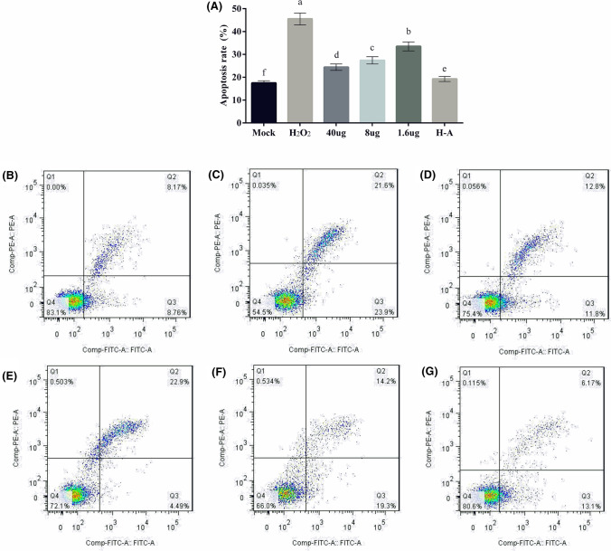
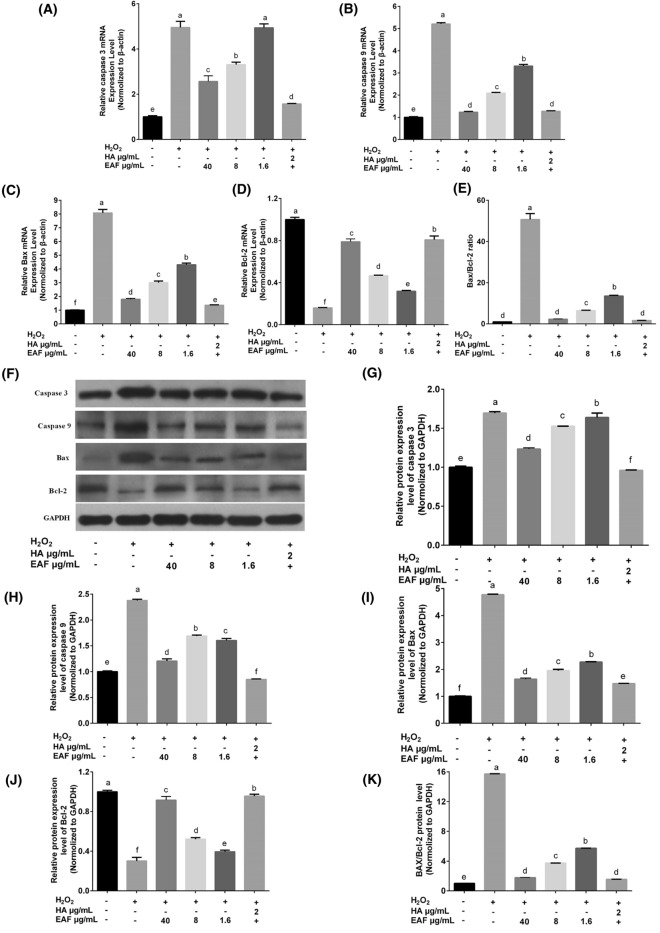
The study aimed to characterize phenolic compounds of the Inonotus sanghuang's ethyl-acetate fraction (EAF) and assess the neuroprotective effect of EAF using the H2O2-treated primary cortical neuronal cells (PCNC) model. Using HPLC-ECD, 5 phenolics were identified and quantified from EAF. H2O2-treated PCNC experiments in vitro showed that pretreatment with EAF increased the GSH-PX and SOD activities and reduced the NO, MDA, and Aβ contents. Furthermore, EAF suppressed the production of IL-1β, IFN-γ, IL-6, and TNF-α in H2O2-treated PCNC. Other mechanisms found that EAF reduced Bax, caspase 9, and caspase 3 expressions at the mRNA and protein levels while increasing Bcl-2 expression at the mRNA and protein levels. These results showed that EAF could serve as potential agents for anti-NDD.
Supplementary information: The online version contains supplementary material available at 10.1007/s10068-022-01107-x.
Keywords: Anti-apoptosis; Anti-inflammatory; Antioxidation; Inonotus sanghuang; Neuroprotective effect.
© The Korean Society of Food Science and Technology 2022.
Conflict of interest statement
Conflict of interestThe authors declare no competing financial interest.
Figures


Similar articles
-
Hypoglycemic and H2O2-induced oxidative injury protective effects and the phytochemical profiles of the ethyl acetate fraction from Radix Paeoniae Alba.Front Nutr. 2023 Feb 24;10:1126359. doi: 10.3389/fnut.2023.1126359. eCollection 2023. Front Nutr. 2023. PMID: 36908916 Free PMC article.
-
Protective effect of a polyphenols-rich extract from Inonotus Sanghuang on bleomycin-induced acute lung injury in mice.Life Sci. 2019 Aug 1;230:208-217. doi: 10.1016/j.lfs.2019.05.074. Epub 2019 May 29. Life Sci. 2019. PMID: 31152815
-
[Effect of germacrone in alleviating HUVECs damaged by H2O2-induced oxidative stress].Zhongguo Zhong Yao Za Zhi. 2017 Sep;42(18):3564-3571. doi: 10.19540/j.cnki.cjcmm.20170731.006. Zhongguo Zhong Yao Za Zhi. 2017. PMID: 29218943 Chinese.
-
Neuroprotective effects of natural compounds on neurotoxin-induced oxidative stress and cell apoptosis.Nutr Neurosci. 2022 May;25(5):1078-1099. doi: 10.1080/1028415X.2020.1840035. Epub 2020 Nov 8. Nutr Neurosci. 2022. PMID: 33164705 Review.
-
Therapeutic potentials of plant iridoids in Alzheimer's and Parkinson's diseases: A review.Eur J Med Chem. 2019 May 1;169:185-199. doi: 10.1016/j.ejmech.2019.03.009. Epub 2019 Mar 8. Eur J Med Chem. 2019. PMID: 30877973 Review.
Cited by
-
Research Progress of Bioactive Components in Sanghuangporus spp.Molecules. 2024 Mar 7;29(6):1195. doi: 10.3390/molecules29061195. Molecules. 2024. PMID: 38542832 Free PMC article. Review.
-
Comparative genomic analysis of Sanghuangporus sanghuang with other Hymenochaetaceae species.Braz J Microbiol. 2024 Mar;55(1):87-100. doi: 10.1007/s42770-023-01212-x. Epub 2023 Dec 15. Braz J Microbiol. 2024. PMID: 38099978 Free PMC article.
-
Fucoidan JHCF4s from Hizikia fusiformis against ethanol-induced damage in vitro and in vivo.Food Sci Biotechnol. 2025 Apr 29;34(10):2295-2306. doi: 10.1007/s10068-025-01865-4. eCollection 2025 Jun. Food Sci Biotechnol. 2025. PMID: 40351724
References
-
- Bucchieri F, Marino Gammazza A, Pitruzzella A, Fucarino A, Farina F, Howarth P, Holgate ST, Zummo G, Davies DE. Cigarette smoke causes caspase-independent apoptosis of bronchial epithelial cells from asthmatic donors. PloS ONE. 2015;10:e0120510. doi: 10.1371/journal.pone.0120510. - DOI - PMC - PubMed
LinkOut - more resources
Full Text Sources
Research Materials