

Cervical tracheal rupture with persistence of a pseudotrachea in a dog
- PMID: 35919475
- PMCID: PMC9281890
Cervical tracheal rupture with persistence of a pseudotrachea in a dog
Abstract
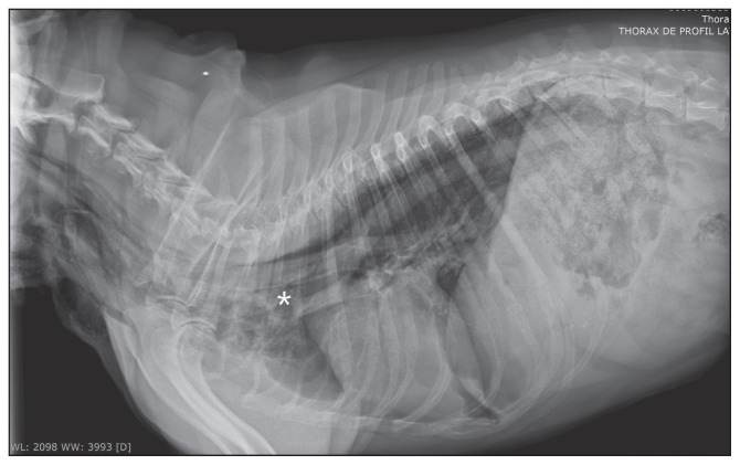
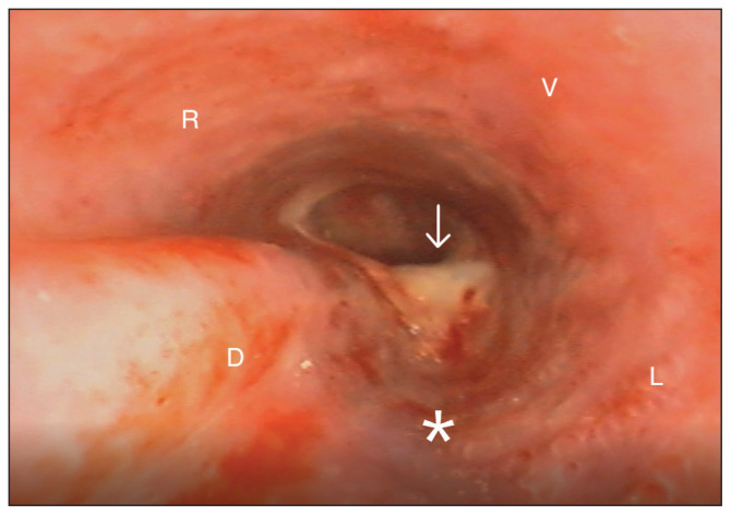
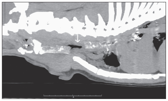
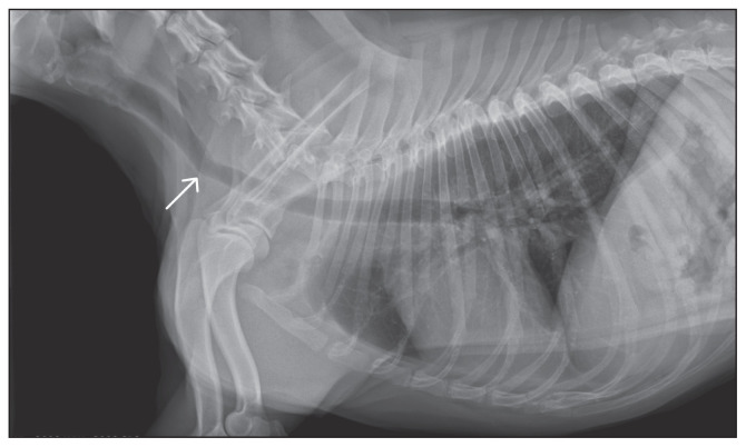
A dog was examined because of acute onset of respiratory distress following a cervical dog bite. Physical examination revealed a deep ventral cervical bite wound associated with localized mild subcutaneous emphysema. Thoracic radiographs showed moderate pneumomediastinum. Medical management consisting of oxygen therapy, antibiotics, and anti-inflammatories was initiated. After 2 days, respiratory distress suddenly worsened. Tracheoscopy showed a discontinuity between the tracheal rings of the cervical trachea; however, the inner tracheal wall appeared intact. Computed tomography scan revealed a ~3-cm complete rupture of all layers of the trachea. Surgical resection and anastomosis of the trachea were performed successfully. Follow-up 15 days after surgery showed complete resolution of respiratory signs, as well as subcutaneous emphysema. A mild ventral angulation of the trachea at the surgical site was noticed on thoracic radiographs. This is the first case report of a pseudotrachea in a dog. Persistence of a pseudotrachea may initially result in only minor clinical signs responsive to medical therapy despite tracheal rupture. In the presence of a pseudotrachea, tracheal rupture may be difficult to identify with tracheoscopy alone. Therefore, CT scan should be proposed in every patient with suspected tracheal trauma. Key clinical message: This case report highlights the importance of including a tracheal rupture in the differential diagnosis of cervical subcutaneous emphysema, even if the amount is small and not associated with significant respiratory signs. The presence of a pseudotrachea may result in less severe clinical signs than expected based on the actual degree of tracheal injury; however, the clinical status may rapidly deteriorate and become life-threatening. This case report also underlines the importance of a CT scan as a complement to tracheoscopy, which may not be sufficient to identify a tracheal rupture in the presence of a pseudotrachea.
Rupture trachéale cervicale avec persistance d’une pseudotrachée chez un chien. Un chien a été présenté pour une dyspnée aiguë modérée consécutive à des morsures cervicales par un autre chien. L’examen clinique révéla une plaie cervicale ventrale profonde associée à un emphysème sous-cutané localisé léger. Les radiographies thoraciques ont montré un pneumomédiastin modéré. Un traitement médical consistant en une oxygénothérapie, des antibiotiques et des anti-inflammatoires a été initié. Après deux jours, la dyspnée s’aggrava brutalement. Une trachéoscopie révéla une discontinuité entre les anneaux trachéaux malgré la persistance d’une paroi trachéale interne intègre. L’examen par tomodensitométrie montra une rupture trachéale cervicale complète dans toute son épaisseur, sur 3 cm de long. Une chirurgie de résection-anastomose de la trachée a été réalisée avec succès.Il s’agit de la première description de pseudotrachée chez un chien. La persistance d’une pseudotrachée peut initialement ne provoquer que des signes cliniques mineurs, notamment un emphysème sous-cutané léger et une dyspnée répondant au traitement médical, malgré une lésion trachéale en réalité importante. Par conséquent, un examen par tomodensitométrie de la trachée doit être envisagé chez tous les patients pour lesquels un traumatisme trachéal est suspecté.Message clinique clé :Ce cas souligne l’importance d’inclure une rupture trachéale dans le diagnostic différentiel de l’emphysème souscutané cervical, et cela même s’il n’est présent qu’en petite quantité et associé à faibles signes cliniques respiratoires. La persistance d’une pseudotrachée peut entraîner des signes cliniques moins importants qu’une rupture trachéale complète, cependant l’état respiratoire de l’animal peut rapidement s’aggraver et devenir une urgence vitale.Ce cas souligne de plus l’importance de l’examen par tomodensitométrie en complément de la trachéoscopie, qui peut parfois s’avérer insuffisante pour le diagnostic des ruptures trachéales, en particulier en présence d’une pseudotrachée.(Traduit par les auteurs).
Copyright and/or publishing rights held by the Canadian Veterinary Medical Association.
Figures
References
-
- Holt D. Tracheal trauma. In: Silverstein DC, Hopper K, editors. Small Animal Critical Care Medicine. 2nd ed. Ames, Iowa: Blackwell Publishing; 2014. pp. 107–110.
-
- White RN, Burton CA. Surgical management of intrathoracic tracheal avulsion in cats: Long-term results in 9 consecutive cases. Vet Surg. 2000;29:430–435. - PubMed
-
- Moreau C, Blanchot D, Jacques D. Rupture trachéale intrathoracique chez un chat. Pratique médicale et chirurgicale de l’animal domestique. 2010;45:73–77.
-
- Mitchell SL, McCarthy R, Rudloff E, Pernell RT. Tracheal rupture associated with intubation in cats: 20 cases (1996–1998) J Am Vet Med Assoc. 2000;216:1592–1595. - PubMed
-
- Basdani E, Papazoglou LG, Patsikas MN, Kazakos GM, Adamama-Moraitou KK, Tsokataridis I. Upper airway injury in dogs secondary to trauma: 10 dogs (2000–2011) J Am Hosp Assoc. 2016;52:291–296. - PubMed
Publication types
MeSH terms
LinkOut - more resources
Full Text Sources