. 2022 Jan 27;2(1):ltac002.
doi: 10.1093/immadv/ltac002. eCollection 2022.

Safety of the use of gold nanoparticles conjugated with proinsulin peptide and administered by hollow microneedles as an immunotherapy in type 1 diabetes

Affiliations

Safety of the use of gold nanoparticles conjugated with proinsulin peptide and administered by hollow microneedles as an immunotherapy in type 1 diabetes

D Tatovic et al. Immunother Adv. .

Abstract

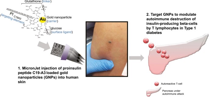
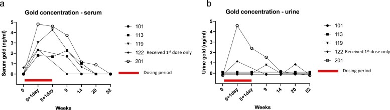
Antigen-specific immunotherapy is an immunomodulatory strategy for autoimmune diseases, such as type 1 diabetes, in which patients are treated with autoantigens to promote immune tolerance, stop autoimmune β-cell destruction and prevent permanent dependence on exogenous insulin. In this study, human proinsulin peptide C19-A3 (known for its positive safety profile) was conjugated to ultrasmall gold nanoparticles (GNPs), an attractive drug delivery platform due to the potential anti-inflammatory properties of gold. We hypothesised that microneedle intradermal delivery of C19-A3 GNP may improve peptide pharmacokinetics and induce tolerogenic immunomodulation and proceeded to evaluate its safety and feasibility in a first-in-human trial. Allowing for the limitation of the small number of participants, intradermal administration of C19-A3 GNP appears safe and well tolerated in participants with type 1 diabetes. The associated prolonged skin retention of C19-A3 GNP after intradermal administration offers a number of possibilities to enhance its tolerogenic potential, which should be explored in future studies.

Keywords: gold nanoparticle; microneedle; peptide immunotherapy; proinsulin; type 1 diabetes.


Figures

None
Graphical abstract
Figure 1.
Figure 1.
Study recruitment and visit schedule. (a) Consort diagram and (b) schedule of the study visits. D, dose; V, visit; W, week.
Figure 2.
Figure 2.
Effect of C19-A3 GNP on β-cell function and metabolic parameters. (a) Serum C-peptide expressed as area under the curve (AUC) over 120 min after mixed-meal challenge; (b) urine C-peptide to creatinine ratio (UCPCR) 2 h after mixed meal; (c) HbA1c; (d) median total daily insulin dose recorded over 3 days before the study visit and normalised for body weight; (e) insulin dose-adjusted A1c (IDAA1c) calculated according to the formula: HbA1c (%) + [4× insulin dose (units per kg per 24 h)]. Each line represents an individual participant with an assigned study number as shown in the legend.
Figure 3.
Figure 3.
Effect of C19-A3 GNP on glucose variability. (a) Time in range (the % of time participants recorded a blood glucose level of 4–10 mmol/l); (b) time above range (the % of time participants recorded a blood glucose level of >10 mmol/l); (c) time below range (the % of time participants recorded a blood glucose level of <4 mmol/l). Recordings were taken for at least 72 h before study visits. Each line represents an individual participant with an assigned study number as shown in the legend.
Figure 4.
Figure 4.
Serum and urine gold concentration following administration of C19-A3 GNP. (a) Serum gold concentration; (b) urine gold concentration. Blood and urine samples for measurement of gold concentration were taken at baseline (week 0), one day after 1st dose (week 0 + 1 day) and one day after 3rd dose (week 8 + 1 day). Detection limit was 0.2 ng/ml and quantitation limit 0.6 ng/ml. Levels below detection and quantitation limit are presented as zero on the graph. Each line represents an individual participant with an assigned study number as shown in the legend.
Figure 5.
Figure 5.
Skin changes after intradermal injection of C19-A3 GNP. (a) Immediately after injection; (b) 5 min after injection; (c) 24 h after injection; (d) 30 days after injection; (e) 2 months after injection (upper left), 1 month after injection (right) and 7 days after injection (lower left); (f) 20 months after injection; (g) close-up of the injection site showing central area of hyperpigmentation and surrounding induration and redness.
Figure 6.
Figure 6.
Histopathology and immunohistochemistry staining of the punch biopsies of the injection sites. (a) Anti-CD3 staining; (b) anti-CD20 staining; (c) anti-CD4 staining; (d) anti-CD8 staining; (e) and (f) gold staining.

References

    1. Foster NC, Beck RW, Miller KM.et al. . State of type 1 diabetes management and outcomes from the T1D exchange in 2016-2018. Diabetes Technol Ther 2019;21:66–72. - PMC - PubMed
    1. Skyler JS. Primary and secondary prevention of Type 1 diabetes. Diabet Med. 2013;30(2):161–9. - PMC - PubMed
    1. Dayan CM, Korah M, Tatovic D.et al. . Changing the landscape for type 1 diabetes: the first step to prevention. Lancet 2019;394:1286–96. - PubMed
    1. Mallone R, Brezar V, Boitard C.. T cell recognition of autoantigens in human type 1 diabetes: clinical perspectives. Clin Dev Immunol 2011;2011:513210. - PMC - PubMed
    1. Tian J, Kaufman DL.. Antigen-based therapy for the treatment of type 1 diabetes. Diabetes 2009;58:1939–46. - PMC - PubMed

LinkOut - more resources