

Review on Hybrid Segmentation Methods for Identification of Brain Tumor in MRI
- PMID: 35919500
- PMCID: PMC9293518
- DOI: 10.1155/2022/1541980
Review on Hybrid Segmentation Methods for Identification of Brain Tumor in MRI
Abstract
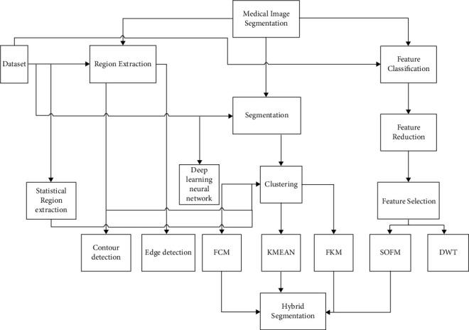
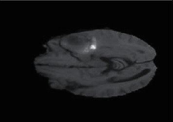
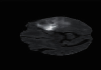
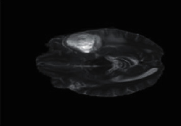
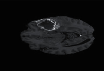
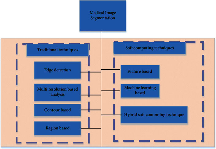
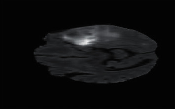
Modalities like MRI give information about organs and highlight diseases. Organ information is visualized in intensities. The segmentation method plays an important role in the identification of the region of interest (ROI). The ROI can be segmented from the image using clustering, features, and region extraction. Segmentation can be performed in steps; firstly, the region is extracted from the image. Secondly, feature extraction performed, and better features are selected. They can be shape, texture, or intensity. Thirdly, clustering segments the shape of tumor, tumor has specified shape, and shape is detected by feature. Clustering consists of FCM, K-means, FKM, and their hybrid. To support the segmentation, we conducted three studies (region extraction, feature, and clustering) which are discussed in the first line of this review paper. All these studies are targeting MRI as a modality. MRI visualization proved to be more accurate for the identification of diseases compared with other modalities. Information of the modality is compromised due to low pass image. In MRI Images, the tumor intensities are variable in tumor areas as well as in tumor boundaries.
Copyright © 2022 Khurram Ejaz et al.
Conflict of interest statement
The authors declare that they have no conflicts of interest.
Figures
Similar articles
-
MRI Brain Tumour Segmentation Using Hybrid Clustering and Classification by Back Propagation Algorithm.Asian Pac J Cancer Prev. 2018 Nov 29;19(11):3257-3263. doi: 10.31557/APJCP.2018.19.11.3257. Asian Pac J Cancer Prev. 2018. PMID: 30486629 Free PMC article.
-
Spectral embedding based active contour (SEAC) for lesion segmentation on breast dynamic contrast enhanced magnetic resonance imaging.Med Phys. 2013 Mar;40(3):032305. doi: 10.1118/1.4790466. Med Phys. 2013. PMID: 23464337 Free PMC article.
-
A mathematical theory of shape and neuro-fuzzy methodology-based diagnostic analysis: a comparative study on early detection and treatment planning of brain cancer.Int J Clin Oncol. 2017 Aug;22(4):667-681. doi: 10.1007/s10147-017-1110-5. Epub 2017 Mar 20. Int J Clin Oncol. 2017. PMID: 28321787
-
Comparative Approach of MRI-Based Brain Tumor Segmentation and Classification Using Genetic Algorithm.J Digit Imaging. 2018 Aug;31(4):477-489. doi: 10.1007/s10278-018-0050-6. J Digit Imaging. 2018. PMID: 29344753 Free PMC article. Review.
-
Recent Advancements in Fuzzy C-means Based Techniques for Brain MRI Segmentation.Curr Med Imaging. 2021;17(8):917-930. doi: 10.2174/1573405616666210104111218. Curr Med Imaging. 2021. PMID: 33397241 Review.
Cited by
-
Learning Architecture for Brain Tumor Classification Based on Deep Convolutional Neural Network: Classic and ResNet50.Diagnostics (Basel). 2025 Mar 5;15(5):624. doi: 10.3390/diagnostics15050624. Diagnostics (Basel). 2025. PMID: 40075870 Free PMC article.
-
Multi class robust brain tumor with hybrid classification using DTA algorithm.Heliyon. 2023 Dec 13;10(1):e23610. doi: 10.1016/j.heliyon.2023.e23610. eCollection 2024 Jan 15. Heliyon. 2023. PMID: 38187263 Free PMC article.
References
-
- Chouhan S. S., Kaul A., Singh U. P. Soft computing approaches for image segmentation: a survey. Multimedia Tools and Applications . 2018;77(21) doi: 10.1007/s11042-018-6005-6.28483 - DOI
-
- Kumar E. P., Kumar V. M., Sumithra M. Tumour detection in brain MRI using improved segmentation algorithm. Proceedings of the 2013 Fourth International Conference on Computing, Communications and Networking Technologies (ICCCNT); July, 2013; Tiruchengode, India. IEEE; - DOI
-
- Aslam A., Khan E., Beg M. S. Improved edge detection algorithm for brain tumor segmentation. Procedia Computer Science . 2015;58:430–437. doi: 10.1016/j.procs.2015.08.057. - DOI
-
- Vishnuvarthanan G., Rajasekaran M. P., Subbaraj P., Vishnuvarthanan A. An unsupervised learning method with a clustering approach for tumor identification and tissue segmentation in magnetic resonance brain images. Applied Soft Computing . 2016;38:190–212. doi: 10.1016/j.asoc.2015.09.016. - DOI
-
- Swamy S., Kulkarni P. Image processing for identifying brain tumor using intelligent system. Int. J. Innov. Res. Sci. Eng. Technol . 2015;4(11)
Publication types
MeSH terms
LinkOut - more resources
Full Text Sources
Medical
