

Computed tomography patterns of intracranial infarcts in a Ghanaian tertiary facility
- PMID: 35919779
- PMCID: PMC9334949
- DOI: 10.4314/gmj.v56i1.5
Computed tomography patterns of intracranial infarcts in a Ghanaian tertiary facility
Abstract
Objective: To determine the Computed Tomography (CT) patterns of intracranial infarcts.
Design: A retrospective cross-sectional study.
Setting: The CT scan unit of the Radiology Department, Cape Coast Teaching Hospital (CCTH), from February 2017 to February 2021.
Participants: One thousand, one hundred and twenty-five patients with non-contrast head CT scan diagnosis of ischaemic strokes, consecutively selected over the study period without any exclusions.
Main outcome measures: Patterns of non-contrast head CT scan of ischaemic strokes.
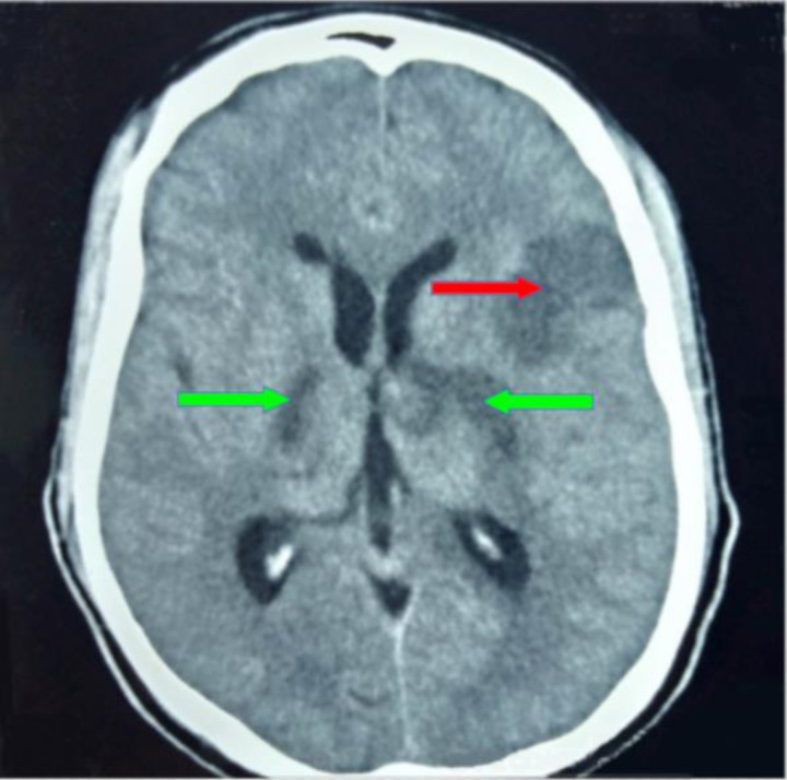
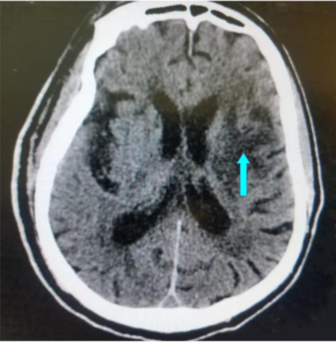
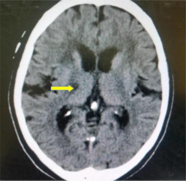
Results: About 50.6% of the study participants were females with an average age of 62.59±13.91 years. Males were affected with ischaemic strokes earlier than females (p<0.001). The risk factors considered were, hyperlipidaemia (59.5%), hypertension (49.0%), Type 2 diabetes mellitus (DM-2) (39.6%) and smoking (3.0%). The three commonest ischaemic stroke CT scan features were wedge-shaped hypodensity extending to the edge of the brain (62.8%), sulcal flattening/effacement (57.6%) and loss of grey-white matter differentiation (51.0%), which were all significantly associated with hypertension. Small deep brain hypodensities, the rarest feature (2.2%), had no significant association with any of the risk factors considered in the study.
Conclusion: Apart from the loss of grey-white matter differentiation, there was no significant association between the other CT scan features and sex. Generally, most of the risk factors and the CT scan features were significantly associated with increasing age.
Funding: None declared.
Keywords: Brain; Computed Tomography; Ghana; Intracranial Infarcts; Patterns.
Copyright © The Author(s).
Conflict of interest statement
Conflict of interest: None declared
Figures
Similar articles
-
Dyslipidaemia as a risk factor in the occurrence of stroke in Nigeria: prevalence and patterns.Pan Afr Med J. 2016 Oct 4;25:72. doi: 10.11604/pamj.2016.25.72.6496. eCollection 2016. Pan Afr Med J. 2016. PMID: 28292035 Free PMC article.
-
CT and clinical correlation of stroke diagnosis, pattern and clinical outcome among stroke patients visting Tikur Anbessa Hospital.Ethiop Med J. 2010 Apr;48(2):117-22. Ethiop Med J. 2010. PMID: 20608015
-
Computed tomography features of spontaneous acute intracranial hemorrhages in a tertiary hospital in Southern Ghana.Pan Afr Med J. 2021 Dec 16;40:226. doi: 10.11604/pamj.2021.40.226.31934. eCollection 2021. Pan Afr Med J. 2021. PMID: 35145588 Free PMC article.
-
In light of recently published clinical trials and their implication for clinical practice, does a large catchment area acute hospital require 24 hour CT neck and head angiography and/or neuro-interventional services in the setting of acute ischaemic stroke?Ir J Med Sci. 2018 May;187(2):351-358. doi: 10.1007/s11845-017-1674-0. Epub 2017 Aug 15. Ir J Med Sci. 2018. PMID: 28812226 Review.
-
[Value of serial CT scanning and intracranial pressure monitoring for detecting new intracranial mass effect in severe head injury patients showing lesions type I-II in the initial CT scan].Neurocirugia (Astur). 2005 Jun;16(3):217-34. Neurocirugia (Astur). 2005. PMID: 16007322 Review. Spanish.
References
-
- World Stroke Organization, author. Facts and Figures about Stroke. 2021. [Cited January 18 2021] Available from: https://www.world-stroke.org/world-stroke-day-campaign/why-stroke-matter....
-
- World Health Organization (WHO), author Stroke, Cerebrovascular Accident. 2021. [Cited January 19 2021] Available from: http://www.emro.who.int/health-topics/stroke-cerebrovascular-accident/in....
-
- Avan A, Digaleh H, Di Napoli M, Stranges S, Behrouz R, Shojaeianbabaei G, et al. Socioeconomic status and stroke incidence, prevalence, mortality, and worldwide burden: an ecological analysis from the Global Burden of Disease Study 2017. BMC Med. 2019 Dec 1;17(1):191. doi: 10.1186/s12916-019-1397-3. - DOI - PMC - PubMed
MeSH terms
LinkOut - more resources
Full Text Sources
Medical
Miscellaneous