. 2022 Aug;12(8):220015.
doi: 10.1098/rsob.220015. Epub 2022 Aug 3.

Plasmodium berghei leucine-rich repeat protein 1 downregulates protein phosphatase 1 activity and is required for efficient oocyst development

Affiliations

Plasmodium berghei leucine-rich repeat protein 1 downregulates protein phosphatase 1 activity and is required for efficient oocyst development

Aline Fréville et al. Open Biol. 2022 Aug.

Abstract

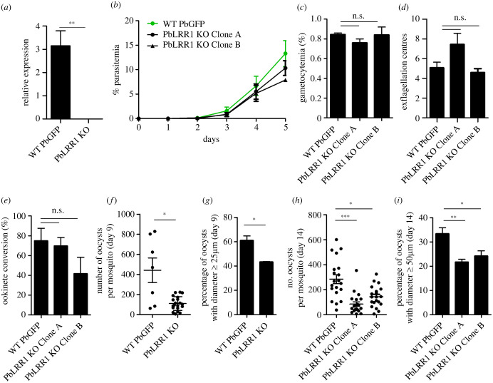
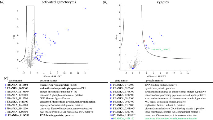
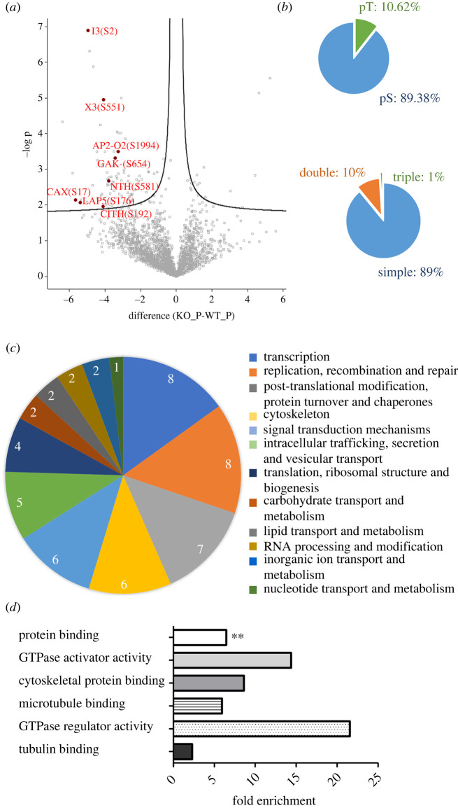
Protein phosphatase 1 (PP1) is a key enzyme for Plasmodium development. However, the detailed mechanisms underlying its regulation remain to be deciphered. Here, we report the functional characterization of the Plasmodium berghei leucine-rich repeat protein 1 (PbLRR1), an orthologue of SDS22, one of the most ancient and conserved PP1 interactors. Our study shows that PbLRR1 is expressed during intra-erythrocytic development of the parasite, and up to the zygote stage in mosquitoes. PbLRR1 can be found in complex with PbPP1 in both asexual and sexual stages and inhibits its phosphatase activity. Genetic analysis demonstrates that PbLRR1 depletion adversely affects the development of oocysts. PbLRR1 interactome analysis associated with phospho-proteomics studies identifies several novel putative PbLRR1/PbPP1 partners. Some of these partners have previously been characterized as essential for the parasite sexual development. Interestingly, and for the first time, Inhibitor 3 (I3), a well-known and direct interactant of Plasmodium PP1, was found to be drastically hypophosphorylated in PbLRR1-depleted parasites. These data, along with the detection of I3 with PP1 in the LRR1 interactome, strongly suggest that the phosphorylation status of PbI3 is under the control of the PP1-LRR1 complex and could contribute (in)directly to oocyst development. This study provides new insights into previously unrecognized PbPP1 fine regulation of Plasmodium oocyst development through its interaction with PbLRR1.

Keywords: PP1; SDS22; inhibitor 3; leucine-rich repeat protein 1; protein phosphatase 1.


Conflict of interest statement

The authors report no conflicts of interest in this work.

Figures

Figure 1.
Figure 1.
Plasmodium berghei Leucine-Rich-Repeat 1 (PbLRR1) LRR domains and structural model. (a) PbLRR1 schematic domain composition showing the 11 predicted Leucine Rich Repeat (LRR) domains fitting the LXXLXLXXNXIXXIXXLXXL/I consensus sequence. Is also represented the LRR cap domain. All predictions were made using Prosite software (). Numbers at the left side indicate the amino acid position at the beginning of each domain. (b) PbLRR1 tertiary structure predicted using I-TASSER and further annotated with Chimera 1.14. Each LRR domain is highlighted with the same colour code used in panel A. This panel shows that PbLRR1 folds into a typical horseshoe shape. (c) PbLRR1/PbPP1c in silico docking analysis. The model was built using HADDOCK 2.2 server (https://milou.science.uu.nl/services/HADDOCK2.2/haddock.php) and the complex displaying the most representative binding mode was kept for further analysis and annotation in Chimera 1.14. The three binding sites are emphasized, as well as the main hydrogen bonds involved in the formation of these sites. (d,e) PbLRR1 sequences corresponding to the PfLRR1 peptides described as able to disrupt PfLRR1/PfPP1c complex by Pierrot et al. [30] are highlighted in red in the PbLRR1 three-dimensional structure model (d) and then placed on the PbLRR1/PbPP1 complex in silico model (highlighted within orange circles (e)). Those binding analyses were carried out using Chimera 1.14.
Figure 2.
Figure 2.
PbLRR1 expression and sub-cellular localization during Plasmodium life cycle. Confocal microscopy images of PbLRR1-AID-3HA (anti-HA mAb, red) expressing parasites in (a) rings, trophozoites, schizont, and in (b) non activated and activated gametocytes (30 min), zygotes (4 h post fertilization) and ookinetes (24 h post fertilization). GFP Parasites (green) nuclei are stained with DAPI (blue). Scale bar, 10 µm. (c) RT-PCR confirming the expression of Pbrr1 mRNA in day 7 and day 9 oocysts (Pr1-Pr2, electronic supplementary material, table S1).
Figure 3.
Figure 3.
Immunoprecipitation assay of the PbLRR1/PbPP1 complex in schizonts, non-activated and activated gametocytes and zygotes. Investigation of the presence of the PbLRR1/PbPP1c complex during various stages of P. berghei development. Protein extracts from schizont, non-activated gametocyte (NAG), activated gametocyte (AG) and zygotes stages expressing mCherry-tagged PbPP1c were immunoprecipitated using anti-RFP antibodies followed by immunoblotting with the same Ab (upper panels) and anti-PfLRR1 antibodies (lower panels). Immunoblots were re-probed after several washings and without stripping.
Figure 4.
Figure 4.
PbLRR1 functional analysis using Xenopus oocytes model. (a) PbLRR1 is expressed in Xenopus oocytes following the micro-injection of its cRNA. Immunoblot analysis of extracts were prepared from micro-injected oocytes with either PbLRR1 cRNA (60 ng in 60 nl, lane 3) or control cRNA (60 ng in 60nl, irrelevant protein, lane 1), or treated by progesterone (4 µg ml−1, PG, lane 2) using an anti-HA mAb. (b) Interaction of PbLRR1 with Xenopus PP1 (XePP1c). Extracts prepared from Xenopus oocytes previously micro-injected with either PbLRR1 cRNA (60 ng in 60nl, lane 3), or control cRNA (60 ng in 60nl, irrelevant protein, lane 1) or treated by a progesterone (4 µg ml−1, PG, lane 2) were immunoprecipitated using an anti-PP1 mAb followed by an immunoblotting using anti-HA mAb (upper panel) and anti-PP1 mAb (lower panel). (c) PbLRR1 can regulate PP1 activity. The appearance of GVBD induced by the micro-injection of PbLRR1 cRNA (60 ng in 60nl, lane 4), irrelevant control protein (60 ng in 60 nl, lane 3, negative control), water for the injection control (60 nl, lane 2) or after a progesterone treatment (4 µg ml−1) for the positive GVBD control (PG, lane 1), was monitored after 15 h. Values are presented as mean percentages and sem (error bars). Each experiment was performed using a set of 20 oocytes and repeated on three animals. (d) Percentage of GVBD induced after the micro-injection in oocyte of the peptide LRR1-1 (VKKKKIKAEIKIIENLQNCKKLRLLELGYNKIRM), LRR1-2 (VKKKKIKAEIKIENYRKTIIHILPQLKQLDAL), the control peptide P0 (VKKKKIKAEIKI), the control peptide P1 (derived from Plasmodium inhibitor 2 PP1 interacting domain ((VKKKKIKREIKKTISWKKTISW)) and its mutated version P10 (VKKKKIKREIKKTASAKKTASA) or with progesterone (PG, as positive control). Each experiment was performed using a set of 8 oocytes and repeated on three animals. (e) Disruption of the PbLRR1/XePP1c interaction after the injections of peptides derived from the PfLRR1 sequence. Oocyte extracts were micro-injected with peptide LRR1-1 (lane 2), LRR1-2 (lane 3), LRR1-1 and LRR1-2 (lane 4), the control peptide P0 (lane 5), or with PBS (lane 6), 1-hour prior PbLRR1 cRNA. Then, the extracts were immunoprecipitated with anti-PP1 mAb and subjected to immunodetection using an anti-HA mAb (upper panel) and anti-PP1 mAb (lower panel). Each experiment was performed using a set of 20 oocytes and repeated on two animals. (f) The same oocyte extracts were immunoprecipitated with an anti-HA and subjected to immunodetection using anti-PP1 mAb (upper panel) and anti-HA mAb (lower panel). (g) The disruption of the PbLRR1/XePP1c complex affect PbLRR1 ability to induce a GVBD. Percentage of GVDB induced by the injection of PbLRR1 cRNA in Xenopus oocytes after the pre-injection of peptides derived from the sequence of PfLRR1 or Pf inhibitor-2 (P1 and P10, used as negative control) as described in (d). The percentages of GVBD induction after peptide pre-injection are: Peptide LRR1-1: 45%, Peptide LRR1-2: 32.50%, Peptides LRR1-1 and LRR1-2: 5.2%, PBS: 85%, Peptide control P0: 85%, Peptide control P1: 80%, Peptide control P10: 80%. *: statistically significant p < 0.05 (Mann–Whitney). Each experiment was performed using a set of 10 oocytes and repeated on three animals.
Figure 5.
Figure 5.
Phenotypic analysis of PbLRR1-depleted (PbLRR1 KO) versus wild-type (WT PbGFP) parasites. (a) pblrr1 mRNA levels in blood stage parasites assessed by RT-qPCR. Values represent mean pblrr1 mRNA levels relative to those of the houskeeping gene arginyl-t RNA ligase (PBANKA_1434200). (b) Asexual blood stage parasite development. (c) Gametocyte development. (d) Male exflagellation. Values represent mean exflagellation centres per field at 15 min post activation (10 fields per experiment). (e) Ookinete conversion. (f) Oocyst numbers at 9 days post mosquito infection (n = 10). (g) Percentage of oocysts with diameters ≥ 25 µm at 9 days post mosquito infection. Based on measurements of 611 oocysts from 3 different mosquitoes (pbGFP), and 464 oocysts from 3 different mosquitoes (PbLRR1KO). (h) Oocysts numbers at 14 days post mosquito infection (n = 20). (i) Percentage of oocysts with diameters ≥ 50 µm at 14 days post mosquito infection. Based on measurements of 3094 oocysts from 11 different mosquitoes (pbGFP), and 1811 oocysts from 20 different mosquitoes (PbLRR1KO). ns: not statistically significant; *: statistically significant p < 0.05; **: statistically significant p < 0.01); ***: statistically significant p < 0.0001) (Mann–Whitney).
Figure 6.
Figure 6.
PbLRR1 interactome analysis. (a) Volcano plot representation of the outcome of the PbLRR1 interactome study carried out in activated gametocytes. The proteins highlighted in blue were identified as PbLRR1 interacting partners (t-test, FDR < 0.01, S0 = 1). (b) Volcano plot representation of the outcome of the PbLRR1 interactome study carried out in zygotes. The proteins highlighted in blue were identified as PbLRR1 interacting partners. The protein highlighted in green was specifically identified at that stage as a PbLRR1 interacting partners (t-test, FDR < 0.01, S0 = 1). The proteins highlighted in brown were identified as partners in the activated gametocytes analysis but not in zygotes. (c) List of PbLRR1 interacting partners with names and PlasmoDB accession numbers. The proteins highlighted in bold were identify as PbLRR1 partners in both analyses. The proteins followed by an asterisk were later identified in the phospho-proteomic analysis (figure 7).
Figure 7.
Figure 7.
PbLRR1KO phospho-proteomic analysis. (a) Volcano plot representation of the outcome of the PbLRR1 KO phospho-proteomic analysis carried out in zygote. A selection of proteins carrying phosphosites with a significant differential phosphorylation status between the KO line and the parental line were highlighted in red (t-test S0 = 0.1, FDR = 0.05). Those proteins have been selected based of their interest in the study and/or their previous characterization in the sexual development of Plasmodium berghei. (b) Distribution of phosphoSer (pS), phospho-Thr (pT) residues and multiplicity of phosphosites detected in the analysis (n = 1138). (c) Functional annotation (based on the COG database) of proteins identified in the analysis with a differential phosphorylation status in the KO line versus parental line. (d) GO terms enrichment analysis. Fold enrichment was performed on the 100 proteins identified as hypophosphorylated in the PbLRR1 phosphoproteome. The x-axis represents the fold enrichment for the indicated biological function (hypergeometric test, Bonferroni correction **: statistically significant p < 0.01).

Similar articles

Cited by

References

    1. Gerald N, Mahajan B, Kumar S. 2011. Mitosis in the human malaria parasite plasmodium falciparum. Eukaryot. Cell 10, 474-482. ( 10.1128/EC.00314-10) - DOI - PMC - PubMed
    1. Francia ME, Striepen B. 2014. Cell division in apicomplexan parasites. Nat. Rev. Microbiol. 12, 125-136. ( 10.1038/nrmicro3184) - DOI - PubMed
    1. White MW, Suvorova ES. 2018. Apicomplexa cell cycles: something old, borrowed, lost, and new. Trends Parasitol. 34, 759-771. ( 10.1016/j.pt.2018.07.006) - DOI - PMC - PubMed
    1. Ruivo MTG, Vera IM, Sales-Dias J, Meireles P, Gural N, Bhatia SN, Mota MM, Mancio-Silva L. 2016. Host AMPK Is a modulator of Plasmodium liver infection. Cell Rep. 16, 2539-2545. ( 10.1016/j.celrep.2016.08.001) - DOI - PMC - PubMed
    1. Baker DA, Matralis AN, Osborne SA, Large JM, Penzo M. 2020. Targeting the malaria parasite cGMP-dependent protein kinase to develop new drugs. Front. Microbiol. 11, 3189. ( 10.3389/fmicb.2020.602803) - DOI - PMC - PubMed

Publication types

Substances