. 2022 Oct;26(4):297.
doi: 10.3892/mmr.2022.12813. Epub 2022 Aug 3.

LncRNA GAS5 promotes spermidine‑induced autophagy through the miRNA‑31‑5p/NAT8L axis in pulmonary artery endothelial cells of patients with CTEPH

Affiliations

LncRNA GAS5 promotes spermidine‑induced autophagy through the miRNA‑31‑5p/NAT8L axis in pulmonary artery endothelial cells of patients with CTEPH

Qinghua Wu et al. Mol Med Rep. 2022 Oct.

Abstract

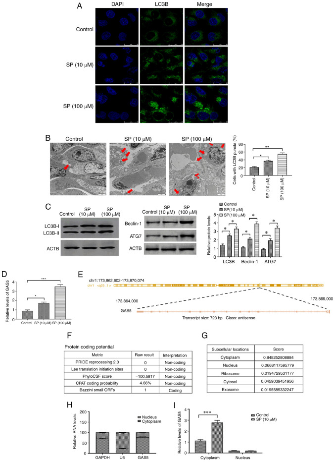
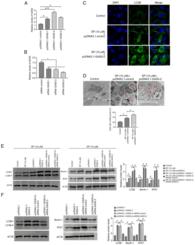
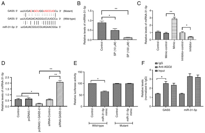
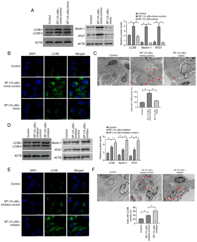
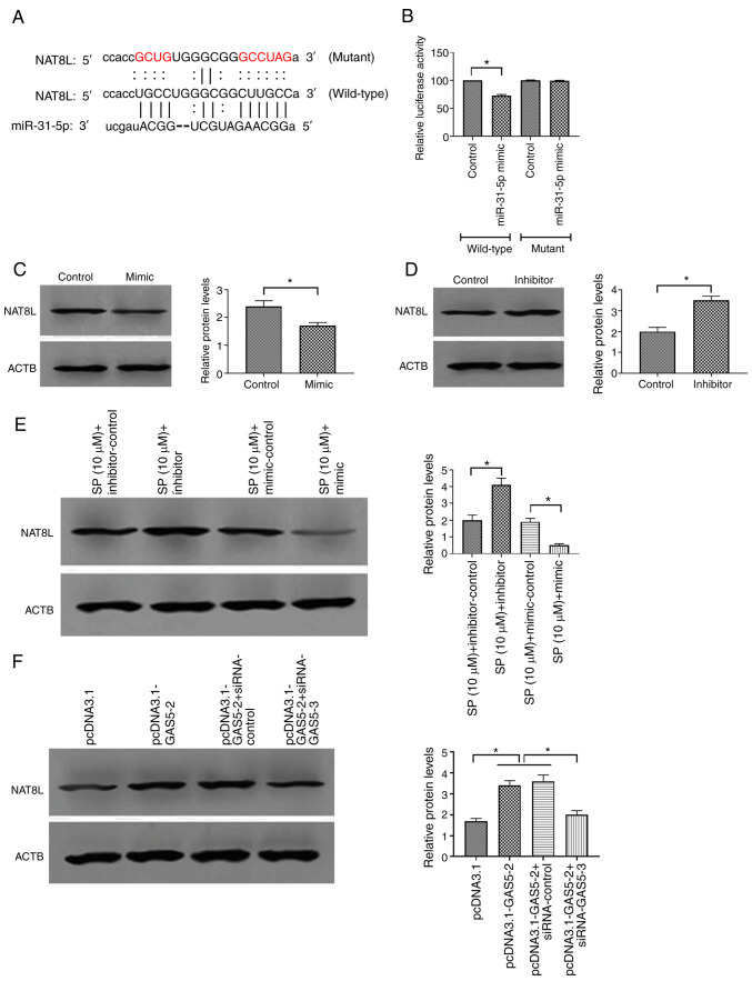
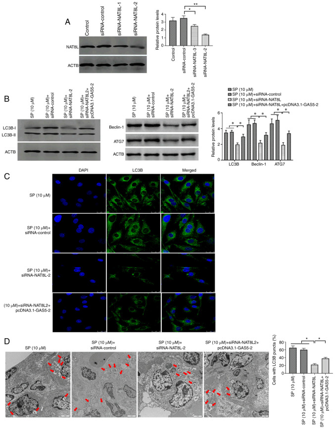
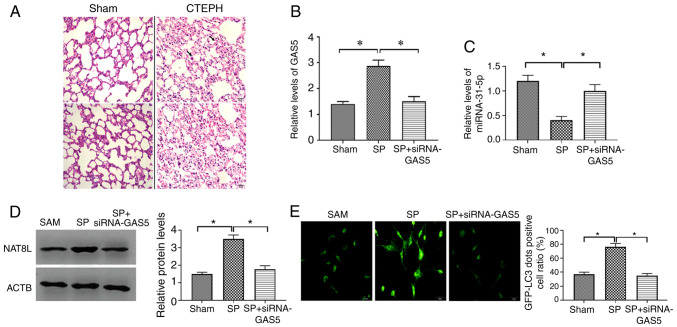
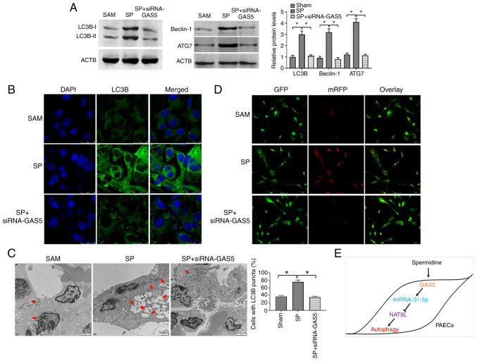
Chronic thromboembolic pulmonary hypertension (CTEPH) is a leading cause of pulmonary hypertension. The present study investigated the mechanisms of long non‑coding RNA growth arrest‑specific transcript 5 (GAS5) on spermidine (SP)‑induced autophagy. Pulmonary artery endothelial cells (PAECs) were collected from patients with CTEPH and the rat model. Immunofluorescence, Western blots, reverse transcription‑quantitative polymerase chain reaction, bioinformatics, rapid amplification of cDNA ends assays, luciferase reporter assays, RNA‑binding protein immunoprecipitation assays, GFP‑LC3 adenoviruses, tfLC3 assays and transmission electron microscopy were performed. The results revealed that SP‑induced autophagy increased GAS5 in PAECs. The upregulation of GAS5 enhanced and the downregulation of GAS5 reversed the roles of SP in PAECs. Furthermore, GAS5 promoted SP‑induced autophagy in PAECs by targeting miRNA‑31‑5p. The miRNA‑31‑5p mimic suppressed and the inhibitor promoted SP‑induced autophagy. Furthermore, N‑Acetyltransferase 8 Like (NAT8L) was a target gene of miRNA‑31‑5p and knockdown of NAT8L inhibited the autophagic levels of PAECs. In vivo, SP treatment decreased miRNA‑31‑5p and increased NAT8L levels, which was reversed by the knockdown of GAS5. The downregulation of GAS5 abolished the stimulatory role of SP in PAECs of CTEPH rats. In conclusion, GAS5 promoted SP‑induced autophagy through miRNA‑31‑5p/NAT8L signaling pathways in vitro and in vivo and GAS5 may be a promising molecular marker for therapies of CTEPH.1.

Keywords: N‑Acetyltransferase 8 Like; autophagy; chronic thromboembolic pulmonary hypertension; lncRNA growth arrest‑specific transcript 5; miRNA‑31‑5p; pulmonary artery endothelial cell; spermidine.


Conflict of interest statement

The authors declare that they have no competining interests.

Figures

Figure 1.
Figure 1.
SP promotes autophagy and increases GAS5 in PAECs. (A) Abundance of LC3B in SP (10 and 100 µM)-treated PAECs, as assessed by immunofluorescence assays (scale bar, 25 µm; magnification, ×20). (B) Autophagic vacuoles in the cellular cytoplasm of SP (10 and 100 µM)-treated PAECs, as evaluated by transmission electron microscopy (scale bar, 2 µm; magnification, ×10,000). Red arrows indicate the autophagic vacuoles. (C) Protein expression of LC3B, Beclin-1 and ATG7 in SP (10 and 100 µM)-treated PAECs were assessed by Western blotting. (D) Expression of GAS5 in PAECs treated with SP (10 and 100 µM). (E) Sequence information of GAS5. (F) Prediction of protein-coding potential of GAS5. (G) Prediction of subcellular location of GAS5. (H) The subcellular location of GAS5 was assessed by nuclear/cytoplasmic RNA separation assays. (I) Effect of SP (10 µM) on the subcellular location of GAS5. *P<0.05; **P<0.01; ***P<0.001. There were at least three replicates in each group available for analysis. SP, spermidine; GAS5, growth arrest-specific transcript 5; PAECs, pulmonary artery endothelial cells.
Figure 2.
Figure 2.
GAS5 promotes SP-induced autophagy in PAECs. (A) Transfection efficiency of pcDNA3.1-GAS5 in PAECs. (B) Transfection efficiency of siRNA-GAS5 in PAECs. (C) The abundance of LC3B in PAECs treated with the combination of SP (10 µM) and pcDNA3.1-GAS5, as assessed by immunofluorescence assays (scale bar, 25 µm; magnification, ×20). (D) Autophagic vacuoles in the cellular cytoplasm of PAECs treated with the combination of SP (10 µM) and pcDNA3.1-GAS5, as evaluated by transmission electron microscopy (scale bar, 2 µm; magnification, ×10,000). Red arrows indicate the autophagic vacuoles. (E) Protein expressions of LC3B, Beclin-1 and ATG7 in SP (10 µM)-treated PAECs transfected with pcDNA3.1-GAS5 and the combination of pcDNA3.1-GAS5 and siRNA-GAS5, as assessed by Western blotting. (F) Abundance of LC3B in PAECs transfected with pcDNA3.1-GAS5 and the combination of pcDNA3.1-GAS5 and siRNA-GAS5, as assessed by Western blotting. *P<0.05. There were at least three replicates in each group available for analysis. GAS5, growth arrest-specific transcript 5; SP, spermidine; PAECs, pulmonary artery endothelial cells; siRNA, small interfering RNA.
Figure 3.
Figure 3.
GAS5 promotes SP-induced autophagy in PAECs through targeting miRNA-31-5p. (A) Putative binding site of miRNA-31-5p in 3′-UTR of GAS5. (B) Effect of SP (10 µM) on the expression of miRNA-31-5p. (C) Transfection efficiency of miRNA-31-5p mimic and inhibitor. (D) Effect of pcDNA3.1-GAS5 and siRNA-GAS5 on the expression of miRNA-31-5p. (E) Relative luciferase activity in PAECs co-transfected with mutant/wild-type plasmids containing the putative binding site of miRNA-31-5p in GAS5 and miRNA-31-5p mimic. (F) Physical communication between GAS5 and miRNA-31-5p in PAECs assessed by RNA binding protein immunoprecipitation assays. *P<0.05; **P<0.01. There were at least three replicates in each group available for analysis. GAS5, growth arrest-specific transcript 5; SP, spermidine; PAECs, pulmonary artery endothelial cells; siRNA, small interfering RNA.
Figure 4.
Figure 4.
MiRNA-31-5p inhibits SP-induced autophagy in PAECs. (A) Abundance of LC3B in PAECs treated the combination of SP (10 µM) and miRNA-31-5p mimic, as assessed by Western blotting. (B) Protein expression of LC3B, Beclin-1 and ATG7 in PAECs treated with the combination of SP (10 µM) and miRNA-31-5p mimic, as assessed by immunofluorescence assays (scale bar, 25 µm; magnification, ×20). (C) Autophagic vacuoles in the cellular cytoplasm of PAECs treated the combination of SP (10 µM) and miRNA-31-5p mimic, as evaluated by transmission electron microscopy (scale bar, 2 µm; magnification, ×10,000). Red arrows indicate the autophagic vacuoles. (D) Protein expression of LC3B, Beclin-1 and ATG7 in PAECs treated with the combination of SP (10 µM) and miRNA-31-5p inhibitor, as assessed by Western blotting. (E) The abundance of LC3B in PAECs treated with the combination of SP (10 µM) and miRNA-31-5p inhibitor, as assessed by immunofluorescence assays (scale bar, 25 µm; magnification, ×20). (F) Autophagic vacuoles in the cellular cytoplasm of PAECs treated with the combination of SP (10 µM) and miRNA-31-5p inhibitor, as evaluated by transmission electron microscopy (scale bar, 2 µm; magnification, ×10,000). Red arrows indicate the autophagic vacuoles. *P<0.05. There were at least three replicates in each group available for analysis. SP, spermidine; PAECs, pulmonary artery endothelial cells.
Figure 5.
Figure 5.
MiRNA-31-5p inhibits SP-induced autophagy in PAECs through targeting NAT8L. (A) The putative binding site of miRNA-31-5p in 3′-UTR of NAT8L. (B) Relative luciferase activity in PAECs co-transfected with mutant/wild-type plasmids containing the putative binding site of miRNA-31-5p in NAT8L and miRNA-31-5p mimic. (C and D) Effect of miRNA-31-5p mimic and inhibitor on the protein expression of NAT8L in control PAECs. (E) Effect of miRNA-31-5p mimic and inhibitor on the protein expression of NAT8L in control PAECs treated with SP (10 µM). (F) Protein expression of NAT8L in PAECs transfected with pcDNA3.1-GAS5 and the combination of pcDNA3.1-GAS5 and siRNA-GAS5, as assessed by Western blotting. *P<0.05. There were at least three replicates in each group available for analysis. SP, spermidine; PAECs, pulmonary artery endothelial cells.
Figure 6.
Figure 6.
NAT8L promotes SP-induced autophagy in PAECs. (A) Transfection efficiency of siRNA-NAT8L in PAECs. (B) Protein expression of LC3B, Beclin-1 and ATG7 in PAECs transfected with siRNA-NAT8L and the combination of siRNA-NAT8L and pcDNA3.1-GAS5, as assessed by Western blotting. (C) The abundance of LC3B in PAECs, as assessed by immunofluorescence assays (scale bar, 25 µm; magnification, ×20). (D) Autophagic vacuoles in the cellular cytoplasm of PAECs, as evaluated by transmission electron microscopy (scale bar, 2 µm; magnification, ×10,000). Red arrows indicate the autophagic vacuoles. *P<0.05; **P<0.01. There were at least three replicates in each group available for analysis. SP, spermidine; PAECs, pulmonary artery endothelial cells; siRNA, small interfering RNA.
Figure 7.
Figure 7.
GAS5/miRNA-31-5p/NAT8L axis is associated with SP-induced autophagy in PAECs in vivo. (A) Representative hematoxylin and eosin staining images of proximal pulmonary vascular tissues. (Scale bar, 20 µm; magnification, ×40). (B) Expression of GAS5 in PAECs of CTEPH rats treated with SP and the combination of SP and siRNA-GAS5. (C) Expression of miRNA-31-5p in PAECs of rats with CTEPH treated with SP and the combination of SP and siRNA-GAS5. (D) Protein expression of NAT8L in PAECs of rats with CTEPH treated with SP and the combination of SP and siRNA-GAS5. (E) The percentage of GFP-LC3 positive PAECs as assessed by GFP-LC3 adenovirus assays. (Scale bar, 50 µm; magnification, ×20). *P<0.05. There were at least three replicates in each group available for analysis. GAS5, growth arrest-specific transcript 5; SP, spermidine; PAECs, pulmonary artery endothelial cells; siRNA, small interfering RNA; CTEPH, chronic thromboembolic pulmonary hypertension.
Figure 8.
Figure 8.
NAT8L regulates SP-induced autophagy in PAECs in vivo. (A) Protein expression of LC3B, Beclin-1 and ATG7 in PAECs of CTEPH rats treated with SP and the combination of SP and siRNA-GAS5. (B) The abundance of LC3B in PAECs of rats with CTEPH treated with SP and the combination of SP and siRNA-GAS5, as assessed by immunofluorescence assays (scale bar: 25 µm; magnification, ×20). (C) Autophagic vacuoles in the cellular cytoplasm of PAECs of CTEPH rats treated with SP and the combination of SP and siRNA-GAS5, as evaluated by transmission electron microscopy (scale bar, 2 µm; magnification, ×10,000). Red arrows indicate the autophagic vacuoles. (D) GFP signals in PAECs as assessed by tfLC3 assays. (Scale bar, 50 µm; magnification, ×20). (E) Schematic of GAS5 regulating PAECs autophagy induced by SP. *P<0.05. There were at least three replicates in each group available for analysis. SP, spermidine; PAECs, pulmonary artery endothelial cells; siRNA, small interfering RNA; CTEPH, chronic thromboembolic pulmonary hypertension.

Similar articles

Cited by

References

    1. Pengo V, Lensing AW, Prins MH, Marchiori A, Davidson BL, Tiozzo F, Albanese P, Biasiolo A, Pegoraro C, Iliceto S, et al. Incidence of chronic thromboembolic pulmonary hypertension after pulmonary embolism. N Engl J Med. 2004;350:2257–2264. doi: 10.1056/NEJMoa032274. - DOI - PubMed
    1. Becattini C, Agnelli G, Pesavento R, Silingardi M, Poggio R, Taliani MR, Ageno W. Incidence of chronic thromboembolic pulmonary hypertension after a first episode of pulmonary embolism. Chest. 2006;130:172–175. doi: 10.1378/chest.130.1.172. - DOI - PubMed
    1. Sakao S, Tatsumi K. Crosstalk between endothelial cell and thrombus in chronic thromboembolic pulmonary hypertension: Perspective. Histol Histopathol. 2013;28:185–193. - PubMed
    1. Lang IM, Pesavento R, Bonderman D, Yuan JX. Risk factors and basic mechanisms of chronic thromboembolic pulmonary hypertension: A current understanding. Eur Respir J. 2013;41:462–468. doi: 10.1183/09031936.00049312. - DOI - PubMed
    1. Jin Y, Choi AM. Cross talk between autophagy and apoptosis in pulmonary hypertension. Pulm Circ. 2012;2:407–414. doi: 10.4103/2045-8932.105029. - DOI - PMC - PubMed