. 2022 Aug;11(8):561-574.
doi: 10.1302/2046-3758.118.BJR-2021-0576.R1.

Tendon healing: a concise review on cellular and molecular mechanisms with a particular focus on the Achilles tendon

Affiliations

Tendon healing: a concise review on cellular and molecular mechanisms with a particular focus on the Achilles tendon

Gundula G Schulze-Tanzil et al. Bone Joint Res. 2022 Aug.

Abstract

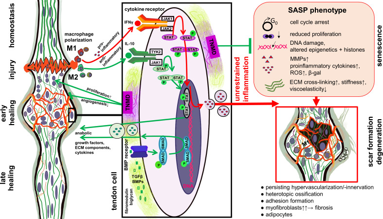
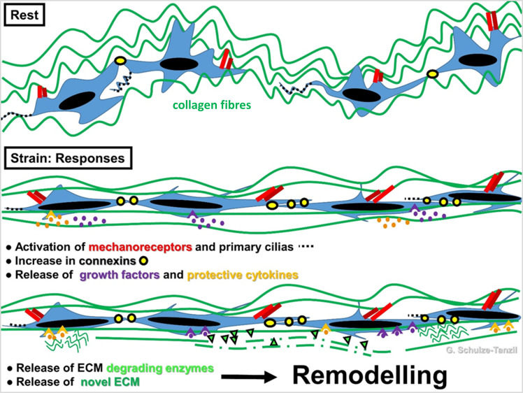
Tendon is a bradytrophic and hypovascular tissue, hence, healing remains a major challenge. The molecular key events involved in successful repair have to be unravelled to develop novel strategies that reduce the risk of unfavourable outcomes such as non-healing, adhesion formation, and scarring. This review will consider the diverse pathophysiological features of tendon-derived cells that lead to failed healing, including misrouted differentiation (e.g. de- or transdifferentiation) and premature cell senescence, as well as the loss of functional progenitors. Many of these features can be attributed to disturbed cell-extracellular matrix (ECM) or unbalanced soluble mediators involving not only resident tendon cells, but also the cross-talk with immigrating immune cell populations. Unrestrained post-traumatic inflammation could hinder successful healing. Pro-angiogenic mediators trigger hypervascularization and lead to persistence of an immature repair tissue, which does not provide sufficient mechano-competence. Tendon repair tissue needs to achieve an ECM composition, structure, strength, and stiffness that resembles the undamaged highly hierarchically ordered tendon ECM. Adequate mechano-sensation and -transduction by tendon cells orchestrate ECM synthesis, stabilization by cross-linking, and remodelling as a prerequisite for the adaptation to the increased mechanical challenges during healing. Lastly, this review will discuss, from the cell biological point of view, possible optimization strategies for augmenting Achilles tendon (AT) healing outcomes, including adapted mechanostimulation and novel approaches by restraining neoangiogenesis, modifying stem cell niche parameters, tissue engineering, the modulation of the inflammatory cells, and the application of stimulatory factors.Cite this article: Bone Joint Res 2022;11(8):561-574.

Keywords: Achilles tendon; Cell plasticity; Tendon healing; Tendon-derived stem cells; extracellular matrix; inflammation; neoangiogenesis; stem cells; stiffness; strength; tendons; tissue engineering.


Figures

Fig. 1
Fig. 1
Macroscopical anatomy of the Achilles tendon (AT). a) Scheme of a dorsal view. b) and c) Dorsolateral views: b) dissection photograph; and c) scheme of the AT, bursae, fat pads, and enthesis zones (inset). a) and c) The images were created by G. G. Schulze-Tanzil using Krita 4.1.7 (Krita Foundation, The Netherlands).
Fig. 2
Fig. 2
Scheme of the microscopic anatomy of the Achilles tendon. The stem cell niches are numbered (1: within the tendon proper; 2: within the epi/paratenon; interfascicular niches comprise 3a: perivascular; and 3b: niches within the wall of small vessels, containing pericytes). The image was created by G. G. Schulze-Tanzil using Krita 4.1.7 (Krita Foundation, The Netherlands).
Fig. 3
Fig. 3
Role of Janus kinase signal transducer and activator of transcription (JAK/STAT) and bone morphogenetic protein (BMP) pathways in tendon healing and degeneration. Simplified scheme of selected signalling pathways involved in tendon healing. The balance of M1 and M2 macrophage polarization plays a central role in resolving the inflammatory phase and affects the outcome of tendon healing. Proinflammatory cytokines released during dominant and prolonged M1 macrophage polarization stimulate molecular factors released in response of the JAK/STAT pathway activation, which can trigger the senescence-associated secretory phenotype (SASP) shift of tendon cells. SASP is associated with degenerative features in healing tendons. Mediators released during M2 macrophage polarization, including anti-inflammatory cytokines such as interleukin (IL)-10, stimulate other parts of the JAK/STAT pathway. Proteoglycans such as biglycan and fibromodulin can bind and stabilize growth factors activating the BMP pathway. Tenomodulin produced by the tendon cells can exert pro-proliferative effect as well as protective roles against cellular senescence and unrestrained angiogenesis. The image was created by G. G. Schulze-Tanzil using Krita 4.1.7 (Krita Foundation, The Netherlands). β-gal, β-galactosidase; ECM, extracellular matrix; IFNγ, interferon γ; MMP, matrix metalloproteinase; ROS, reactive oxygen species; TGFβ, transforming growth factor β; TNMD, tenomodulin.
Fig. 4
Fig. 4
Scheme of tenocyte mechanoresponse. Stretching at a physiological level leads to activation of mechanoreceptor and primary cilias. Crimping of the collagen fibre bundles disappears (in biomechanical measurement: toe region of a stress-stain curve). Connexin expression is elevated and hence, cell–cell signalling via gap junctions is also elevated. Protective cytokines and anabolic growth factors are released followed by de novo extracellular matrix (ECM) synthesis and ECM-degrading enzyme release, which mediate reorganization of ECM by a remodelling process to adapt the ECM biomechanics according to the stretch direction. The image was created by G. G. Schulze-Tanzil using Krita 4.1.7 (Krita Foundation, The Netherlands).

Similar articles

Cited by

References

    1. O’Brien M . The anatomy of the achilles tendon . Foot Ankle Clin . 2005. ; 10 ( 2 ): 225 – 238 . 10.1016/j.fcl.2005.01.011 - DOI - PubMed
    1. Ballal MS , Walker CR , Molloy AP . The anatomical footprint of the achilles tendon: a cadaveric study . Bone Joint J . 2014. ; 96-B ( 10 ): 1344 – 1348 . 10.1302/0301-620X.96B10.33771 - DOI - PubMed
    1. Doral MN , Alam M , Bozkurt M , et al. . Functional anatomy of the Achilles tendon . Knee Surg Sports Traumatol Arthrosc . 2010. ; 18 ( 5 ): 638 – 643 . 10.1007/s00167-010-1083-7 - DOI - PubMed
    1. Szaro P , Witkowski G , Smigielski R , Krajewski P , Ciszek B . Fascicles of the adult human achilles tendon - an anatomical study . Ann Anat . 2009. ; 191 ( 6 ): 586 – 593 . 10.1016/j.aanat.2009.07.006 - DOI - PubMed
    1. Hess GW . Achilles tendon rupture: a review of etiology, population, anatomy, risk factors, and injury prevention . Foot Ankle Spec . 2010. ; 3 ( 1 ): 29 – 32 . 10.1177/1938640009355191 - DOI - PubMed