

Structural insights into the assembly and activation of the IL-27 signaling complex
- PMID: 35920255
- PMCID: PMC9535766
- DOI: 10.15252/embr.202255450
Structural insights into the assembly and activation of the IL-27 signaling complex
Abstract
Interleukin 27 (IL-27) is a heterodimeric cytokine that elicits potent immunosuppressive responses. Comprised of EBI3 and p28 subunits, IL-27 binds GP130 and IL-27Rα receptor chains to activate the JAK/STAT signaling cascade. However, how these receptors recognize IL-27 and form a complex capable of phosphorylating JAK proteins remains unclear. Here, we used cryo electron microscopy (cryoEM) and AlphaFold modeling to solve the structure of the IL-27 receptor recognition complex. Our data show how IL-27 serves as a bridge connecting IL-27Rα (domains 1-2) with GP130 (domains 1-3) to initiate signaling. While both receptors contact the p28 component of the heterodimeric cytokine, EBI3 stabilizes the complex by binding a positively charged surface of IL-27Rα and Domain 1 of GP130. We find that assembly of the IL-27 receptor recognition complex is distinct from both IL-12 and IL-6 cytokine families and provides a mechanistic blueprint for tuning IL-27 pleiotropic actions.
Keywords: GP130; IL-27; cryo electron microscopy; cytokine.
© 2022 The Authors. Published under the terms of the CC BY 4.0 license.
Figures
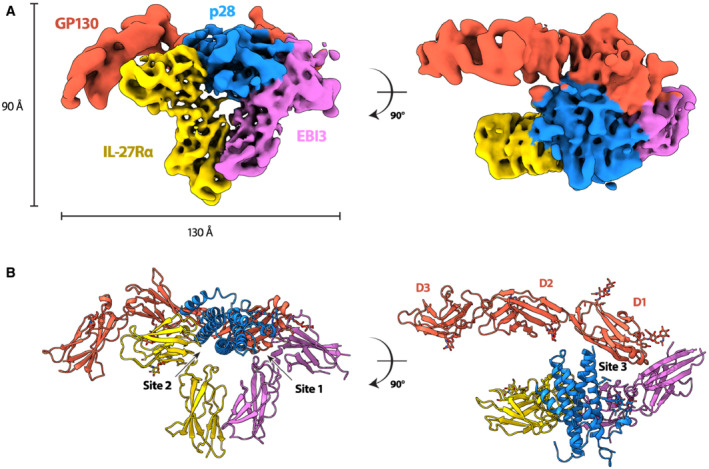
- A, B
CryoEM reconstruction (A) and atomic model (B) of the IL‐27 receptor recognition complex. The complex consists of the IL‐27 heterodimeric cytokine, p28 (blue) and EBI3 (purple), bound to two signaling receptors, GP130 (red) and IL‐27Rα (yellow). EBI3 occupies site 1 on p28, while IL‐27Rα occupies site 2. GP130 binds p28 at site 3 via domains 1 and 2 (D1, D2). The canonical cytokine recognition site on GP130 between domains D2 and D3 is unoccupied.
- A
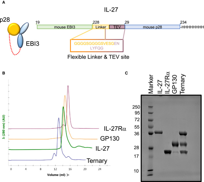
Schematic representation of the single chain IL‐27 heterodimer.
- B
Chromatogram overlay from size exclusion chromatography of the different IL‐27 complex components.
- C
Coomassie‐stained SDS‐PAGE analysis under reducing conditions of the individual components plus the IL‐27:IL‐27Rα:GP130 complex used for cryoEM studies.
- A
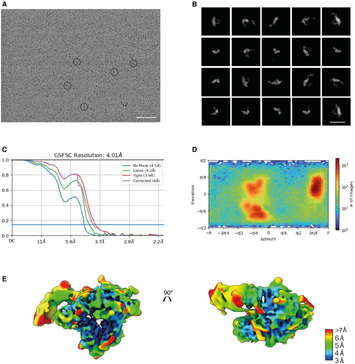
Raw cryoEM micrograph with examples of the IL‐27 receptor complex circled. Scale bar 50 nm.
- B
A subset of representative 2D class averages. Scale bar 110 Å.
- C
Gold standard Fourier shell correlation (GSFSC) for the final reconstruction. The resolution at the 0.143 cutoff is reported.
- D
Angular distribution plot.
- E
IL‐27 receptor recognition reconstruction filtered according to local resolution ranging from 3 Å (blue) to 7–15 Å (red).
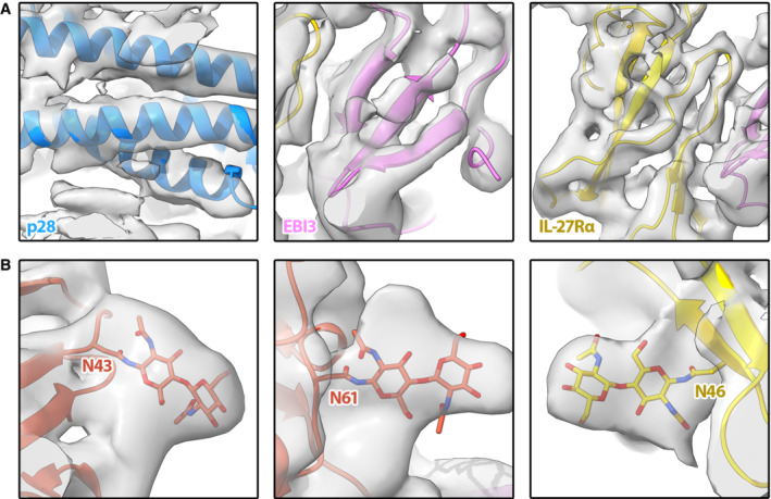
- A
Map/model overlays for density corresponding to p28 (left panel), EBI3 (middle panel), and IL‐27Rα (right panel).
- B
Representative glycan density in the map corresponding to known glycosylation sites on GP130 (left and middle panels) and IL‐27Rα (right panel).
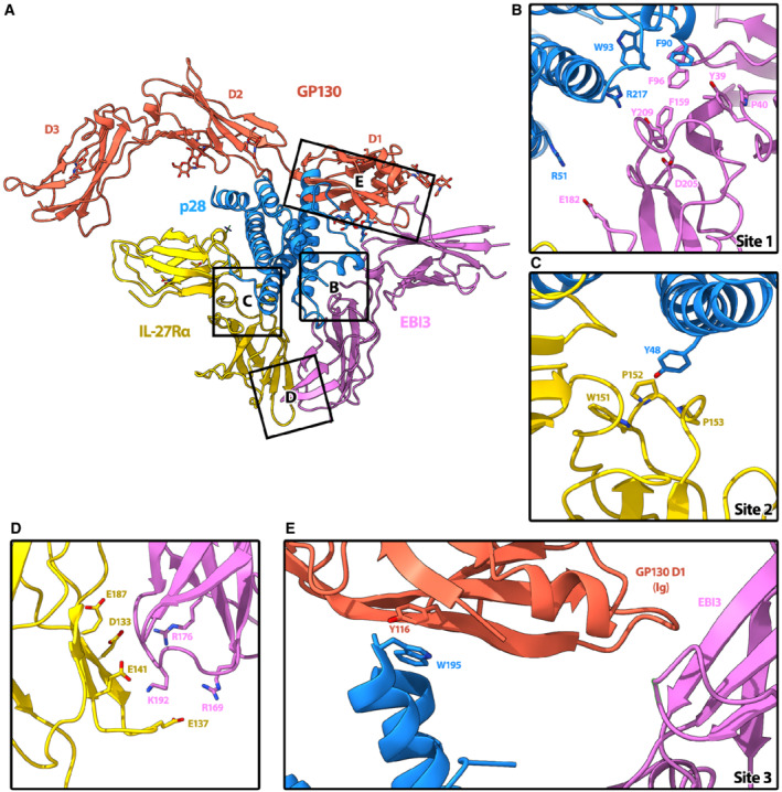
- A
Overview of IL‐27 interactions with signaling receptors. Ribbon representation of the complex colored according to protein components: p28 (blue), EBI3 (purple), IL‐27Rα (yellow), GP130 (red). Individual domains of GP130 (D1, D2, and D3) are labeled.
- B
The hinge between the two CHR domains of EBI3 form a hydrophobic groove (Y39, P40, F96, F159, and Y209) that is filled by W93 of p28. Residue EBI3: D205, which is important for assembly of the heterodimeric cytokine (Rousseau et al, 2010), is also present in the binding interface and could form a salt bridge with p28:R217.
- C
IL‐27Rα binds site 2 of p28 at the apex of the elbow between its two CHR domains. The loops of IL‐27Rα form a pocket comprised of residues IL‐27Rα: W151, P152, and P153 in which the aromatic side chain of p28:Y48 could slot into.
- D
The orientation of IL‐27Rα at site 2 is stabilized by a second interaction interface with EBI3, which is dominated by electrostatic complementarity between the two domains (IL‐27Rα: E187, D133, E141, E137, and EBI3:R176, R169, K192).
- E
Site 3 of p28 is occupied by the bend between the CHR domain 2 (D2) and the immunoglobulin (Ig) domain D1 of GP130. While this interface is not well resolved, p28:W195, which is essential for GP130‐mediated signaling (Rousseau et al, 2010), is facing D1. The key residues that mediate the interactions at interfaces are shown as sticks.
- A–E
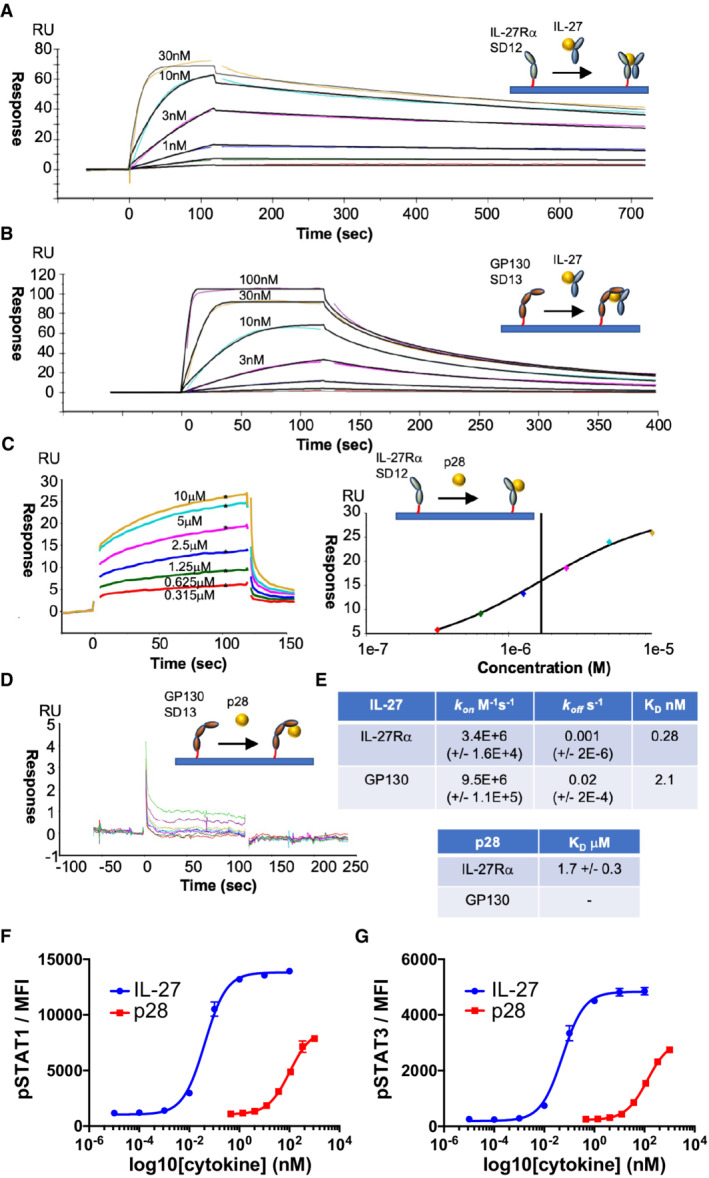
For SPR measurements, IL‐27Rα or GP130 was immobilized on the chip surface by biotin–streptavidin interaction and IL‐27 or p28 was flowed across the chip in solution. SPR data are representative of three biological replicates. Kinetic charts for IL‐27Rα (A) and GP130 (B). Concentrations used are shown on the curves. Data traces were fitted using a 1:1 interaction model (black) to quantify the kinetics (kon, koff) and binding affinity (KD) of the interactions. (C, D) Equilibrium chart for p28 binding to IL‐27Rα (C, left panel) and curve‐fitting to data points generated at various concentrations of p28 (C, right panel). (D) Equilibrium chart for p28 binding GP130. (E) Table presenting kinetically derived (IL‐27) and thermodynamically derived (p28) binding constants. Standard Error values (SEs) are shown in parentheses.
- F, G
Dose response curves for pSTAT1 (F) and pSTAT3 (G) in resting mouse CD8+ T cells. Cells were stimulated with mIL‐27 or p28 for 15 min with the indicated doses. Data shown are the mean of four biological replicates with error bars depicting standard error of the mean.
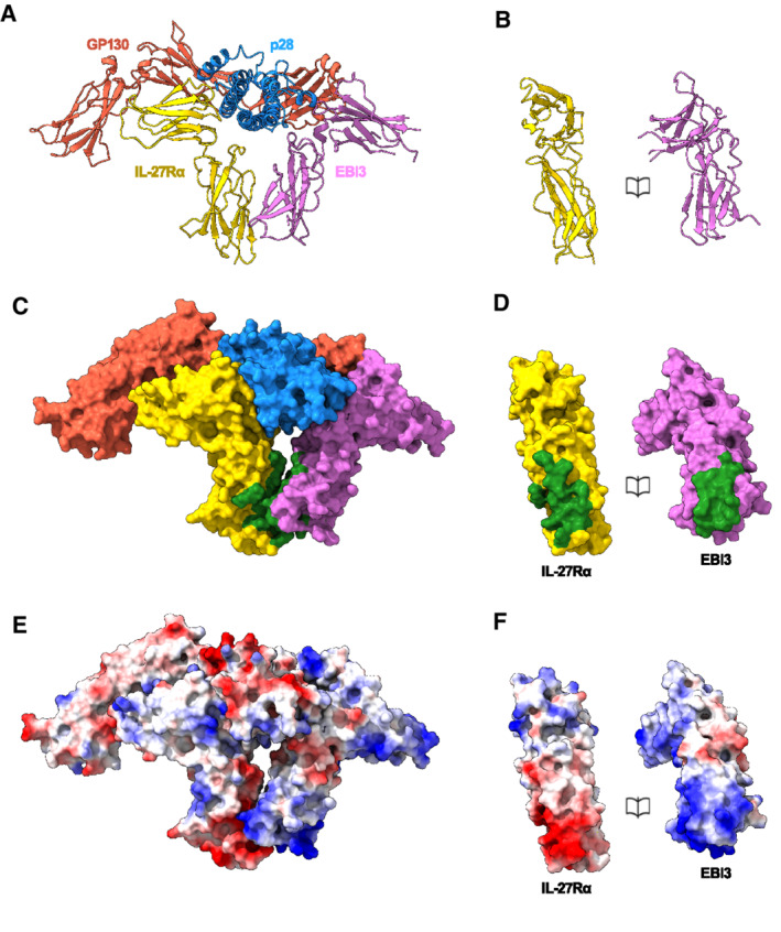
- A–F
Ribbon representation of the IL‐27 receptor recognition complex (A) and book representation of the EBI3:IL‐27Rα interface (B). Individual proteins colored: EBI3 (purple), IL‐27Rα (yellow), p28 (blue), GP130 (red). Surface representation of the complex (C) and book representation of the interface (D). Proteins colored as in (A) with EBI3: IL‐27Rα interface residues in green. Coulombic electrostatic potential ranging from −10 (red) to 10 (blue) kcal/(mole) calculated from the models in A (E) and corresponding book representation of the interface (F).
- A
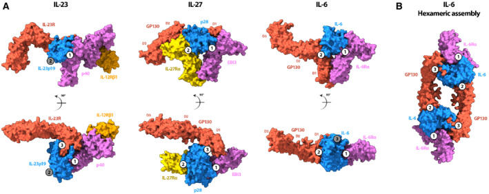
(left panel) Surface rendering of the IL‐23 receptor recognition complex (Data ref: Glassman et al, 2021a) colored according to protein components: IL‐23 receptor (red), IL‐12β (p40) (purple), IL‐23p19 (blue), IL‐12Rβ1 (orange). A comparison with IL‐27 receptor complex (middle panel) shows that both GP130 (red) and IL‐23 receptor occupy site 3 of their respective cytokines, orienting domains 2 and 3 similarly towards the membrane. Both EBI3 (purple) and the other component of the IL‐23 heterocytokine, p40, occupy the site 1 epitope. Surface rendering of the IL‐6 receptor recognition complex (Data ref: Boulanger et al, c) colored according to protein components: IL‐6 (blue), IL‐6Rα (purple), GP130 (red; right panel). A comparison with the IL‐27 receptor complex shows that the nonsignaling components of both complexes (EBI3 and IL‐6Rα) occupy site 1, while GP130 binds in a different way. For IL‐6, GP130 binds site 2 through an interaction site located between the CHR domains, D2 and D3. Instead, GP130 binds site 3 of the IL‐27 cytokine between domains D1 and D2, leaving the canonical cytokine binding site unoccupied.
- B
Surface rendering of the hexameric IL‐6 assembly (PDB: 1P9M) colored according to protein components: IL‐6 (blue), IL‐6Rα (purple), GP130 (red). GP130 bound to site 2 of one IL‐6 molecule bridges a second cytokine by binding at site 3 to stabilize the complex.
References
-
- Boulanger MJ, Bankovich AJ, Kortemme T, Baker D, Garcia KC (2003a) Convergent mechanisms for recognition of divergent cytokines by the shared signaling receptor gp130. Mol Cell 12: 577–589 - PubMed
-
- Boulanger MJ, Chow D, Brevnova EE, Garcia KC (2003b) Hexameric structure and assembly of the interleukin‐6/IL‐6 α‐receptor/gp130 complex. Science 300: 2101–2104 - PubMed
Publication types
MeSH terms
Substances
Associated data
- Actions
- Actions
Grants and funding
LinkOut - more resources
Full Text Sources
Other Literature Sources
Molecular Biology Databases
