Loop nerve graft prefabrication for peripheral nerve defect reconstruction
- PMID: 35920436
- PMCID: PMC10315981
- DOI: 10.14744/tjtes.2022.68353
Loop nerve graft prefabrication for peripheral nerve defect reconstruction
Abstract
Background: Delayed autologous nerve graft reconstruction is inevitable in devastating injuries. Delayed or prolonged repair time has deleterious effects on nerve grafts. We aimed improving and accelerating nerve graft reconstruction process in a rat long nerve defect model with loop nerve graft prefabrication particularly to utilize for injuries with tissue loss.
Methods: Twenty-four Sprague-Dawley rats were allocated into three groups. 1.5 cm long peroneal nerve segment was excised, reversed in orientation, and used as autologous nerve graft. In conventional interpositional nerve graft group (Group 1), nerve defects were repaired in single-stage. In loop nerve graft prefabrication group (Group 2), grafts were sutured end-to-end (ETE) to the proximal peroneal nerve stumps. Distal ends of the grafts were sutured end-to-side to the peroneal nerve stumps 5 mm proximal to the ETE repair sites in first stage. In second stage, distal ends of the prefabricated grafts were transposed and sutured to distal nerve stumps. In staged conventional interpositional nerve graft group (Group 3), grafts were sutured ETE to proximal peroneal nerve stumps in first stage. Distal ends of the grafts and nerve stumps were tacked to the surrounding muscles until the final repair in second stage. Follow-up period was 4 weeks for each stage in Groups 2 and 3, and 8 weeks for Group 1. Peroneal function index (PFI), electrophysiology, and histological assessments were conducted after 8 weeks. P<0.05 was considered significant for statistical analysis.
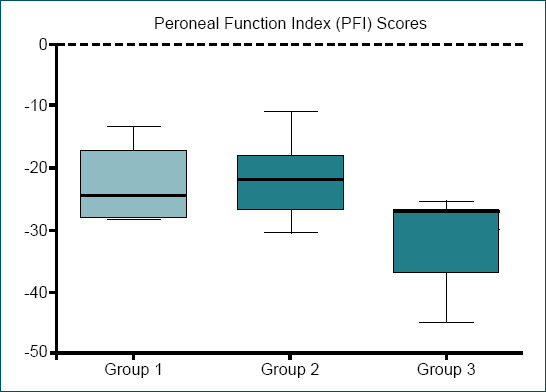
Results: PFI results of Group 1 (-22.75±5.76) and 2 (-22.08±6) did not show statistical difference (p>0.05). Group 3 (-33.64±6.4) had a statistical difference compared to other groups (p<0.05). Electrophysiology results of Group 1 (16.19±2.15 mV/1.16±0.21 ms) and 2 (15.95±2.82 mV/1.17±0.16 ms) did not present statistical difference (p>0.05), whereas both groups had a statistical difference compared to Group 3 (10.44±1.96 mV/1.51±0.15 ms) (p<0.05). Axon counts of Group 1 (2227±260.4) and 3 (2194±201.1) did not have statistical difference (p>0.05), whereas both groups had significantly poor axon counts compared to Group 2 (2531±91.18) (p<0.05).
Conclusion: Loop nerve graft prefabrication improved axonal regeneration without delay. Loop prefabrication can accelerate prolonged regeneration time for the injuries indicating a delayed nerve reconstruction. Higher axon counts derived with loop nerve prefabrication may even foster its investigation in immediate long nerve defect reconstructions in further studies.
Conflict of interest statement
Figures


Similar articles
-
Targeted mesenchymal stem cell and vascular endothelial growth factor strategies for repair of nerve defects with nerve tissue implanted autogenous vein graft conduits.Microsurgery. 2016 Oct;36(7):578-585. doi: 10.1002/micr.22401. Epub 2015 Apr 13. Microsurgery. 2016. PMID: 25867169
-
Comparison of regeneration results of prefabricated nerve graft, autogenous nerve graft, and vein graft in repair of nerve defects.Microsurgery. 2009;29(2):138-43. doi: 10.1002/micr.20586. Microsurgery. 2009. PMID: 18942646
-
End-to-Side vs. Free Graft Nerve Reconstruction-Experimental Study on Rats.Int J Mol Sci. 2023 Jun 21;24(13):10428. doi: 10.3390/ijms241310428. Int J Mol Sci. 2023. PMID: 37445608 Free PMC article.
-
A rat study of the use of end-to-side peripheral nerve repair as a "babysitting" technique to reduce the deleterious effect of chronic denervation.J Neurosurg. 2018 Sep 14;131(2):622-632. doi: 10.3171/2018.3.JNS172357. J Neurosurg. 2018. PMID: 30215557
-
Pedicled Vascularized Common Peroneal Nerve Graft in Sciatic Nerve Reconstruction With Involvement of Inner Pelvic Lumbar and Sacral Nerve Roots: A Case Report and Literature Review.Microsurgery. 2025 May;45(4):e70064. doi: 10.1002/micr.70064. Microsurgery. 2025. PMID: 40285651 Review.
References
-
- Valero-Cabré A, Tsironis K, Skouras E, Navarro X, Neiss WF. Peripheral and spinal motor reorganization after nerve injury and repair. J Neurotrauma. 2004;21:95–108. - PubMed
-
- Eren F, Öksüz S, Küçükodaci Z, Kendirli MT, Cesur C, Alarçin E, et al. Targeted mesenchymal stem cell and vascular endothelial growth factor strategies for repair of nerve defects with nerve tissue implanted autogenous vein graft conduits. Microsurgery. 2016;36:578–85. - PubMed
-
- Siemionow M, Bozkurt M, Zor F. Regeneration and repair of peripheral nerves with different biomaterials:Review. Microsurgery. 2010;30:574–88. - PubMed
-
- Mackinnon SE, Colbert SH. Principles and techniques of peripheral nerve repair, grafts, and transfers. In: Thorne CH, editor. Grabb and Smith's Plastic Surgery. 7th ed. Philadelphia: Lippincott Williams &Wilkins; 2014. pp. 77–86.
-
- Birch R. Nerve repair. In: Wolfe SW, editor. Green's operative hand surgery. 6th ed. Philadelphia: Churchill Livingstone; 2011. pp. 1035–74.
MeSH terms
LinkOut - more resources
Full Text Sources