

Six-year post-surgical evaluation in the treatment protocols in the dental arches of children with oral cleft: longitudinal study
- PMID: 35920507
- PMCID: PMC9586431
- DOI: 10.1590/1678-7757-2022-0120
Six-year post-surgical evaluation in the treatment protocols in the dental arches of children with oral cleft: longitudinal study
Abstract
Objective: Oral cleft surgical repairs are performed using different techniques worldwide. To evaluate and compare the development of the dental arches of children with unilateral cleft lip and palate before and after the primary surgeries performed with different techniques at the first months and six years of life.
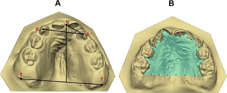
Methodology: This is a retrospective longitudinal study. The sample comprised 56 dental casts divided int the following groups: Group 1 (G1) - cheiloplasty (Millard technique) at three months and one-step palatoplasty (von Langenbeck technique) at 12 months; and Group 2 (G2) - cheiloplasty (Millard technique) and two-step palatoplasty: anterior hard palate closure (Hans Pichler technique) at three months and posterior soft palate closure (Sommerlad technique) at 12 months. The digitized dental casts were evaluated at three months - pre-surgical (T1) and six years of life- post-surgical (T2). The following linear measurements were analyzed: intercanine (C-C'), intertuberosity (T-T') distances; anterior dental arch (I-CC'), anterior intersegment (I-C'), and total arch (I-TT') lengths. The palate area was also measured. Parametric and non-parametric tests were applied (p<0.05).
Results: In G1, the intragroup comparison showed statistically significant smaller I-CC' and I-C' at T2 (p=0.001 and p<0.001, respectively), while T-T', I-TT', and area comparisons were significantly greater (p<0.001, p=0.002, and p<0.001, respectively). In G2, the intragroup comparison exhibited statistically significant smaller C-C' and I-C' at T2 (p=0.004, for both), whereas T-T', I-TT' and area comparisons were significantly greater (p<0.001, p=0.004, and p<0.001, respectively). At T2, the intergroup analysis revealed that G1 had a statistically significant smaller I-CC' (p=0.014). The analysis of the intergroup differences (∆=T2-T1) showed that G1 had a statistically smaller I-CC' (p=0.043).
Conclusion: The two-step palatoplasty showed a more favorable prognosis for the maxillary growth than one-step palatoplasty in children with oral clefts.
Conflict of interest statement
Conflict of interest
The authors declare no conflict of interest.
Figures


References
-
- Freitas JA, Neves LT, Almeida AL, Garib DG, Trindade-Suedam IK, Yaedú RY, et al. Rehabilitative treatment of cleft lip and palate: experience of the Hospital for Rehabilitation of Craniofacial Anomalies/USP (HRAC/USP)--Part 1: overall aspects. J Appl Oral Sci. 2012;20(1):9-15. doi: 10.1590/s1678-7757201200010000
-
- Hoffmannova E, Moslerová V, Dupej J, Borský J, Bejdová Š, Velemínská J. Three-dimensional development of the upper dental arch in unilateral cleft lip and palate patients after early neonatal cheiloplasty. Int J Pediatr Otorhinolaryngol. 2018;109:1-6. doi: 10.1016/j.ijporl.2018.03.009 - PubMed
-
- Haque S, Khamis MF, Alam MK, Ahmad WM. Effects of multiple factors on treatment outcome in the three-dimensional maxillary arch morphometry of children with unilateral cleft lip and palate. J Craniofac Surg. 2020;31(6):e534-8. doi: 10.1097/SCS.0000000000006464 - PubMed
-
- Ambrosio EC, Sforza C, Menezes M, Carrara CF, Soares S, Machado MA, et al. Prospective cohort 3D study of dental arches in children with bilateral orofacial cleft: assessment of volume and superimposition. Int J Paediatr Dent. 2021;31(5):606-12. doi: 10.1111/ipd.12731 - PubMed
MeSH terms
LinkOut - more resources
Full Text Sources
Medical
