. 2022 Aug 24;96(16):e0070322.
doi: 10.1128/jvi.00703-22. Epub 2022 Aug 3.

Passive Immunization with a Single Monoclonal Neutralizing Antibody Protects against Cutaneous and Mucosal Mouse Papillomavirus Infections

Affiliations

Passive Immunization with a Single Monoclonal Neutralizing Antibody Protects against Cutaneous and Mucosal Mouse Papillomavirus Infections

Sarah A Brendle et al. J Virol. .

Abstract

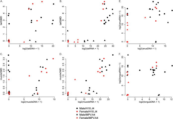
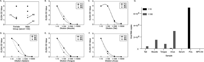
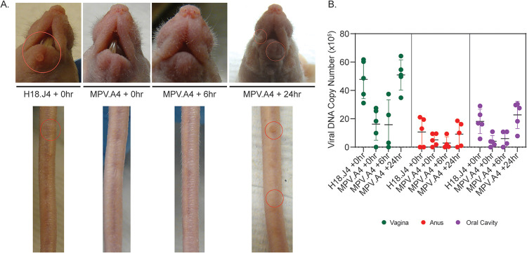
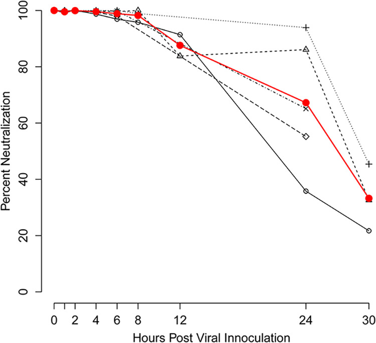
We have established a mouse papillomavirus (MmuPV1) model that induces both cutaneous and mucosal infections and cancers. In the current study, we use this model to test our hypothesis that passive immunization using a single neutralizing monoclonal antibody can protect both cutaneous and mucosal sites at different time points after viral inoculation. We conducted a series of experiments involving the administration of either a neutralizing monoclonal antibody, MPV.A4, or control monoclonal antibodies to both outbred and inbred athymic mice. Three clinically relevant mucosal sites (lower genital tract for females and anus and tongue for both males and females) and two cutaneous sites (muzzle and tail) were tested. At the termination of the experiments, all tested tissues were harvested for virological analyses. Significantly lower levels of viral signals were detected in the MPV.A4-treated female mice up to 6 h post-viral inoculation compared to those in the isotype control. Interestingly, males displayed partial protection when they received MPV.A4 at the time of viral inoculation, even though they were completely protected when receiving MPV.A4 at 24 h before viral inoculation. We detected MPV.A4 in the blood starting at 1 h and up to 8 weeks postadministration in some mice. Parallel to these in vivo studies, we conducted in vitro neutralization using a mouse keratinocyte cell line and observed complete neutralization up to 8 h post-viral inoculation. Thus, passive immunization with a monoclonal neutralizing antibody can protect against papillomavirus infection at both cutaneous and mucosal sites and is time dependent. IMPORTANCE This is the first study testing a single monoclonal neutralizing antibody (MPV.A4) by passive immunization against papillomavirus infections at both cutaneous and mucosal sites in the same host in the mouse papillomavirus model. We demonstrated that MPV.A4 administered before viral inoculation can protect both male and female athymic mice against MmuPV1 infections at cutaneous and mucosal sites. MPV.A4 also offers partial protection at 6 h post-viral inoculation in female mice. MPV.A4 can be detected in the blood from 1 h to 8 weeks after intraperitoneal (i.p.) injection. Interestingly, males were only partially protected when they received MPV.A4 at the time of viral inoculation. The failed protection in males was due to the absence of neutralizing MPV.A4 at the infected sites. Our findings suggest passive immunization with a single monoclonal neutralizing antibody can protect against diverse papillomavirus infections in a time-dependent manner in mice.

Keywords: anogenital tract; cutaneous; cutaneous infection; in situ analysis; in vivo; monoclonal antibody; mouse papillomavirus (MmuPV1); mucosal; mucosal infections; neutralizing; oral cavity; papillomavirus; passive immunization; sex difference; sexually transmitted diseases.


Conflict of interest statement

The authors declare no conflict of interest.

Figures

FIG 1
FIG 1
MPV.A4 provided complete protection at both cutaneous and mucosal sites. HSD:NU nude mice were i.p. injected with either an anti-MmuPV1 monoclonal antibody, MPV.A4, or the control antibody H11.B2 24 h before viral inoculation. Complete protection was observed in the MPV.A4-treated mice at cutaneous tail and muzzle sites (A), and significantly lower viral DNA copy numbers were detected at all three tested mucosal sites (B) (P < 0.05, Wilcoxon rank sum tests). Photographs and lavages were conducted biweekly, and the data shown are from week 6 after viral inoculation. (C) Tissues were collected at week 6 after viral inoculation. Viral DNA was detected in vaginal, tongue, and anal tissues of the control group by ISH (×20;, scale bar = 100 μm), while no viral DNA was detected in the MPV.A4-treated mice (not shown). A representative ISH tissue section is presented.
FIG 2
FIG 2
Absence of viral activities was found in the inoculated cutaneous and mucosal sites of the female mice treated with MPV.A4 at the time of inoculation. Female Hsd:NU nude mice were injected intraperitoneally with MPV.A4 or control antibody (H18.J4) at the time of viral inoculation. Both cutaneous (tail and muzzle) and mucosal (vagina, anus and tongue) tissues were further analyzed for viral RNA(A)/DNA(B) by qPCR and in situ assays. No viral RNA (A) or DNA (B) was detected by qPCR in MPV.A4-treated mice. Three out of three females treated with MPV.A4 (D) at the time of inoculation were absent of visible muzzle and tail lesions, while three out of three H18.J4-treated mice developed visible lesions at both muzzle and tail (C). Viral E4 proteins (E, F) were absent in both cutaneous and mucosal tissues of MPV.A4-treated female mice (F), while these viral proteins were readily detected in both cutaneous and mucosal tissues of the control (H18.J4)-treated mice (E).
FIG 3
FIG 3
MPV.A4 provided minimal protection for males at both cutaneous and mucosal sites. Male Hsd:NU mice were injected intraperitoneally with anti-MmuPV1 monoclonal antibody MPV.A4 or an isotype control (H18.J4) at the time of viral inoculation. Two out of three males treated with MPV.A4 (B) at the time of inoculation were absent of visible muzzle and tail lesions, while three out of three H18.J4-treated mice developed visible lesions at both muzzle and tail (A). The experiment was repeated, and two out of three males receiving MPV.A4 were absent of visible lesions at the tail and muzzle while all H18.J4-treated mice developed visible lesions. Significantly smaller lesions were found in the infected tissues of MPV.A4-treated when compared to those of H18.J4-treated male mice (C, P < 0.05, Wilcoxon rank sum tests). Viral RNA (E) and viral DNA (F) were quantitated by qPCR in harvested tissues at the termination of the experiment (week 5 after viral inoculation). Squares represent experiment 1, and circles represent experiment 2. The tongue tissues showed very low viral RNA/DNA by qPCR in the H18.J4 mice. To further determine the viral presence in these mice, we conducted additional in situ hybridization on the second half of infected tongue and detected viral DNA, RNA, and E4 protein by in situ hybridization, RNA in situ hybridization, and immunohistochemistry, respectively (D).
FIG 4
FIG 4
Correlation among viral RNA, DNA levels, and lesion size. Viral RNA and DNA levels in the infected tail (A and B) and muzzle (D and E) tissues were tested against the lesion size. As shown in panels A and D, viral DNA levels in tail and muzzle were higher in large lesions. Viral RNA levels (B and E) correlate well with lesion size and better than viral DNA (A and D). However, RNA and DNA did not correlate highly in both tail (C) and muzzle (F) tissues as well as those in mucosal sites (data not shown).
FIG 5
FIG 5
Antibody detection after intraperitoneal delivery in the Hsd:NU athymic nude mice. Five female (red) and five male (blue) Hsd:NU athymic nude mice received 0.2 mg MPV.A4 and 0.04 mg H18.J4 mixture intraperitoneally. At 1, 6, and 24 h postadministration, blood samples were collected for MPV.A4 and H18.J4 detection by ELISA. All five females and four out of five males were positive for both MPV.A4 (A) and H18.J4 (B) at 1 h postinjection, suggesting the antibodies were transferred to the circulating system quickly.
FIG 6
FIG 6
Antibody detection in wounded tissues after intraperitoneal delivery. We further tested the serum (A) and local wounded tissues (tail, muzzle, tongue, and anus) as well as the spleens. They were collected at 24 h posttreatment for three animals (F3, M1, and M5). Two positive serum samples (F3 and M5) showed comparable MPV.A4 levels in all tested tissues (B to F), while the negative sample, M1, was also negative for MPV.A4 in all tested tissues. (G) Viral RNA was absent completely in most tested sites at a higher concentration (1:10) except the anus, which was also the least protected site among all.
FIG 7
FIG 7
A lower dose of MPV.A4 only provided partial protection at both cutaneous and mucosal sites of females. Three groups of Hsd:NU nude females (n = 3) were injected at the tail vein with MPV.A4 (high dose, 0.3 mg/mouse, and low dose, 0.03 mg/mouse) or an isotype control (H18.J4, 0.3 mg/mouse) at the time of viral inoculation. No visible lesions were observed in three out of three females in both the high-dose (A and B) and low-dose (C and D) MPV.A4-treated mice, while all control mice grew visible lesions at both muzzle and tail sites (E and F). Viral E4 proteins were detected in the control tail (G) and vaginal (H) tissues of H18.J4-treated mice and absent in corresponding tissues of both high-dose (0.3 mg) and low-dose (0.03 mg) MPV.A4-treated mice (I). Lower but not significantly lower levels of viral DNA were detected at all mucosal sites (vagina, anus, and tongue), even in the lower-dose group, than in the control group (P > 0.05, Wilcoxon rank sum tests). (J) Viral RNA was quantitated by qPCR in harvested tissues, and significantly lower levels of viral RNA were not found in the low-dose MPV.A4-treated groups at two cutaneous sites and vaginal tissues compared with those in the H18.J4-treated groups (P > 0.05, Wilcoxon rank sum tests). The tongue and anal tissues showed very low levels of viral RNA in both MPV.A4 and control mice.
FIG 8
FIG 8
Partial protection was observed in the MPV.A4-treated group up to 6 h post-viral inoculation. NU/J nude mice (n = 5) were injected in the tail vein with MPV.A4 or an isotype control (H18.J4) at 0, 6, and 24 h after viral inoculation. (A) Visible lesions were observed at tail and muzzle sites of the H18.J4 control group, but not in the MPV.A4-treated animals at 5 weeks post-viral inoculation. (B) Viral DNA from the lavage fluid collected at week 5 from individual animals was detected at three mucosal sites. A significant difference was found between the control and 0- and 6-h post-viral inoculation groups at the vaginal and oral sites (P < 0.05, Wilcoxon rank sum tests). No significant difference was detected at the anal sites (P > 0.05, Wilcoxon rank sum tests).
FIG 9
FIG 9
Significantly lower levels of viral RNA and DNA were detected in tissues of the MPV.A4-treated group up to 6 h post-viral inoculation. Viral RNA (A) and viral DNA (B) in the infected tissues were quantified by qPCR, and MAb MPV.A4-treated animals were compared to Mab H18.J4 (control)-treated animals. Significance among the groups was calculated based on Wilcoxon rank sum tests, and a P value of <0.05 was considered significant.
FIG 10
FIG 10
MPV.A4 protected mouse keratinocytes against MmuPV1 inoculation up to 8 h post-viral inoculation. In vitro postattachment neutralization of MmuPV1 by MPV.A4 was conducted in mouse keratinocyte cell line K38. K38 cells were infected with MmuPV1 at 4°C. At different time points (1 to 30 h post-viral inoculation), K38 cells were washed, and 10 μg/mL MPV.A4 or negative controls was added (H16.V5 or H11.B2). The cells were harvested at 48 to 72 h post-viral inoculation, and RNA was extracted for MmuPV1 detection. E1^E4 transcripts were quantitated by qRT-PCR and plotted as a percentage of neutralization of MmuPV1 controls.

Similar articles

Cited by

  • Monitoring mouse papillomavirus-associated cancer development using longitudinal Pap smear screening.
    Atkins HM, Uslu AA, Li JJ, Shearer DA, Brendle SA, Han C, Kozak M, Lopez P, Nayar D, Balogh KK, Abendroth C, Copper J, Cheng KC, Christensen ND, Zhu Y, Avril S, Burgener AD, Murooka TT, Hu J. Atkins HM, et al. mBio. 2024 Aug 14;15(8):e0142024. doi: 10.1128/mbio.01420-24. Epub 2024 Jul 16. mBio. 2024. PMID: 39012151 Free PMC article.
  • Immune Responses in Oral Papillomavirus Clearance in the MmuPV1 Mouse Model.
    Brendle SA, Li JJ, Walter V, Schell TD, Kozak M, Balogh KK, Lu S, Christensen ND, Zhu Y, El-Bayoumy K, Hu J. Brendle SA, et al. Pathogens. 2023 Dec 14;12(12):1452. doi: 10.3390/pathogens12121452. Pathogens. 2023. PMID: 38133335 Free PMC article.
  • Intraperitoneal delivery of cannabidiol (CBD) and Δ9-tetrahydocannabinol (THC) promotes papillomavirus infections in athymic nude mice.
    Brendle SA, Li J, Sun D, Zhu J, Henderson-Redmond AN, Morgan DJ, Balogh KK, Covington D, Shearer DA, Hu J. Brendle SA, et al. Tumour Virus Res. 2025 Jun;19:200307. doi: 10.1016/j.tvr.2024.200307. Epub 2024 Dec 16. Tumour Virus Res. 2025. PMID: 39694192 Free PMC article.
  • FLT3L governs the development of partially overlapping hematopoietic lineages in humans and mice.
    Momenilandi M, Lévy R, Sobrino S, Li J, Lagresle-Peyrou C, Esmaeilzadeh H, Fayand A, Le Floc'h C, Guérin A, Della Mina E, Shearer D, Delmonte OM, Yatim A, Mulder K, Mancini M, Rinchai D, Denis A, Neehus AL, Balogh K, Brendle S, Rokni-Zadeh H, Changi-Ashtiani M, Seeleuthner Y, Deswarte C, Bessot B, Cremades C, Materna M, Cederholm A, Ogishi M, Philippot Q, Beganovic O, Ackermann M, Wuyts M, Khan T, Fouéré S, Herms F, Chanal J, Palterer B, Bruneau J, Molina TJ, Leclerc-Mercier S, Prétet JL, Youssefian L, Vahidnezhad H, Parvaneh N, Claeys KG, Schrijvers R, Luka M, Pérot P, Fourgeaud J, Nourrisson C, Poirier P, Jouanguy E, Boisson-Dupuis S, Bustamante J, Notarangelo LD, Christensen N, Landegren N, Abel L, Marr N, Six E, Langlais D, Waterboer T, Ginhoux F, Ma CS, Tangye SG, Meyts I, Lachmann N, Hu J, Shahrooei M, Bossuyt X, Casanova JL, Béziat V. Momenilandi M, et al. Cell. 2024 May 23;187(11):2817-2837.e31. doi: 10.1016/j.cell.2024.04.009. Epub 2024 May 3. Cell. 2024. PMID: 38701783 Free PMC article.

References

    1. Benevolo M, Dona MG, Ravenda PS, Chiocca S. 2016. Anal human papillomavirus infection: prevalence, diagnosis and treatment of related lesions. Expert Rev Anti Infect Ther 14:465–477. 10.1586/14787210.2016.1174065. - DOI - PubMed
    1. Shigeishi H, Sugiyama M. 2016. Risk factors for oral human papillomavirus infection in healthy individuals: a systematic review and meta-analysis. J Clin Med Res 8:721–729. 10.14740/jocmr2545w. - DOI - PMC - PubMed
    1. Gravitt PE, Winer RL. 2017. Natural history of HPV infection across the lifespan: role of viral latency. Viruses 9:267. 10.3390/v9100267. - DOI - PMC - PubMed
    1. Khoury R, Sauter S, Butsch Kovacic M, Nelson AS, Myers KC, Mehta PA, Davies SM, Wells SI. 2018. Risk of human papillomavirus infection in cancer-prone individuals: what we know. Viruses 10:47. 10.3390/v10010047. - DOI - PMC - PubMed
    1. Roden RBS, Stern PL. 2018. Opportunities and challenges for human papillomavirus vaccination in cancer. Nat Rev Cancer 18:240–254. 10.1038/nrc.2018.13. - DOI - PMC - PubMed

Publication types

LinkOut - more resources