. 2022 Aug;76(4):153-163.
doi: 10.1007/s10858-022-00402-3. Epub 2022 Aug 3.

The measurement of binding affinities by NMR chemical shift perturbation

Affiliations

The measurement of binding affinities by NMR chemical shift perturbation

Billy Hobbs et al. J Biomol NMR. 2022 Aug.

Abstract

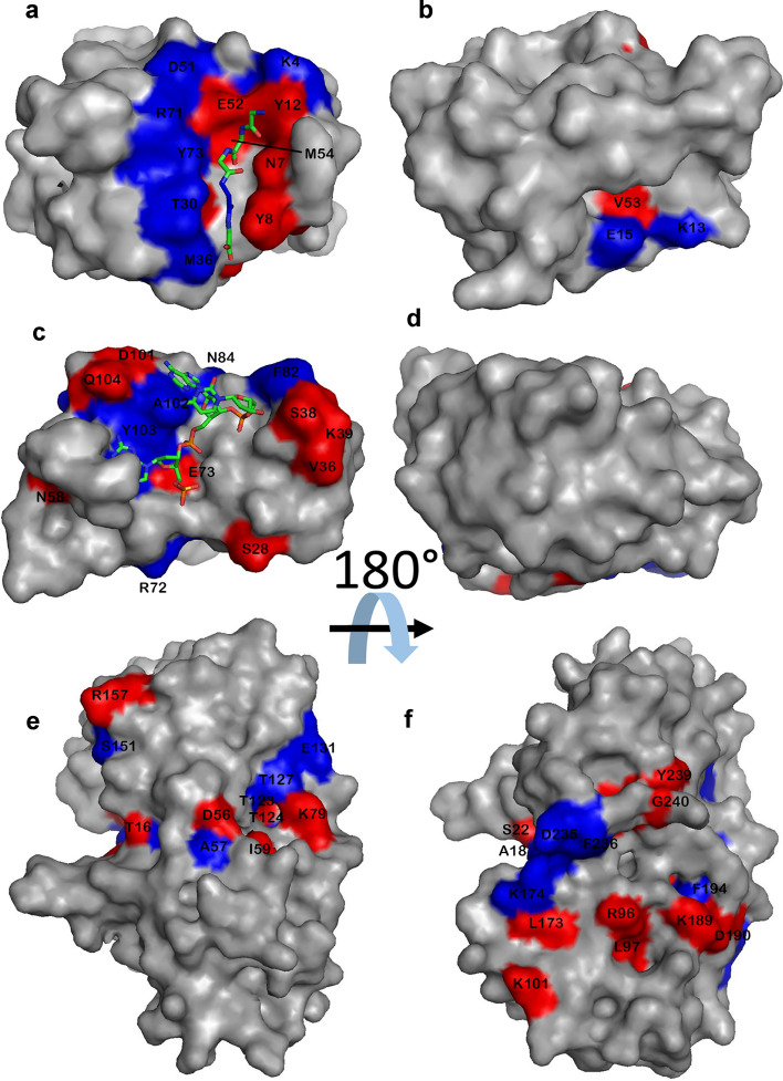
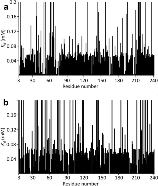
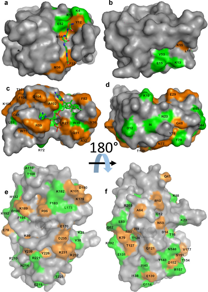
We have carried out chemical shift perturbation titrations on three contrasting proteins. The resulting chemical shifts have been analysed to determine the best way to fit the data, and it is concluded that a simultaneous fitting of all raw shift data to a single dissociation constant is both the most accurate and the most precise method. It is shown that the optimal weighting of 15N chemical shifts to 1H chemical shifts is protein dependent, but is around the consensus value of 0.14. We show that chemical shift changes of individual residues can be fit to give residue-specific affinities. Residues with affinities significantly stronger than average are found in close contact with the ligand and are suggested to form a rigid contact surface, but only when the binding involves little conformational change. This observation may be of value in analysing binding and conformational change.

Keywords: Affinity; Binding; Chemical shift; Conformational change; Dissociation constant; NMR.


Conflict of interest statement

The authors declare that they have no financial interests. The datasets generated during the current study are available from the corresponding author on reasonable request.

Figures

Fig. 1
Fig. 1
15N HSQC titration data for a SH3b b Barnase c HisJ. In each case, peaks are colored from red to violet with addition of ligand. Signals undergoing large shift changes are labelled
Fig. 2
Fig. 2
The locations of large chemical shift changes on titration of a, b SH3b c, d barnase e, f HisJ with their ligands. In each case, the right panel is a rotation of 180° around a horizontal axis. Blue is used to denote negative (upfield) chemical shift changes in H or N, and red for positive (downfield) changes. For SH3b and barnase, the ligand is denoted by sticks. For HisJ the ligand is completely buried and is in the center of the protein. The shift changes shown include approximately 20% of the amino acids in the protein that have reliably fitted shift changes. Residues undergoing large shift changes are labelled
Fig. 3
Fig. 3
Typical chemical shift titration data: HisJ, showing change in chemical shift with addition of the ligand lysine. The curves are individual best fits to the data. a Shift changes in 1H. Data are shown for T77 (red: fitted to Kd of 62 ± 6 μM), D14 (blue: fitted to Kd of 49 ± 6 μM), and G129 (black, fitted to Kd of 54 ± 12 μM). b Shift changes in 15N. Data are shown for F194 (red: fitted to Kd of 56 ± 5 μM), G171 (blue: fitted to Kd of 60 ± 11 μM), and T198 (black: fitted to Kd of 58 ± 13 μM). All data are shown as positive shift changes for ease of presentation. The actual shift changes for T77, D14, F194 and T198 are negative
Fig. 4
Fig. 4
Kd values fitted for HisJ binding to lysine, for a 1H b 15N. Data are shown for all residues that could be fitted. A small number are not shown, mainly because of overlap or because they are prolines. Note that the Kd values are truncated at 0.2 mM: many of the truncated residues are much larger than this
Fig. 5
Fig. 5
Kd values fitted for HisJ binding to lysine, after filtering out unreliable values, for a 1H b 15N. Nuclei were removed if they had a fitted Kd of > 200 μM, total 1H shift changes of < 0.03 ppm, or total 15N changes of < 0.15 ppm. It was not necessary to filter out very small Kd values because these were removed by the chemical shift limits
Fig. 6
Fig. 6
Residues fitting to Kd values significantly different from the mean (defined as outside the 99% confidence intervals), for a SH3b b barnase, c HisJ. Residues with Kd values significantly smaller (stronger) than the mean are in orange, and residues significantly larger (weaker) are in green, and are labelled. In each case, the right panel is a rotation of 180° around a horizontal axis. For SH3b and barnase the ligand is shown as sticks; for HisJ the protein surface is drawn slightly transparent, allowing the bound histidine ligand to be seen in the center of the protein. For SH3b the ligand shown is GGGGG, using the crystal structure with PDB ID 5leo. The ligand used here is YGGGGG, where the additional tyrosine is at the N-terminal (top, in the view shown in a) end of the peptide
Fig. 7
Fig. 7
A simple model for flexible protein/ligand binding. Both the protein and the ligand may have internal flexibility (a). When they bind, the resulting complex has lost this flexibility (d). The process of binding may thus be separated conceptually into a rigid docking of part of the ligand (a, b), with an affinity K1, followed by an induced-fit type rearrangement accompanied by loss of flexibility (bd). The equilibrium constant for this process is K2, which has values between 0 and 1, but may typically be expected to be greater than 0.5. Alternatively, the binding can be modelled as a loss of internal degrees of freedom (ac) followed by a rigid docking (c, d)

References

    1. Arai M, Ferreon JC, Wright PE. Quantitative analysis of multisite protein-ligand interactions by NMR: Binding of intrinsically disordered p53 transactivation subdomains with the TAZ2 domain of CBP. J Am Chem Soc. 2012;134:3792–3803. doi: 10.1021/ja209936u. - DOI - PMC - PubMed
    1. Buckle AM, Fersht AR. Subsite binding in an RNAse: structure of a barnase tetranucleotide complex at 1.76 Å resolution. Biochemistry. 1994;33:1644–1653. doi: 10.1021/bi00173a005. - DOI - PubMed
    1. Bye JW, Baxter NJ, Hounslow AM, Falconer RJ, Williamson MP. Molecular mechanism for the Hofmeister effect derived from NMR and DSC measurements on barnase. ACS Omega. 2016;1:669–679. doi: 10.1021/acsomega.6b00223. - DOI - PMC - PubMed
    1. Cioffi M, Hunter CA, Pandya M, Packer MJ, Williamson MP. Use of quantitative 1H NMR chemical shift changes for ligand docking into barnase. J Biomol NMR. 2009;43:11–19. doi: 10.1007/s10858-008-9286-7. - DOI - PubMed
    1. Eaton JD, Williamson MP (2017) Multi-site binding of epigallocatechin gallate to human serum albumin measured by NMR and isothermal titration calorimetry. Biosci Rep. 10.1042/BSR20170209 - PMC - PubMed

MeSH terms