. 2022 Sep 1;79(9):907-919.
doi: 10.1001/jamapsychiatry.2022.2075.

Exploring Links Between Psychosis and Frontotemporal Dementia Using Multimodal Machine Learning: Dementia Praecox Revisited

Collaborators, Affiliations

Exploring Links Between Psychosis and Frontotemporal Dementia Using Multimodal Machine Learning: Dementia Praecox Revisited

Nikolaos Koutsouleris et al. JAMA Psychiatry. .

Abstract

Importance: The behavioral and cognitive symptoms of severe psychotic disorders overlap with those seen in dementia. However, shared brain alterations remain disputed, and their relevance for patients in at-risk disease stages has not been explored so far.

Objective: To use machine learning to compare the expression of structural magnetic resonance imaging (MRI) patterns of behavioral-variant frontotemporal dementia (bvFTD), Alzheimer disease (AD), and schizophrenia; estimate predictability in patients with bvFTD and schizophrenia based on sociodemographic, clinical, and biological data; and examine prognostic value, genetic underpinnings, and progression in patients with clinical high-risk (CHR) states for psychosis or recent-onset depression (ROD).

Design, setting, and participants: This study included 1870 individuals from 5 cohorts, including (1) patients with bvFTD (n = 108), established AD (n = 44), mild cognitive impairment or early-stage AD (n = 96), schizophrenia (n = 157), or major depression (n = 102) to derive and compare diagnostic patterns and (2) patients with CHR (n = 160) or ROD (n = 161) to test patterns' prognostic relevance and progression. Healthy individuals (n = 1042) were used for age-related and cohort-related data calibration. Data were collected from January 1996 to July 2019 and analyzed between April 2020 and April 2022.

Main outcomes and measures: Case assignments based on diagnostic patterns; sociodemographic, clinical, and biological data; 2-year functional outcomes and genetic separability of patients with CHR and ROD with high vs low pattern expression; and pattern progression from baseline to follow-up MRI scans in patients with nonrecovery vs preserved recovery.

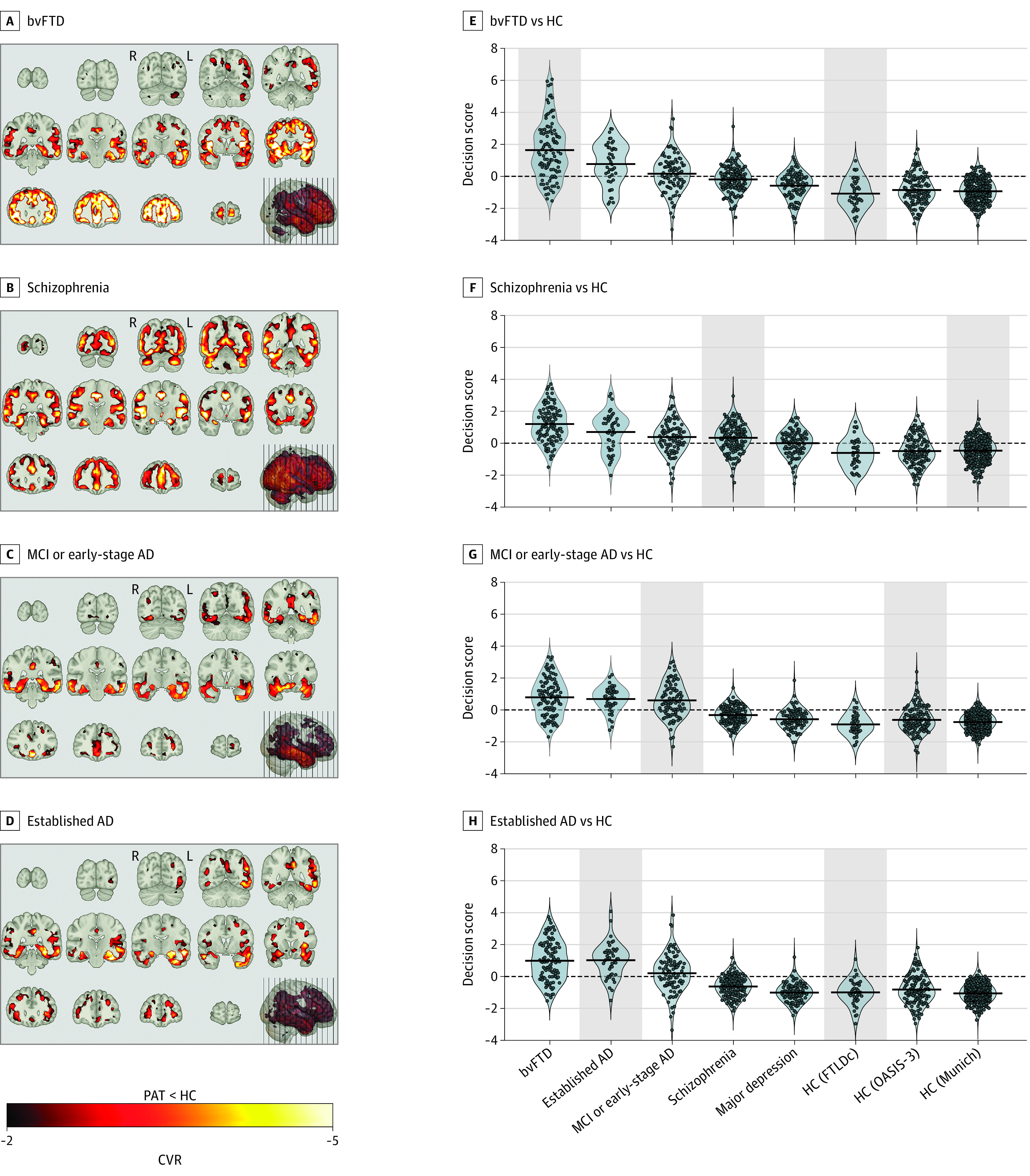
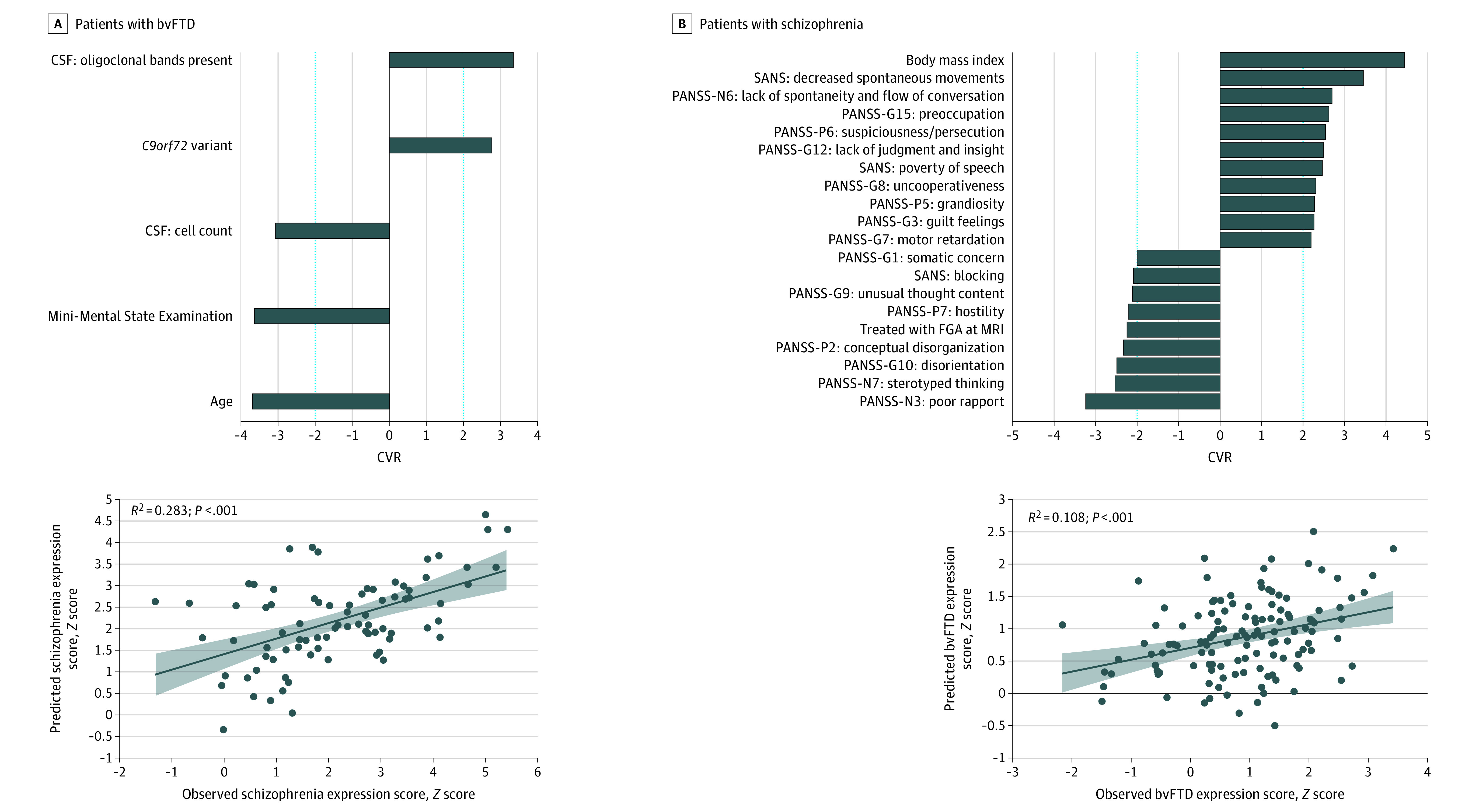
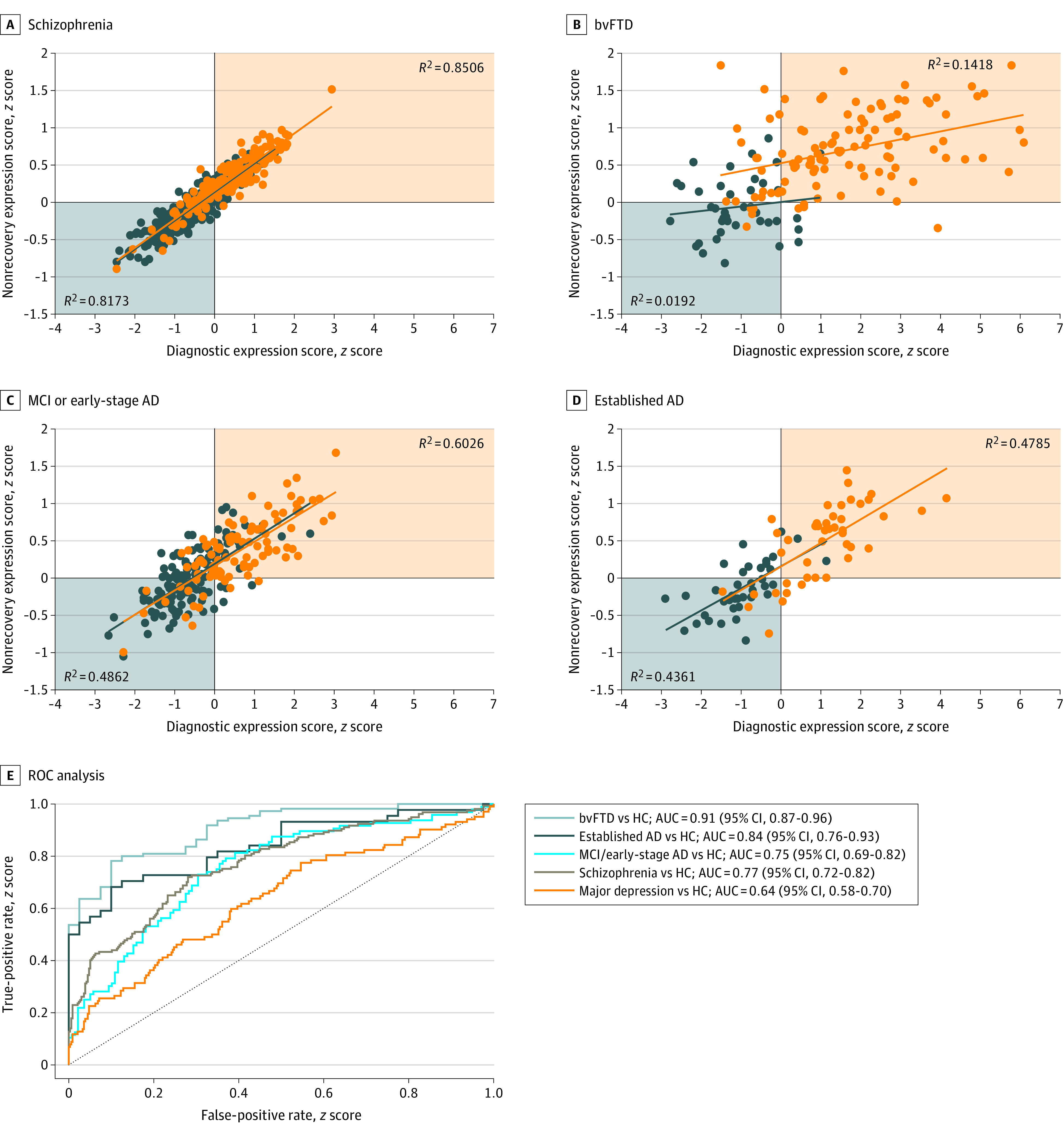
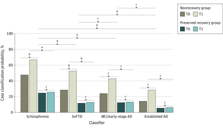
Results: Of 1870 included patients, 902 (48.2%) were female, and the mean (SD) age was 38.0 (19.3) years. The bvFTD pattern comprising prefrontal, insular, and limbic volume reductions was more expressed in patients with schizophrenia (65 of 157 [41.2%]) and major depression (22 of 102 [21.6%]) than the temporo-limbic AD patterns (28 of 157 [17.8%] and 3 of 102 [2.9%], respectively). bvFTD expression was predicted by high body mass index, psychomotor slowing, affective disinhibition, and paranoid ideation (R2 = 0.11). The schizophrenia pattern was expressed in 92 of 108 patients (85.5%) with bvFTD and was linked to the C9orf72 variant, oligoclonal banding in the cerebrospinal fluid, cognitive impairment, and younger age (R2 = 0.29). bvFTD and schizophrenia pattern expressions forecasted 2-year psychosocial impairments in patients with CHR and were predicted by polygenic risk scores for frontotemporal dementia, AD, and schizophrenia. Findings were not associated with AD or accelerated brain aging. Finally, 1-year bvFTD/schizophrenia pattern progression distinguished patients with nonrecovery from those with preserved recovery.

Conclusions and relevance: Neurobiological links may exist between bvFTD and psychosis focusing on prefrontal and salience system alterations. Further transdiagnostic investigations are needed to identify shared pathophysiological processes underlying the neuroanatomical interface between the 2 disease spectra.


Conflict of interest statement

Conflict of Interest Disclosures: Dr Koutsouleris has patent US20160192889A1 issued. Dr Pantelis has received grants from the Australian National Health & Medical Research Council during the conduct of the study as well as grants from Lundbeck Foundation and personal fees from Lundbeck Australia outside the submitted work. Dr Upthegrove has received grants from European Union FP7 during the conduct of the study; grants from Medical Research Council and National Institute for Health Research; and personal fees from Sunovion and Vivalyfe outside the submitted work. Dr Lencer has received personal fees from Laboratorios Farmacéuticos ROVI, S.A., and Johnson & Johnson outside the submitted work. Dr Nöthen has received grants from the European Commission during the conduct of the study as well as personal fees from Life & Brain GmbH and HMG Systems Engineering GmbH outside the submitted work. Dr Jahn has received grants from German Federal Ministry of Education and Research during the conduct of the study. Dr Kornhuber has received grants from German Federal Ministry of Education and Research during the conduct of the study. Dr Landwehrmeyer has received grants from CHDI Foundation during the conduct of the study; grants from Bundesministerium für Bildung und Forschung (BMBF), Deutsche Forschungsgemeinschaft, and the European Commission outside the submitted work; and serves on scientific advisory boards for Hoffmann-LaRoche, Novartis, PTC Therapeutics, Teva, and Triplet Therapeutics. Dr Wiltfang has received personal fees from Abbott, Biogen, Boehringer-Ingelheim, Immungenetics, Janssen, Lilly, Merck Sharp & Dohme, Pfizer, Roche, Actelion, Amgen, Beejing Yibai Science and Technology, and Roboscreen outside the submitted work and has a patent for PCT/EP 2011 001724 issued and a patent for PCT/EP 2015 052945 issued. Dr Diehl-Schmid has received grants from German Ministry for Education and Research during the conduct of the study. Dr Meisenzahl has a patent for US 2016/0192889 A1. Dr Schroeter has received grants from the German Consortium for Frontotemporal Lobar Degeneration, funded by the German Federal Ministry of Education and Research (grant FKZ01GI1007A) during the conduct of the study as well as grants from German Research Foundation and eHealthSax Initiative of the Sächsische Aufbau bank outside the submitted work. No other disclosures were reported.

Figures

Figure 1.
Figure 1.. Classifier Signatures, Pattern Score Distributions, and Predicted Case Label Probabilities of Patient (PAT) and Healthy Control (HC) Samples
A-D, Cross-validation ratio (CVR) (eMethods in Supplement 1) maps overlaid on the MNI single-individual neuroanatomical template indicate patterns of reliable volume reductions (from top to bottom) in the 4 patient samples vs HC. E-H, Violin plots of pattern score distributions produced by the cross-validated within-group application of classification models (gray background) or the crossover application of models to the other diagnostic groups. Decision score refers to the mean output across all support-vector machine classifiers in the given classification analysis produced for the patients/controls in the given sample. Additionally, the Table describes each study group’s probability of being assigned to the respective patient class by the respective classifier. AD indicates Alzheimer disease; bvFTD, behavioral-variant frontotemporal dementia; MCI, mild cognitive impairment; FTLDc, the German Frontotemporal Lobar Degeneration Consortium.
Figure 2.
Figure 2.. Support-Vector Regression Models Predicting the Neuroanatomical Expression of the Schizophrenia Signature in Patients With Behavioral-Variant Frontotemporal Dementia (bvFTD) and the bvFTD Signature in Patients With Schizophrenia
Bar plots show the ranked reliability (cross-validation ratio [CVR]) (eMethods in Supplement 1) of features informing the support-vector regression models’ predictions at |CVR| ≥ 2. Positive and negative CVR values indicate positive and negative predictive associations between features and observed scores. Scatterplots with linear fits, 95% CIs, and coefficients of determination (R2) describe the accuracy of the respective models in predicting neuroanatomical expression scores. CSF indicates cerebrospinal fluid; FGA, first-generation antipsychotics; MRI, magnetic resonance imaging; PANSS, Positive and Negative Symptom Scale; SANS, Scale for Assessment of Negative Symptoms.
Figure 3.
Figure 3.. Associations of the Nonrecovery Prediction Model and the 4 Diagnostic Classifiers
Orange indicates the patient group; blue, the healthy control group. Results were obtained after applying the nonrecovery classifier to patients with behavioral-variant frontotemporal dementia (bvFTD), established Alzheimer disease (AD), mild cognitive impairment (MCI) or early-stage AD, and schizophrenia and the respective healthy control (HC) samples. Scatterplots (A-D) describe the associations between the nonrecovery classifier and the respective diagnostic classifier’s diagnostic expression score in the given derivation cohort. E, The receiver operating characteristic curve analysis displays the separability of patients and HCs in the given diagnostic sample based on the prognostic score produced by the PRONIA nonrecovery classifier for the given sample. See eTable 10 in Supplement 1 for a tabular representation of the nonrecovery classifier prognostic performance and eFigures 24 to 26 in Supplement 1 for the visualization, topographical comparison, and spatial specificity test of the prognostic signature. AUC indicates area under the receiver operating characteristic curve.
Figure 4.
Figure 4.. PRONIA Longitudinal Magnetic Resonance Imaging Analysis Describing the Development of Case Classification Likelihoods Between the Baseline and Follow-up Magnetic Resonance Imaging Data of the PRONIA Nonrecovery and Recovery Samples
Likelihood changes over time were compared by means of generalized estimating equations including Brain Age Gap Estimation as a covariate. Results of estimated marginal means analyses conducted for the functional trajectory, time point, and classifier factors were visualized. See eTable 11 in Supplement 1 for a tabular representation of results. AD indicates Alzheimer disease; bvFTD, behavioral-variant frontotemporal dementia; MCI, mild cognitive impairment; T0, baseline visit; T1, 1-year follow-up visit. aP < .01. bP < .05. cNot significant. dP < .001.

References

    1. Bleuler E. Dementia Praecox Oder Gruppe Der Schizophrenien. Deuticke; 1911. - PubMed
    1. Kraepelin E. Dementia Praecox and Paraphrenia. Chicago Medical Book Co; 1919.
    1. Velthorst E, Fett AJ, Reichenberg A, et al. . The 20-year longitudinal trajectories of social functioning in individuals with psychotic disorders. Am J Psychiatry. 2017;174(11):1075-1085. doi:10.1176/appi.ajp.2016.15111419 - DOI - PMC - PubMed
    1. Plum F. Prospects for research on schizophrenia. 3. neurophysiology. neuropathological findings. Neurosci Res Program Bull. 1972;10(4):384-388. - PubMed
    1. Ribe AR, Laursen TM, Charles M, et al. . Long-term risk of dementia in persons with schizophrenia: a Danish population-based cohort study. JAMA Psychiatry. 2015;72(11):1095-1101. doi:10.1001/jamapsychiatry.2015.1546 - DOI - PubMed

Publication types