. 2022 Nov 1;28(21):4820-4831.
doi: 10.1158/1078-0432.CCR-22-1362.

Phototherapy with Cancer-Specific Nanoporphyrin Potentiates Immunotherapy in Bladder Cancer

Affiliations

Phototherapy with Cancer-Specific Nanoporphyrin Potentiates Immunotherapy in Bladder Cancer

Zheng Zhu et al. Clin Cancer Res. .

Abstract

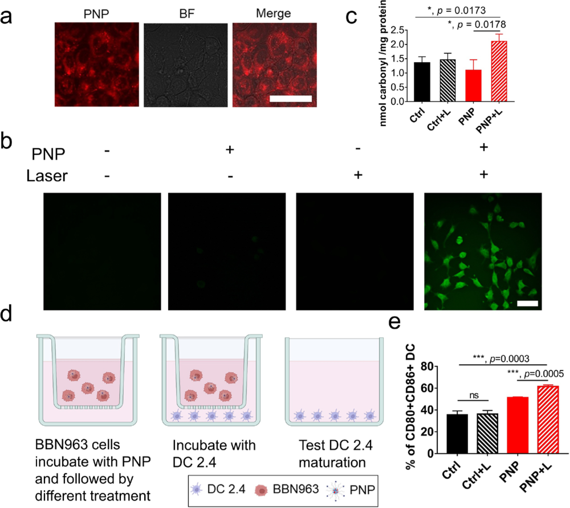
Purpose: Immune checkpoint inhibitors (ICI) in general have shown poor efficacy in bladder cancer. The purpose of this project was to determine whether photodynamic therapy (PDT) with bladder cancer-specific porphyrin-based PLZ4-nanoparticles (PNP) potentiated ICI.

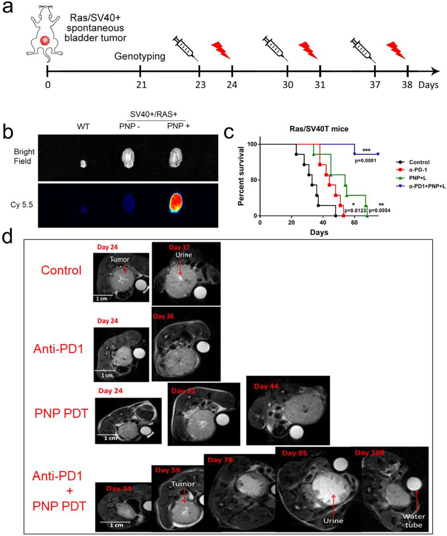
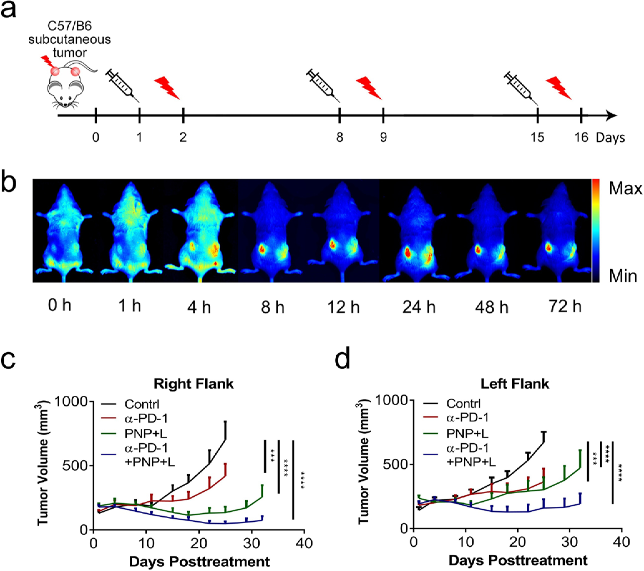
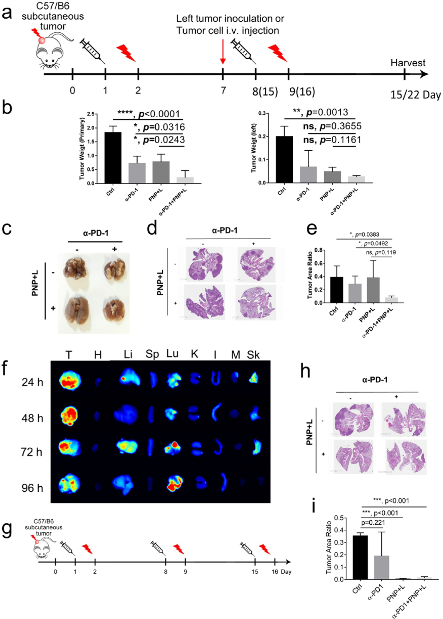
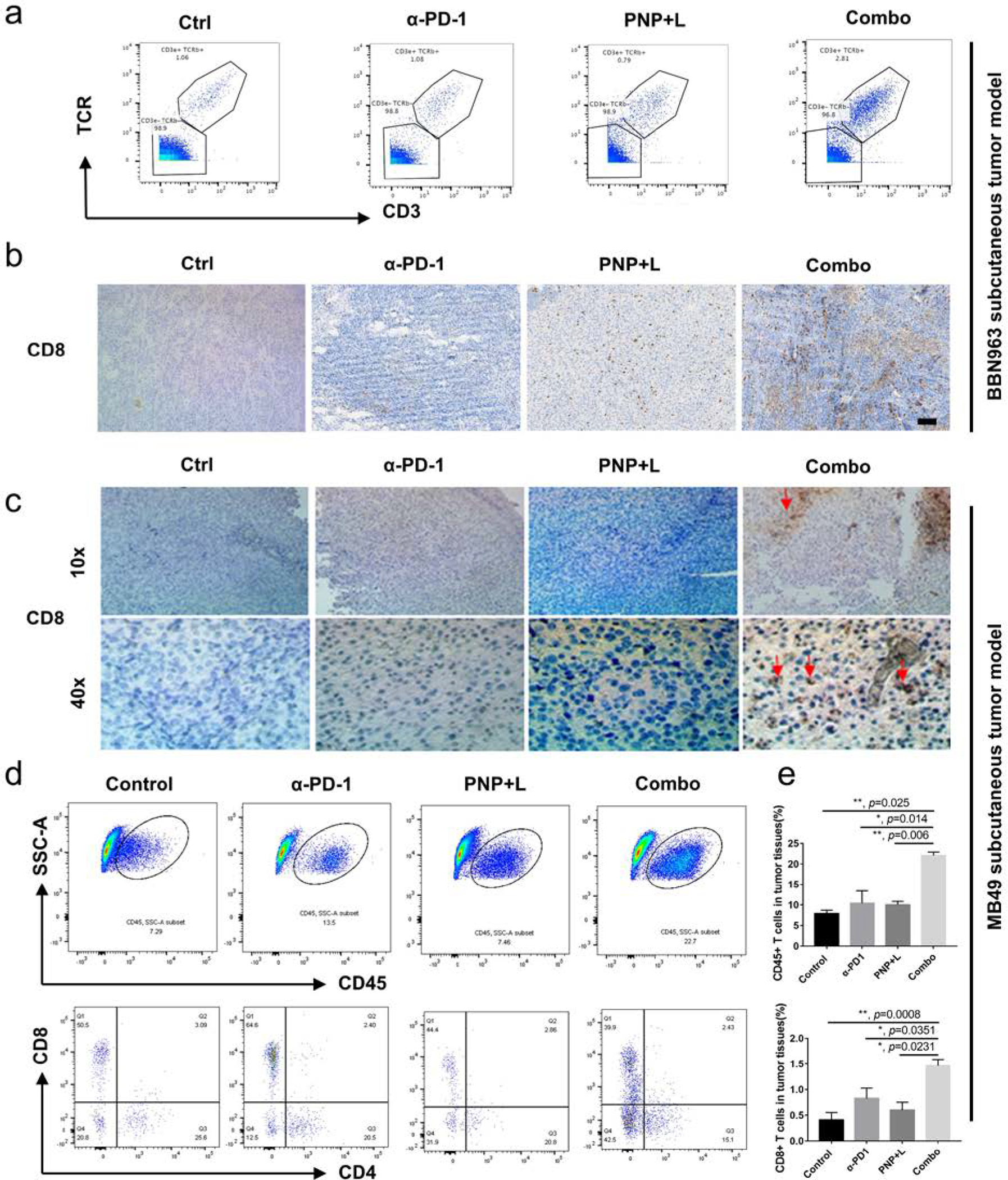
Experimental design: SV40 T/Ras double-transgenic mice bearing spontaneous bladder cancer and C57BL/6 mice carrying syngeneic bladder cancer models were used to determine the efficacy and conduct molecular correlative studies.

Results: PDT with PNP generated reactive oxygen species, and induced protein carbonylation and dendritic cell maturation. In SV40 T/Ras double-transgenic mice carrying spontaneous bladder cancer, the median survival was 33.7 days in the control, compared with 44.8 (P = 0.0123), 52.6 (P = 0.0054), and over 75 (P = 0.0001) days in the anti-programmed cell death-1 antibody (anti-PD-1), PNP PDT, and combination groups, respectively. At Day 75 when all mice in other groups died, only 1 in 7 mice in the combination group died. For the direct anti-tumor activity, compared with the control, the anti-PD-1, PNP PDT, and combination groups induced a 40.25% (P = 0.0003), 80.72% (P < 0.0001), and 93.03% (P < 0.0001) tumor reduction, respectively. For the abscopal anticancer immunity, the anti-PD-1, PNP PDT, and combination groups induced tumor reduction of 45.73% (P = 0.0001), 54.92% (P < 0.0001), and 75.96% (P < 0.0001), respectively. The combination treatment also diminished spontaneous and induced lung metastasis. Potential of immunotherapy by PNP PDT is multifactorial.

Conclusions: In addition to its potential for photodynamic diagnosis and therapy, PNP PDT can synergize immunotherapy in treating locally advanced and metastatic bladder cancer. Clinical trials are warranted to determine the efficacy and toxicity of this combination.


Conflict of interest statement

Conflicts of interest

Drs. Pan, Lam and Zhang are co-inventors of the PLZ4 peptide used for this study; Drs. Lam, Li, Pan and Lin are co-inventors of nanoporphyrin; Drs. Pan, Lam, Li and Lin are co-founders of LP Therapeutics that licensed PLZ4 therapeutics. Dr. Lam is the founder of LamnoTherapeutics that licenses nanoporphyrin. The remaining authors declare no potential conflicts of interest.

Figures

Figure 1:
Figure 1:. ROS production, protein carbonylation and stimulation of dendritic cells during PDT with PNP.
a) Confocal images of PNP uptake in BBN963 cells (scale bar=50μm), BF: bright field. b) ROS production after different treatments as determined by the DCFH-DA assay as determined by confocal laser scanning microscopy (scale bar=50μm). Green fluorescence indicates ROS production. Significantly more abundant ROS production was observed in the cells treated with PNP plus light. c) Protein carbonylation by ROS in BBN963 cells. d) A transwell assay to determine dendritic cell maturation. e) Flow cytometry analysis of CD80 and CD86 expression to determine dendritic cell maturation after different treatments for 24h in the transwell system. In the PNP + Light treatment group, 61.5%±2.04% cells expressed CD80 and CD86, compared to 35.63%±1.89%, (p=0.0003), 36.23%±0.48% (p=0.0003) and 51.27%±0.89% (p=0.0005) of the control, control plus light and PNP groups, respectively.
Figure 2:
Figure 2:. Synergistic anti-tumor activity between PDT and immunotherapy in treating localized spontaneous bladder cancer.
a) Schema showing the experimental design for a SV40T/RAS* double transgenic spontaneous bladder cancer model. SV40T/RAS* double transgenic mice develop localized spontaneous bladder cancer at 20–30 days of age. b) Cancer-specific delivery of PNP to the mouse bladder cancer. The bladder and spontaneous bladder cancer were collected at 24 hours after intravenous injection of PNP for near-infrared imaging. Red fluorescence indicates PNP accumulation. No significant PNP accumulation in the control mice without bladder cancer. c) Survival curves of SV40T/RAS* double transgenic mice with different treatments. Compared to the median survival of 33.7 days in the control group, the median survival of the anti-P1, PNP + light and combination groups were 44.8 (p=0.0123), 52.6 (p=0.0054) and over 75 days (p=0.0001), respectively. Only 1 out of 7 mice died in the combination group at Day 75 when all mice in other groups died and the study ended. d) MRI image of mouse tumors after different treatments. T2 images were shown here. The white circle in each image is a water tube used as an internal control as mice and bladders changed in size over time when mice grew. The white irregular area at the central of each image is urine, especially as shown in the image taken on Day 95. In the mouse of the combination group, the effective bladder cavity area (irregular white area in the middle of the bladder representing urine) significantly increased at Day 78 and 95 and the bladder overall volume decreased while the bladder volume increased over time in the other three groups. This mouse died at Day 158.
Figure 3:
Figure 3:. Synergistic local direct and abscopal anti-tumor activity of PDT and immunotherapy.
a) Experimental design for the bilateral tumor model. C57BL/6 mice bearing BBN963 subcutaneous tumors at both flanks were used in this experiment. When tumors reached the size of around 100 mm3, tumors on the right side were treated with light for PDT and monitored for local direct anti-tumor activity. The tumors at the left flank were not treated with light and monitored for abscopal effects. b) In vivo fluorescence imaging to show PNP biodistribution at different time points of a C57BL/6 mouse bearing BBN963 tumors. Starting at 8 hours after administration, fluorescence from PNP accumulated at tumor sites with very little fluorescence at any other sites in the body. c) Tumor growth curves of the tumors at the right flank which were treated with light, showing the direct anti-tumor activities of PBS, anti-PD-1, PNP+L, or anti-PD-1+PNP+L (n=8). At day 25, compared to the control, the right-site-tumors (treated, direct anti-tumor effect) of the anti-PD-1, PDT and combination groups had a reduction of 40.25% (p=0.0003), 80.72% (p<0.0001) and 93.03% (p<0.0001), respectively. d) Tumor growth curves of the tumors at the left flank which were not treated with light, showing the abscopal anti-tumor activities. At Day 25, compared to the control, the tumor reduction of the left-site-tumors (measuring the abscopal effect) in the anti-PD-1, PDT and combination groups was 45.73% (p=0.0001), 54.92% (p<0.0001) and 75.96% (p<0.0001), respectively. The tumor reduction between the PDT with PNP and anti-PD-1 therapy groups was not statistically different (p=0.8402).
Figure 4:
Figure 4:. Synergistic PNP and immunotherapy for in vivo suppression of distant tumors and lung metastasis.
a) Experimental design to determine whether PDT and immunotherapy could prevent development of induced distant metastasis. MB49(1 × 10⁶ cells) tumor implants were first established at the right flank of C57BL/6 mice. When tumors reached the size of 100 mm3, mice received PBS or PNP (Day 1) followed by light treatment at the right flank 24 hours later (Day 2), and anti-PD-1 treatment was given twice a week. The same set of treatment was repeated once in the groups mimicking distant metastasis and twice in the groups mimicking lung metastasis. MB49 cells (1 × 10⁶ cells) were injected to the left flank on Day 7 to determine abscopal effects. To mimic lung metastasis, MB49 cells (1 × 10⁵ cells) were injected via tail vein on Day 7. b) The tumors on the right side were designated as primary tumors treated with light, and those on left side were designated as distant tumor (n=4). Tumors were harvested 15 days after the first treatment. Statistical analyses were performed with tumor weights on primary site and distant site. For the right (treated) site, tumor weight was 0.21±0.13 g in the combination treatment group, compared 1.83±0.12 g in the control group (p<0.0001), 0.72±0.13 g in the anti-PD-1 group (p=0.0316), and 0.78±0.14 g in the PNP+ PDT group (p=0.0243), respectively (left panel). The distant tumor in the combination group was also much smaller than that in the control (0.02 ± 0.001 gram versus 0.2 ± 0.02 gram, p=0.0013) even though not much smaller than the other two groups (right panel). c-e Lungs were harvested 22 days after the first treatment in the groups mimicking lung metastasis. c) Gross appearance of tumor nodules in the lungs. d) Hematoxylin and eosin staining of lungs. e) Statistical analysis of tumor area. The tumor/lung ratio in the combination group was smaller than the control (p=0.0383) and anti-PD-1 (p=0.0492) groups, but not the PNP plus light treatment group (p=0.119). f) fluorescent signal showing PNP accumulation in tumor and lung, suggesting development of spontaneous lung metastasis. (T: tumor, H: heart, Li: Liver, Sp: Spleen, Lu: Lung, K: Kidney, I: Intestine, M: Muscle, Sk: Skin). g) Schema showing the design to determine whether PDT and immunotherapy could prevent development of spontaneous lung metastasis. h) Hematoxylin and eosin staining of lungs. i) Statistical analysis of tumor area. Compared to the control, the PNP plus light treatment and the combination groups significantly decreased spontaneous lung metastasis (p<0.001).
Figure 5
Figure 5. Synergistic effects of PNP and immunotherapy in activating systematic anti-tumor immunity.
a) CyTOF analysis of left tumor in BBN963 subcutaneous model. Of all nucleated cells in the tumors, the CD3e+TCRb+T cells in the combination treatment group was 2.81% compared to 1.06%, 1.08%, and 0.79% in the control, anti-PD-1 and PDT groups, respectively. b) IHC showing CD8+ T cell infiltration into left tumors (scale bar = 100 μm) of BBN963 tumors. c) CD8+ T cell infiltration into MB49 subcutaneous tumors. Red arrow indicates the CD8+ T cells. (Upper panel, 10X; Lower panel, 40X). In both tumor models, significantly more T cell infiltration was observed in the combination groups. d) Representative flow-cytometry plots showing tumor-infiltrating leucocyte cells, including CD45+ cells and CD8+ T cells, in distant sites of the MB49 model. The numbers at the lower panel of the flow cytometry plots are percentage of CD4/CD8 cells among CD3+ cells. e) statistical analysis of CD45+ cells (upper panel) and CD8+ T cells of all cells in the distant tumors. The percentage of CD45+ leucocytes in the PDT with PNP plus anti-PD-1 treatment group was 22.60% in the distant tumors of the left flank that were not treated with light (abscopal effects), compared to 8.63% (p=0.025), 9.80 (p=0.014) and 9.97 (p=0.006) in the control, anti-PD-1 and PNP groups, respectively (upper panel). The percentage of CD45+CD3+CD8+ cells among all cells in the tumor in the PDT with PNP plus anti-PD-1 treatment group was 1.463% of all cells in tumor, compared to 0.408 (p=0.0008), 0.826(p=0.0351) and 0.598 (p=0.0231) in the control group, anti-PD-1 and PNP PDT groups, respectively (lower panel).

References

    1. Siegel RL, et al., Cancer statistics, 2022. CA Cancer J Clin, 2022. 72(1): p. 7–33. - PubMed
    1. Balar AV, et al., Pembrolizumab monotherapy for the treatment of high-risk non-muscle-invasive bladder cancer unresponsive to BCG (KEYNOTE-057): an open-label, single-arm, multicentre, phase 2 study. Lancet Oncol, 2021. 22(7): p. 919–930. - PubMed
    1. Powles T, et al., Avelumab Maintenance Therapy for Advanced or Metastatic Urothelial Carcinoma. N Engl J Med, 2020. 383(13): p. 1218–1230. - PubMed
    1. Powles T, et al., MPDL3280A (anti-PD-L1) treatment leads to clinical activity in metastatic bladder cancer. Nature, 2014. 515(7528): p. 558–62. - PubMed
    1. Rosenberg JE, et al., Atezolizumab in patients with locally advanced and metastatic urothelial carcinoma who have progressed following treatment with platinum-based chemotherapy: a single-arm, multicentre, phase 2 trial. Lancet, 2016. 387(10031): p. 1909–20. - PMC - PubMed

Publication types

Substances