. 2022 Oct;33(10):1927-1941.
doi: 10.1681/ASN.2022030234. Epub 2022 Aug 3.

Prognostic Implications of a Morphometric Evaluation for Chronic Changes on All Diagnostic Native Kidney Biopsies

Affiliations

Prognostic Implications of a Morphometric Evaluation for Chronic Changes on All Diagnostic Native Kidney Biopsies

Aleksandar Denic et al. J Am Soc Nephrol. 2022 Oct.

Abstract

Background: Semiquantitative visual inspection for glomerulosclerosis, interstitial fibrosis, and arteriosclerosis is often used to assess chronic changes in native kidney biopsies. Morphometric evaluation of these and other chronic changes may improve the prognostic assessment.

Methods: We studied a historical cohort of patients who underwent a native kidney biopsy between 1993 and 2015 and were followed through 2021 for ESKD and for progressive CKD (defined as experiencing 50% eGFR decline, temporary dialysis, or ESKD). Pathologist scores for the percentages of globally sclerosed glomeruli (GSG), interstitial fibrosis and tubular atrophy (IFTA), and arteriosclerosis (luminal stenosis) were available. We scanned biopsy sections into high-resolution images to trace microstructures. Morphometry measures were percentage of GSG; percentage of glomerulosclerosis (percentage of GSG, ischemic-appearing glomeruli, or segmentally sclerosed glomeruli); percentage of IFTA; IFTA foci density; percentage of artery luminal stenosis; arteriolar hyalinosis counts; and measures of nephron size. Models assessed risk of ESKD or progressive CKD with biopsy measures adjusted for age, hypertension, diabetes, body mass index, eGFR, and proteinuria.

Results: Of 353 patients (followed for a median 7.5 years), 75 developed ESKD and 139 experienced progressive CKD events. Visually estimated scores by pathologists versus morphometry measures for percentages of GSG, IFTA, and luminal stenosis did not substantively differ in predicting outcomes. However, adding percentage of glomerulosclerosis, IFTA foci density, and arteriolar hyalinosis improved outcome prediction. A 10-point score using percentage of glomerulosclerosis, percentage of IFTA, IFTA foci density, and any arteriolar hyalinosis outperformed a 10-point score based on percentages of GSG, IFTA, and luminal stenosis >50% in discriminating risk of ESKD or progressive CKD.

Conclusion: Morphometric characterization of glomerulosclerosis, IFTA, and arteriolar hyalinosis on kidney biopsy improves prediction of long-term kidney outcomes.

Keywords: chronic renal disease; end stage kidney disease; interstitial fibrosis; kidney biopsy; morphometry; renal pathology.


Figures

None
Graphical abstract
Figure 1.
Figure 1.
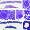
Representative images of kidney biopsy chronic changes and morphometric measures. (A) An example of a PAS-stained biopsy section used in morphometric analysis of native kidney biopsies. (B–H) Examples of (B) nonischemic nonsclerosed glomerulus (traced in red in (A)), (C) glomerulus with a segmental scar, (D) ischemic-appearing glomerulus (traced in blue in (A)), (E) GSG (traced in pink in (A)), (F) AH affecting whole circumference, and (G) partial or nodular AH (a nodule pointed by an arrowhead). (H) An example of a sequential TRI-stained biopsy section of the same case. In both sections, cortex was traced in green. Areas with distinct inflammatory infiltrate were traced on periodic acid–Schiff-stained sections (orange), and areas with distinct IFTA foci were traced on TRI-stained section (black). (I) An example of a small to medium artery, where media to intima boundary (red trace) and intima to lumen boundary (yellow trace) were used to quantify percentage of luminal stenosis due to intimal thickening.
Figure 2.
Figure 2.
Selection of study sample.
Figure 3.
Figure 3.
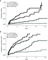
The probability of ESKD and CKD with morphometry measure of IFTA density. Higher IFTA foci density (per mm2 cortex) is predictive of ESKD (A) and progressive CKD (B).
Figure 4.
Figure 4.
Risk of ESKD and progressive CKD based on three different renal chronicity scores. Risk of ESKD and progressive CKD based on severity of pathologist renal chronicity score (A and B), morphometry renal chronicity score (C and D), and modified morphometry renal chronicity score (E and F). Pathologist renal chronicity score was generated by sum of the %GSG score (0–3), two times the %IFTA score (0–3), and arteriosclerosis score (0–1). Morphometric renal chronicity score was generated by sum of the morphometric %GSG score (0–3), two times the morphometric %IFTA score (0–3), and morphometric arteriosclerosis score (0–1). A modified morphometric renal chronicity score was generated by sum of the %glomerulosclerosis score (0–3), morphometric %IFTA score (0–3), IFTA foci density score (0–3), and morphometric AH score (0–1). All chronicity scores (0–10) were then converted into four levels (0–1, 2–4, 5–7, and 8–10).
Figure 5.
Figure 5.
A biologic conceptual model for the interrelationship of prognostic chronic changes found in this study across multiple disease pathways. Biopsy findings included in the modified morphometric renal chronicity score are highlighted in bold.

Similar articles

Cited by

References

    1. Fufaa GD, Weil EJ, Lemley KV, Knowler WC, Brosius FC 3rd, Yee B, et al. : Structural predictors of loss of renal function in American Indians with type 2 diabetes. Clin J Am Soc Nephrol 11: 254–261, 2016 - PMC - PubMed
    1. Mottl AK, Gasim A, Schober FP, Hu Y, Dunnon AK, Hogan SL, et al. : Segmental sclerosis and extracapillary hypercellularity predict diabetic ESRD. J Am Soc Nephrol 29: 694–703, 2018 - PMC - PubMed
    1. Chen T, Li X, Li Y, Xia E, Qin Y, Liang S, et al. : Prediction and risk stratification of kidney outcomes in IgA nephropathy. Am J Kidney Dis 74: 300–309, 2019 - PubMed
    1. Ford SL, Polkinghorne KR, Longano A, Dowling J, Dayan S, Kerr PG, et al. : Histopathologic and clinical predictors of kidney outcomes in ANCA-associated vasculitis. Am J Kidney Dis 63: 227–235, 2014 - PubMed
    1. Leatherwood C, Speyer CB, Feldman CH, D’Silva K, Gómez-Puerta JA, Hoover PJ, et al. : Clinical characteristics and renal prognosis associated with interstitial fibrosis and tubular atrophy (IFTA) and vascular injury in lupus nephritis biopsies. Semin Arthritis Rheum 49: 396–404, 2019 - PMC - PubMed

Publication types