. 2022 Aug 3;13(1):4496.
doi: 10.1038/s41467-022-32147-w.

Dynamic actuation enhances transport and extends therapeutic lifespan in an implantable drug delivery platform

Affiliations

Dynamic actuation enhances transport and extends therapeutic lifespan in an implantable drug delivery platform

William Whyte et al. Nat Commun. .

Abstract

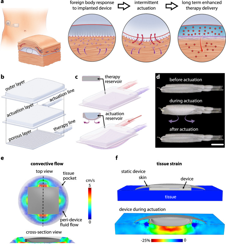
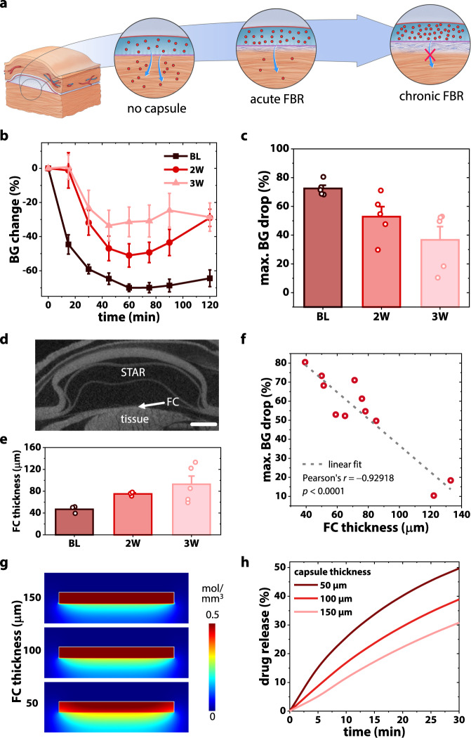
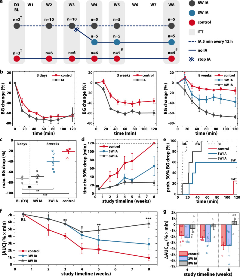
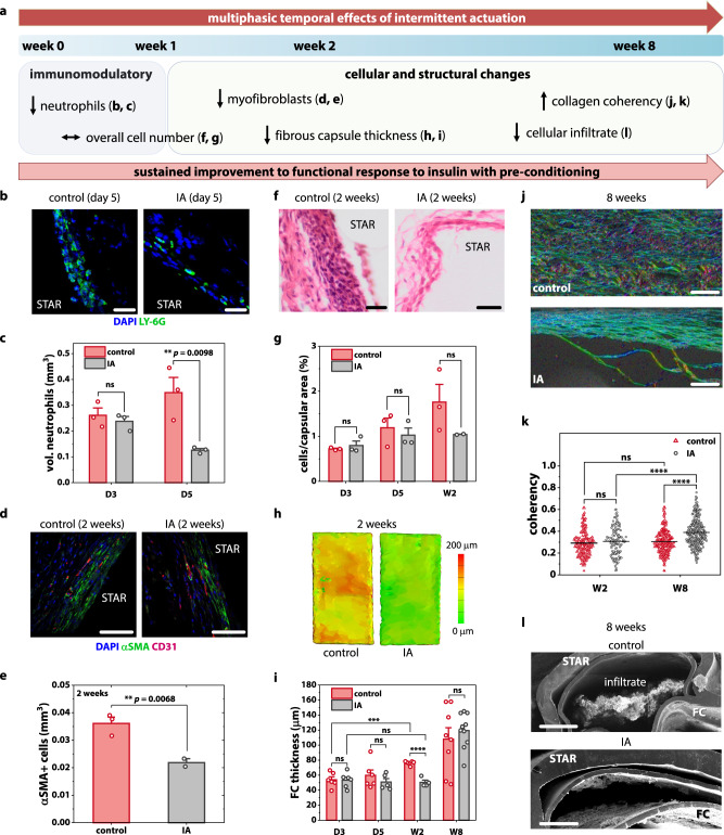
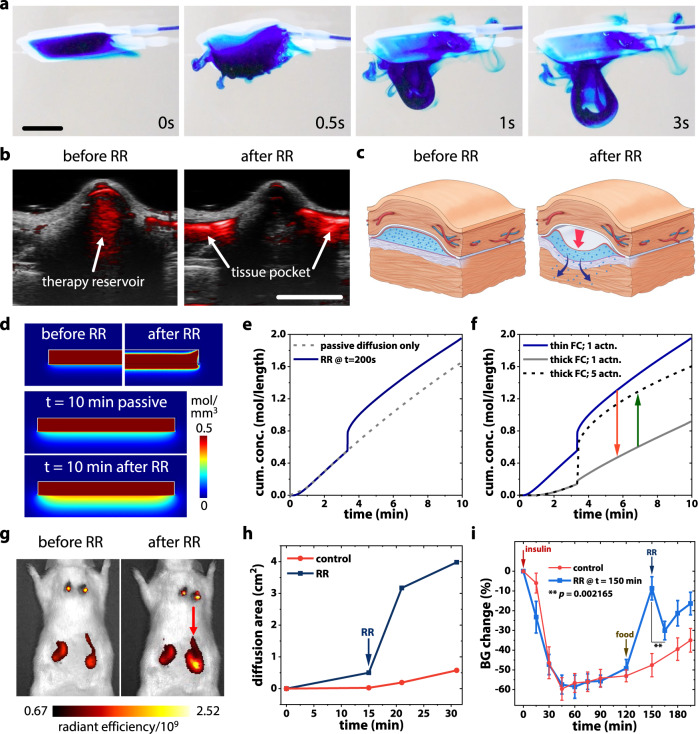
Fibrous capsule (FC) formation, secondary to the foreign body response (FBR), impedes molecular transport and is detrimental to the long-term efficacy of implantable drug delivery devices, especially when tunable, temporal control is necessary. We report the development of an implantable mechanotherapeutic drug delivery platform to mitigate and overcome this host immune response using two distinct, yet synergistic soft robotic strategies. Firstly, daily intermittent actuation (cycling at 1 Hz for 5 minutes every 12 hours) preserves long-term, rapid delivery of a model drug (insulin) over 8 weeks of implantation, by mediating local immunomodulation of the cellular FBR and inducing multiphasic temporal FC changes. Secondly, actuation-mediated rapid release of therapy can enhance mass transport and therapeutic effect with tunable, temporal control. In a step towards clinical translation, we utilise a minimally invasive percutaneous approach to implant a scaled-up device in a human cadaveric model. Our soft actuatable platform has potential clinical utility for a variety of indications where transport is affected by fibrosis, such as the management of type 1 diabetes.


Conflict of interest statement

W.W., S.T.R., K.L.M., C.E.V., G.P.D., E.B.D., and E.T.R. are inventors on a pending patent application related to the device described here. The other authors declare no competing interests.

Figures

Fig. 1
Fig. 1. Design of soft transport augmenting reservoir (STAR).
a Proposed mechanism of STAR: intermittent actuation attenuates the foreign body response, creating a favourable environment for the rapid long-term transport of macromolecular drug therapy. b Exploded view showing the different layers comprising STAR. c Deflection of the actuation and porous layers during actuation. d A prototype of STAR showing the deflection of the porous layer during an actuation cycle. Scale bar is 5 mm. e FE model showing peri-implant fluid velocity of convective flow during actuation. f FE model estimating maximum principal tissue strain induced by actuation.
Fig. 2
Fig. 2. Development of a pre-clinical model to monitor the effect of FBR on therapy transport longitudinally.
a Schematic demonstrating detrimental effect of fibrous capsule (FC) formation on therapy delivery with time. b Blood glucose (BG) response to human insulin delivered via STAR, measured over 120 min at baseline (BL, day 3), week 2 and week 3. n = 5 mice at each time point. c Temporal evolution of the maximum BG % drop (denoting functional effect), calculated from b. d Representative 2D µCT slice of STAR with fibrous encapsulation. Scale bar is 1 mm. e Average FC thickness encapsulating STAR at baseline (day 3), week 2 and week 3 following implantation. n = 3 mice at baseline and 2 weeks, 5 mice at 3 weeks. Data are means ± standard error of mean. f Relationship between FC thickness and maximum effect of insulin measured by reduction in blood glucose level. g COMSOL Multiphysics simulations showing spatial drug diffusion through FCs of varying thicknesses. h Temporal evolution of drug release percentage for varying FC thicknesses.
Fig. 3
Fig. 3. Intermittent actuation (IA) improves long-term macromolecule delivery.
a Preclinical study timeline used to evaluate the effect of IA on insulin transport through a fibrous capsule. b Blood glucose (BG) response to human insulin, measured over 120 min at day 3 (baseline), week 3 and week 8. c Maximum BG% change at the 8-week timepoint. d Time to achieve a 30% drop in BG level, measured longitudinally. e Cumulative incidence curves demonstrating probability of achieving a 30% BG drop over 120 min for all groups at both baseline (3 days) and 8-week timepoints. f Area under BG % curve (AUC) denoting overall functional effect mapped over study duration. Statistical comparisons w.r.t. control group. g Change in AUC from 3-week timepoint. Data are means ± standard error of mean; *p < 0.05, **p < 0.01, ***p < 0.001. See Supplementary Note 1 for detailed statistical analyses. † Baseline study was performed post-hoc with separate mice. # Control mice removed from study at intermediate time points due to self-inflicted device damage with subsequent decrease in n.
Fig. 4
Fig. 4. Multiphasic temporal effects of intermittent actuation (IA).
a Timeline of multiphasic cellular and fibrous capsule (FC) changes induced by IA. b Representative fluorescent images of the FC stained with Ly-6G + (green) and DAPI (blue). Scale bars are 20 µm. c Quantification of neutrophils present within FC+/− IA at day 3 and 5. d Representative fluorescent images of the FC stained with α-SMA (green) and CD31 (red). Scale bars are 50 µm. e Quantification of myofibroblasts present within FC+/− IA at 2 weeks. f Representative histologic images of the FC stained with haematoxylin and eosin. Scale bars are 20 µm. g Quantification of total cells/capsular area +/− IA at day 3, day 5, and 2 weeks. h Representative topographical reconstructions of µCT images showing the differences in FC thickness+/− IA at 2 weeks. i Average FC thickness of the control and actuated groups at day 3, day 5, 2 weeks, and 8 weeks with two measurements taken per animal. j Representative polarised light microscopy images of the FC obtained after picrosirius red staining at 8 weeks. Scale bars are 100 µm. k, Quantification of the FC collagen fibre orientation by optical coherency with 60 ROIs per animal. l Representative SEM images demonstrating reduced cellular invasion with actuation at the 8-week timepoint. Scale bars are 500 µm. n = 2–6 animals per group; data are means ± standard error of mean; *p < 0.05, **p < 0.01, ***p < 0.001, ****p < 0.0001. See Supplementary Note 1 for detailed statistical analyses.
Fig. 5
Fig. 5. On demand, actuation-mediated rapid release (RR) of drug using STAR.
a RR enables convective flow of a model drug, methylene blue, from the therapy reservoir of STAR. Scale bar is 5 mm. b Photoacoustic images showing subcutaneously implanted STAR in a rat model: RR enables convective flow of drug analogue (red) from the therapy reservoir into the surrounding tissue pocket. c Schematic showing actuation-mediated RR overcoming fibrous encapsulation. d Snapshots from COMSOL Multiphysics simulation showing the rate-limiting diffusion barrier created by a FC and the ability to improve transport using RR. e Concentration of drug outside the FC comparing passive diffusion alone to RR at 200 s. f Concentration of drug outside the FC for a thin (100 μm) or thick (200 μm) FC with 1 or 5 RR actuation cycles. g In vivo images of rat model with two STAR devices implanted. Fluorescence shows the distribution of drug analogue Genhance 750. Red arrow indicates the device after undergoing RR actuation. h Temporal evolution of the drug diffusion area of Genhance 750 in passive (control) and RR actuated STAR, quantified by fluorescent IVIS imaging. i Blood glucose response to insulin in control (passive diffusion only) and in RR actuated (at t = 150 min) STAR devices, at 2 weeks following implantation. n = 4 animals per group; data represents means ± standard error of mean. p value calculated from paired one-tailed t-test.
Fig. 6
Fig. 6. Minimally invasive surgical implantation in a human cadaver model.
a Location of the transversus abdominis plane in the anterior abdominal wall. b Ultrasound guided needle access to the desired tissue intermuscular plane in the anterior abdominal wall and hydro-dissection to generate a potential space (EOM: external oblique muscle, IOM: internal oblique muscle, TAM: transversus abdominis muscle). c The Seldinger technique was used to get needle access to the transversus abdominis plane and a 5 Fr sheath was exchanged over a guide wire to maintain durable access to the tissue plane. d A commercially available dilator set is used to expand the space to accommodate positioning of the deployment sheath. e STAR advancement through sheath into tissue space. f, g An echogenic contrast agent was used to inflate the deployment channel to ensure complete unfolding of the STAR device within the plane using ultrasound guidance.

Similar articles

Cited by

References

    1. Corradetti, B. The Immune Response to Implanted Materials and Devices: The Impact of the Immune System on the Success of an Implant. 10.1007/978-3-319-45433-7 (Springer International Publishing, 2016.)
    1. Anderson JM, Rodriguez A, Chang DT. Foreign body reaction to biomaterials. Semin. Immunol. 2008;20:86–100. doi: 10.1016/j.smim.2007.11.004. - DOI - PMC - PubMed
    1. Scuderi GJ, Vaccaro AR, Fitzhenry LN, Greenberg S, Eismont F. Long-term clinical manifestations of retained bullet fragments within the intervertebral disk space. J. Spinal Disord. 2004;17:108–111. doi: 10.1097/00024720-200404000-00005. - DOI - PubMed
    1. Ward, W. K. A review of the foreign-body response to subcutaneously-implanted devices: the role of macrophages and cytokines in biofouling and fibrosis. J. Diabetes Sci. Technol.2, 768–777 (2008). - PMC - PubMed
    1. Coulter, F. B. et al. Additive manufacturing of multi‐scale porous soft tissue implants that encourage vascularization and tissue ingrowth. Adv. Healthc. Mater. 10, 2100229 (2021). - PMC - PubMed

Publication types