Review
. 2022 Oct;20(10):608-620.
doi: 10.1038/s41579-022-00767-0. Epub 2022 Aug 3.

The biofilm life cycle: expanding the conceptual model of biofilm formation

Affiliations
Review

The biofilm life cycle: expanding the conceptual model of biofilm formation

Karin Sauer et al. Nat Rev Microbiol. 2022 Oct.

Abstract

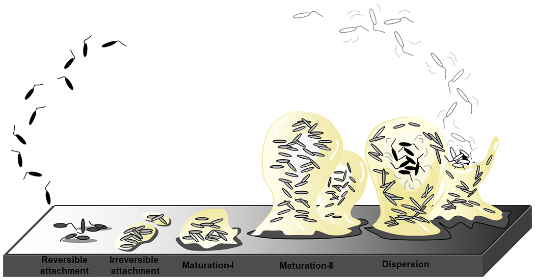
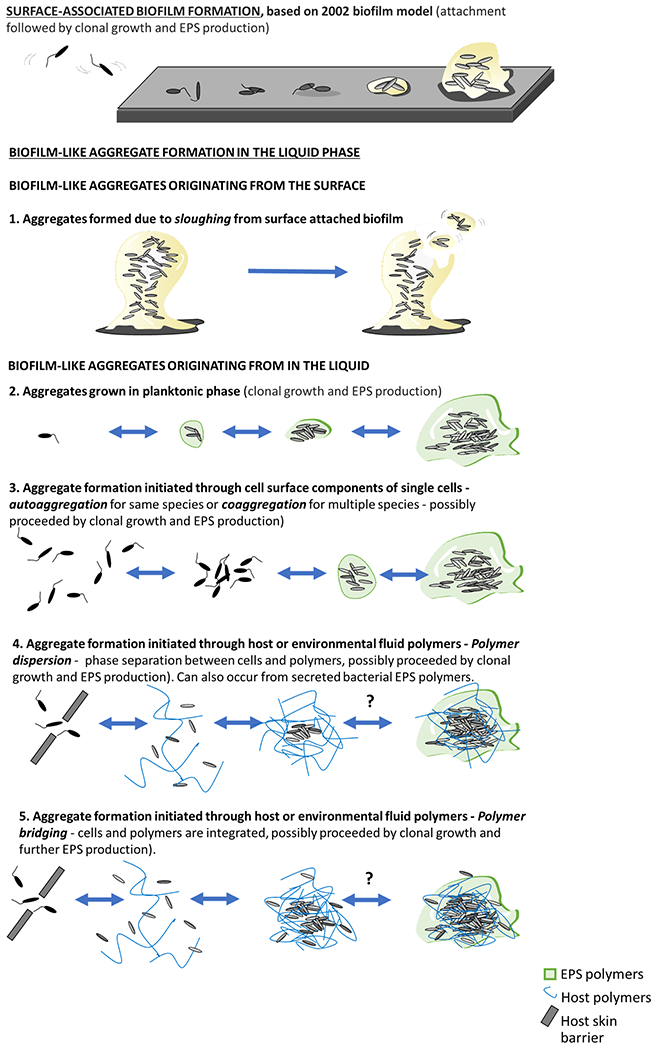
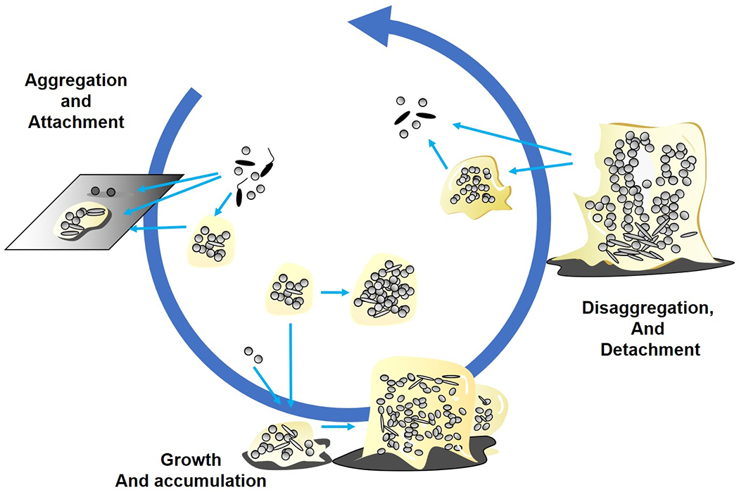
Bacterial biofilms are often defined as communities of surface-attached bacteria and are typically depicted with a classic mushroom-shaped structure characteristic of Pseudomonas aeruginosa. However, it has become evident that this is not how all biofilms develop, especially in vivo, in clinical and industrial settings, and in the environment, where biofilms often are observed as non-surface-attached aggregates. In this Review, we describe the origin of the current five-step biofilm development model and why it fails to capture many aspects of bacterial biofilm physiology. We aim to present a simplistic developmental model for biofilm formation that is flexible enough to include all the diverse scenarios and microenvironments where biofilms are formed. With this new expanded, inclusive model, we hereby introduce a common platform for developing an understanding of biofilms and anti-biofilm strategies that can be tailored to the microenvironment under investigation.


Figures

Figure 1
Figure 1
The stages of biofilm development as depicted in (26). The formation of biofilms is a cyclic process that occurs in a stage-specific and progressive manner. The process is initiated following surface contact by single planktonic cells. Several developmental steps are discernable as reversible attachment, irreversible attachment and biofilm maturation (maturation-I and -II)(26, 43). During reversible attachment, bacteria attach to the substratum via the cell pole or via the flagellum (step I), followed by longitudinal attachment. Transition to the irreversible coincides with a reduction in flagella reversal rates, reduction in flagella gene expression and the production of biofilm matrix components. This stage is also characterized by attached cells demonstrating drug tolerance(44). Biofilm maturation stages are characterized by the appearance of cell clusters that are several cells thick and are embedded in the biofilm matrix (maturation-I stage) which subsequently fully mature into microcolonies (maturation-II stage)(26, 43). Dispersion has been reported to coincide with the decrease in and degradation of matrix components, with dispersed cells being motile and demonstrating increased drug susceptibility relative to biofilm cells. The biofilm matrix is shown in beige.
Figure 2,
Figure 2,
P. aeruginosa grown in flow cells under flow conditions but with different carbon sources shows remarkably different three-dimensional architectures (Sauer, K 2021)
Figure 3.
Figure 3.
Variety of biofilm structures underscoring differences between in vitro and in vivo or environmental biofilms. Original images are shown in the left column and a schematic drawing of the structure and its organization in the right column with shading denoting water (blue), aggregated microbial cells (dark green) and their extracellular polymeric substances (light green), host cells and other material including mucus or tissue (red), and attachment surface (hatched grey). A: Mushroom structure of Pseudomonas aeruginosa biofilm in vitro in a flow cell. B: Mucus embedded aggregates of P. aeruginosa surrounded by polymorphonuclear leukocytes in a cystic fibrosis lung (108) C: Wound-embedded aggregates of P. aeruginosa surrounded by polymorphonuclear leukocytes(109). D: Aerobic granules from a full-scale AquaNereda® wastewater treatment process (image courtesy of Kylie Bodle and Cat Kirkland). E: Striated microbial mat from a Brazilian lake(110). (Jill Story assisted with figure preparation).
Figure 4.
Figure 4.
Microbial aggregate formation mechanisms. The top panel shows the “standard” model for biofilm formation starting from the attachment of single planktonic cells to a smooth surface followed by cell division and production of EPS to form 3D surface attached aggregate structures. Below are different mechanisms for generating free floating biofilm-like aggregates. The first is detachment of aggregates from attached biofilms. The second is from clonal growth (division) in the liquid, which may occur with or without facilitation by bacterially produced EPS matrices The third is aggregation of individual cells in a process called autoaggregation for a single species or coaggregation for multiple species, in which bacteria attach to each other through mutual attraction of surface molecules such as adhesins or EPS bridging interactions. The fourth is bridging aggregation which can also be mediated by host polymers, as appears to be the case in synovial fluid (140). Another mechanism of aggregation, the fifth,, is “polymer depletion aggregation” when bacteria are in the presence of non-absorbing polymers (141) and is due to entropic ordering of the colloidal system. Polymer depletion aggregation can be facilitated through bacterially produced EPS or host derived polymers (136).
Figure 5,
Figure 5,
Inclusive model showing the three main events in biofilm formation encompassing in vitro, in situ and in vivo systems. Aggregation and attachment: In this event bacteria aggregate to each other or attach to surfaces, being both biotic and abiotic. Growth and accumulation: In this event, aggregated and attached bacterial colonies expand by growth and recruitment of surrounding cells. Disaggregation and detachment: In this event bacteria can leave the biofilm as aggregates and as single cells depending on the mechanism. These three events characterize and represent most if not all biofilm scenarios independently of time and maturity.

References

    1. Costerton JW, Geesey GG, Cheng KJ. How bacteria stick. Sci Am. 1978;238(1):86–95. - PubMed
    1. McCoy WF, Bryers JD, Robbins J, Costerton JW. Observations of fouling biofilm formation. Can J Microbiol. 1981;27(9):910–7. - PubMed
    1. Lebeaux D, Chauhan A, Rendueles O, Beloin C. From in vitro to in vivo models of bacterial biofilm-related infections. Pathogens. 2013(2):288–356. - PMC - PubMed
    1. Hoiby N, Bjarnsholt T, Moser C, Bassi GL, Coenye T, Donelli G, et al. ESCMID guideline for the diagnosis and treatment of biofilm infections 2014. Clin Microbiol Infect. 2015;21 Suppl 1:S1–25. - PubMed
    1. Kolpen M, Kragh KN, Enciso JB, Faurholt-Jepsen D, Lindegaard B, Egelund GB, et al. Bacterial biofilms predominate in both acute and chronic human lung infections. Thorax. 2022. - PMC - PubMed