. 2022 Aug 3;12(1):13351.
doi: 10.1038/s41598-022-17315-8.

Positron Emission Tomography reveals age-associated hypothalamic microglial activation in women

Affiliations

Positron Emission Tomography reveals age-associated hypothalamic microglial activation in women

Tracy Butler et al. Sci Rep. .

Abstract

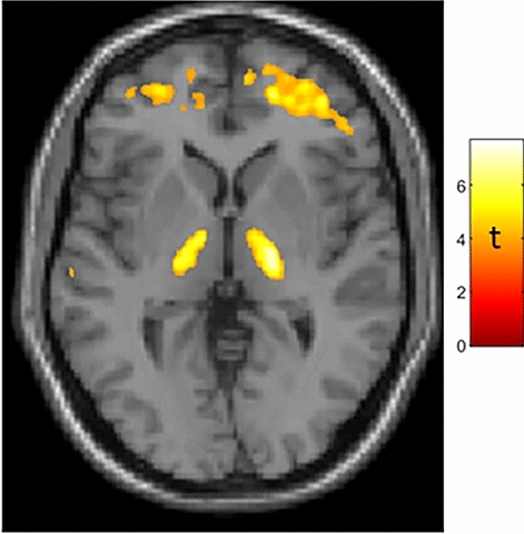
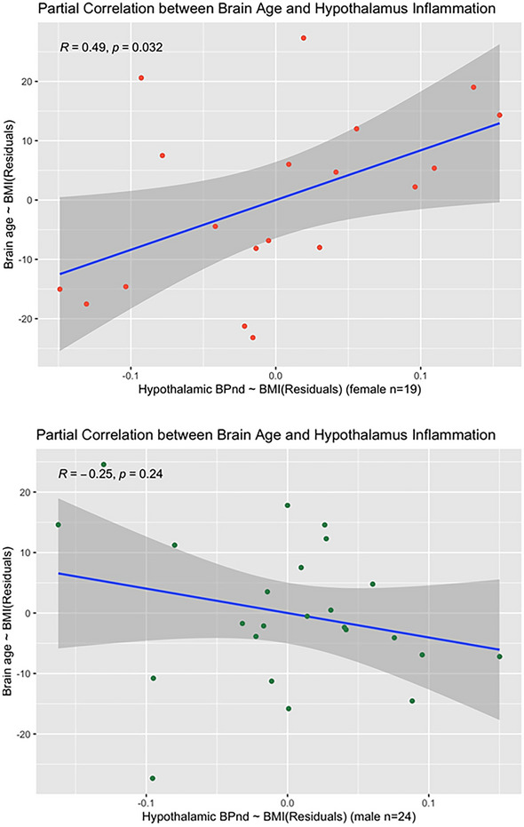
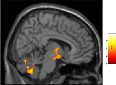
In rodents, hypothalamic inflammation plays a critical role in aging and age-related diseases. Hypothalamic inflammation has not previously been assessed in vivo in humans. We used Positron Emission Tomography (PET) with a radiotracer sensitive to the translocator protein (TSPO) expressed by activated microglia, to assess correlations between age and regional brain TSPO in a group of healthy subjects (n = 43, 19 female, aged 23-78), focusing on hypothalamus. We found robust age-correlated TSPO expression in thalamus but not hypothalamus in the combined group of women and men. This pattern differs from what has been described in rodents. Prominent age-correlated TSPO expression in thalamus in humans, but in hypothalamus in rodents, could reflect evolutionary changes in size and function of thalamus versus hypothalamus, and may be relevant to the appropriateness of using rodents to model human aging. When examining TSPO PET results in women and men separately, we found that only women showed age-correlated hypothalamic TSPO expression. We suggest this novel result is relevant to understanding a stark sex difference in human aging: that only women undergo loss of fertility-menopause-at mid-life. Our finding of age-correlated hypothalamic inflammation in women could have implications for understanding and perhaps altering reproductive aging in women.


Conflict of interest statement

The authors declare no competing interests.

Figures

Figure 1
Figure 1
Age-correlated TSPO expression in bilateral thalamus in a mixed-sex group of 43 subjects. Whole-brain statistical parametric analysis with a machine-learning derived measure of brain age as the regressor of interest, controlling for sex and BMI, shows robust results in bilateral thalamus (peak MNI coordinates: X = 20, Y = − 14, Z = 4 [t = 7.05]; X = 17, Y = − 18, Z = − 4 [t = 6.1]) as well as frontal subcortical regions. T-map is displayed at puncorected < 0.001. Results were similar when using chronologic age, which was strongly correlated with brain age (r = 0.819, p < 0.001) as the regressor of interest. Additional results of SPM analysis are presented in Supplementary Fig. 1 and Table S2.
Figure 2
Figure 2
Partial correlation plots showing sex differences in the partial correlation (controlling for BMI) between brain age and hypothalamic TSPO expression, with the hypothalamus segmented in subjects’ native space using a deep learning technique. Top Panel: Women show a significant positive partial correlation between brain age and hypothalamic TSPO expression (r = 0.49; p = 0.03). Bottom Panel: Men show a nonsignificant negative correlation (r = − 0.25, p = 0.24). Fisher Z-comparison showed that these correlations between brain age and hypothalamic TSPO expression different significantly between women and men (p = 0.009).
Figure 3
Figure 3
Sagittal view (right of midline; X = 8) of whole-brain statistical z-map showing regions where brain age-correlated TSPO expression was greater in women than men, with prominent results in a region spanning both hypothalamus and thalamus (hypothalamus peak: X = 11, Y = − 7, Z = − 13 [z = 4.2]; thalamus peak: X = 4,Y = − 6, Z = 0 [z = 5.1]; center of gravity in hypothalamus: X = 10, Y = − 5, Z = − 9; additional results of SPM analysis are presented in Supplementary Fig. 2 and Table 5). This sex difference corresponds to significant age-correlated TSPO expression in hypothalamus in women (peak: X = 8, Y = 0, Z = − 12 [t = 3.3; pcorrected = 0.002]) but not men. There were no areas of greater age-correlated TSPO expression in men as compared to women.

References

    1. Swaab DF, Fliers E. A sexually dimorphic nucleus in the human brain. Science. 1985;228:1112–1115. - PubMed
    1. Cai D, Khor S. “Hypothalamic microinflammation” paradigm in aging and metabolic diseases. Cell Metab. 2019;30:19–35. - PubMed
    1. Ishii M, Iadecola C. Metabolic and non-cognitive manifestations of Alzheimer’s disease: the hypothalamus as both culprit and target of pathology. Cell Metab. 2015;22:761–776. - PMC - PubMed
    1. Sadagurski M, Cady G, Miller RA. Anti-aging drugs reduce hypothalamic inflammation in a sex-specific manner. Aging Cell. 2017;16:652–660. - PMC - PubMed
    1. Liu T, Xu Y, Yi C-X, Tong Q, Cai D. The hypothalamus for whole-body physiology: from metabolism to aging. Protein Cell. 2021;39:394–421. - PMC - PubMed

Publication types