Review
. 2022 Oct;18(10):611-627.
doi: 10.1038/s41581-022-00601-z. Epub 2022 Aug 3.

Cellular senescence: the good, the bad and the unknown

Affiliations
Review

Cellular senescence: the good, the bad and the unknown

Weijun Huang et al. Nat Rev Nephrol. 2022 Oct.

Abstract

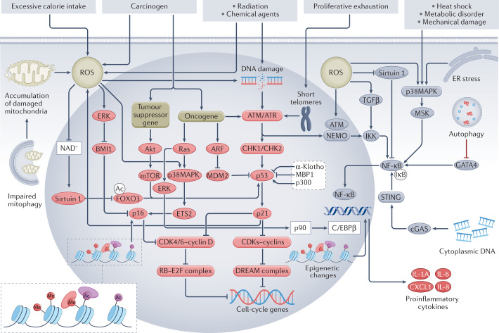
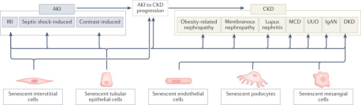
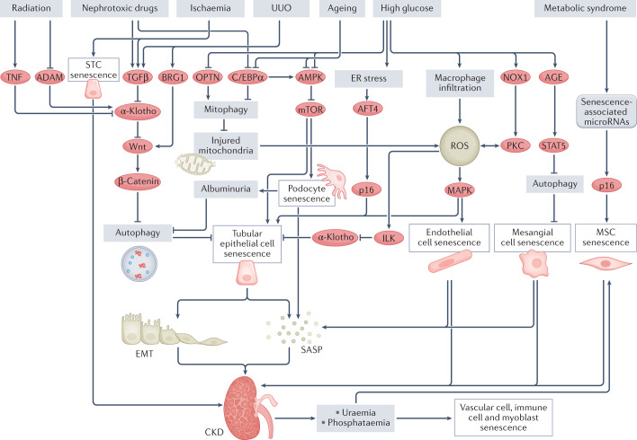
Cellular senescence is a ubiquitous process with roles in tissue remodelling, including wound repair and embryogenesis. However, prolonged senescence can be maladaptive, leading to cancer development and age-related diseases. Cellular senescence involves cell-cycle arrest and the release of inflammatory cytokines with autocrine, paracrine and endocrine activities. Senescent cells also exhibit morphological alterations, including flattened cell bodies, vacuolization and granularity in the cytoplasm and abnormal organelles. Several biomarkers of cellular senescence have been identified, including SA-βgal, p16 and p21; however, few markers have high sensitivity and specificity. In addition to driving ageing, senescence of immune and parenchymal cells contributes to the development of a variety of diseases and metabolic disorders. In the kidney, senescence might have beneficial roles during development and recovery from injury, but can also contribute to the progression of acute kidney injury and chronic kidney disease. Therapies that target senescence, including senolytic and senomorphic drugs, stem cell therapies and other interventions, have been shown to extend lifespan and reduce tissue injury in various animal models. Early clinical trials confirm that senotherapeutic approaches could be beneficial in human disease. However, larger clinical trials are needed to translate these approaches to patient care.


Conflict of interest statement

L.O.L. is an adviser to AstraZeneca, CureSpec, Beren, Ribocure Pharmaceuticals and Butterfly Biosciences. Patents on senolytic drugs and their uses are held by the Mayo Clinic. This research has been reviewed by the Mayo Clinic Conflict of Interest Review Board and was conducted in compliance with Mayo Clinic Conflict of Interest policies. The other authors declare no conflicts of interest.

Figures

Fig. 1
Fig. 1. Mechanisms of cellular senescence and the SASP.
DNA damage secondary to radiation, chemical agents or accumulation of reactive oxygen species (ROS) is the main cause of cellular senescence-inducing stress. Proliferation-induced telomere shortening can also activate the DNA damage response, which in turn leads to activation of the p53–p21 pathway, inhibition of cyclin-dependent kinase (CDK)–cyclin complexes and formation of the DREAM complex, which represses cell-cycle genes, leading to cell-cycle arrest and senescence. Activated p21 can also induce further ROS production, forming a vicious circle. In addition, impaired mitophagy can lead to mitochondrial dysfunction and excessive ROS production. ROS can induce senescence independently of the DNA damage response by activating the p16–RB pathway via activation of ERK, which inhibits BMI1 and thereby enables activation of p16, and by activating p38MAPK signalling, which upregulates ETS2 and in turn activates p16, and by inhibiting NAD+, which leads to reduced expression of sirtuin 1 and activation of FOXO3, which activates p53. P16 inhibits the formation of CDK4/6–cyclin D complexes and thereby promotes formation of the RB–2F complex, which inhibits the transcription of cell-cycle genes. Oncogene activation can activate the p53–p21 pathway not only via the DNA damage response, but also via the ARF–MDM2 signalling. In addition, oncogene activation can activate the p16–RB pathway via p38MAPK signalling. Loss of tumour suppressor genes induces senescence via Akt–mTOR signalling, which activates p53. Other factors that regulate the p53–p21 pathway include α-Klotho, MBP1 and p300. Epigenetic alterations such as methylation (Me) and acetylation (Ac) can induce senescence through the p16–RB pathway. NF-κB signalling regulates the senescence-associated secretory phenotype (SASP) and together with the transcription factor C/EBPβ, co-activates promoters of SASP genes, such as those that encode pro-inflammatory cytokines. The DNA damage response protein ATM together with NEMO activates the NF-κB–C/EBPβ signalling pathway. ROS can activate the SASP not only by promoting nuclear translocation of NEMO and activation of ATM, but also by inhibiting sirtuin 1 and activating p38MAPK and TGFβ, which in turn activate NF-κB. Heat shock, metabolic disorders, mechanical damage and endoplasmic reticulum (ER) stress can also activate the NF-κB–C/EBPβ pathway via p38MAPK signalling. Cytoplasmic DNA accumulation can trigger aberrant activation of cGAS-STING cytoplasmic DNA sensors and promote the SASP via activation of NF-κB. Impairment of autophagy hampers degradation of the transcription factor GATA4, which activates NF-κB and leads to initiation of the SASP. Notably, oncogenic Ras can activate C/EBPβ via ERK–p90 signalling and histone epigenetic changes can regulate the SASP independently of the NF-κB–C/EBPβ pathway. ARF, ADP-ribosylation factor; BMI1, B lymphoma Mo-MLV insertion region 1 homologue; cGAS, cyclic GMP–AMP synthase; CHK, checkpoint kinase; ERK, extracellular regulated protein kinases; ETS2, E26 transformation-specific proto-oncogene 2; E2F, early 2 factor; FOXO3, forkhead box protein O3; IKK, IκB kinase; MDM, mouse double minute 2; MSK, mitogen- and stress-activated protein kinase; NEMO, NF-κB essential modulator; STING, stimulator of interferon genes.
Fig. 2
Fig. 2. Cellular senescence in cancer.
Most carcinogenic mutations induce senescence through the p53–p21 and p16–RB pathways, although some mutations activate p21 directly. Reactive oxygen species (ROS)-induced senescence in cancer cells is mainly dependent on p16, p21 and/or p27. Senescence-induced cell-cycle arrest can prevent mutations from being passed on to the next generation of cells and accelerate immune clearance, resulting in suppression of tumurigenesis. However, the senescence-associated secretory phenotype (SASP) also contributes to a pro-inflammatory and growth-stimulatory microenvironment that can promote tumour development.
Fig. 3
Fig. 3. Cellular senescence in kidney diseases.
Cellular senescence is involved in the pathogenesis of acute kidney injury (AKI) and many types of chronic kidney disease (CKD). Data from animal models suggest that senescent interstitial and tubular epithelial cells contribute to ischaemia–reperfusion injury (IRI), septic shock-induced AKI and contrast-induced AKI, as well as to AKI-to-CKD progression. Tubular epithelial cell senescence has also been detected in many forms of CKD, including obesity-related nephropathy, membranous nephropathy, lupus nephritis, minimal change disease (MCD), unilateral ureteral obstruction (UUO), IgA nephropathy (IgAN) and diabetic kidney disease (DKD). Endothelial cell, podocyte and mesangial cell senescence might also contribute to DKD.
Fig. 4
Fig. 4. Mechanisms of cellular senescence in chronic kidney disease.
Several stimuli can trigger the senescence of various kidney cell types through different pathways. High glucose levels result in macrophage infiltration into the kidney, mitochondrial dysfunction and activation of NOX1–PKC signalling, which lead to an increase in reactive oxygen species (ROS) and senescence of tubular epithelial cells and endothelial cells. High glucose can also induce senescence of tubular epithelial cells by provoking endoplasmic reticulum (ER) stress, which activates ATF4–p16 signalling. In addition, high glucose induces mesangial cell senescence via AGE–STAT5 signalling, which leads to the inhibition of autophagy and therefore results in the accumulation of injured mitochondrial and ROS (not shown). Nephrotoxic drugs and ageing can induce podocyte and tubular epithelial cell senescence via inhibition of C/EBPα, which leads to a reduction in AMPK–mTOR signalling. Ageing and high glucose can also induce tubular epithelial cell senescence by inhibiting AMPK. Nephrotoxic drugs, ischaemia, radiation and unilateral ureteral obstruction (UUO) activate Wnt–β-catenin signalling, which inhibits autophagy and therefore induces the senescence of tubular epithelial cells. Notably, ischaemia and metabolic syndrome can induce the senescence of kidney scattered tubule-like cells (STCs) and mesenchymal stem cells (MSCs), respectively, resulting in impairment of kidney repair, which promotes progression to chronic kidney disease (CKD). Chronic kidney cell senescence promotes epithelial-to-mesenchymal transition (EMT) and results in the senescence-associated secretory phenotype (SASP), which increases inflammation, eventually leading to the development of fibrosis and CKD. Furthermore, CKD can lead to uraemia-induced senescence of immune cells, endothelial cells, vascular smooth muscle cells and MSCs. CKD is also associated with hyperphosphataemia, which induces senescence in myoblasts, endothelial cells and vascular smooth muscle cells and thereby contributes to sarcopenia and vascular calcification. ADAM, A-disintegrin and metalloproteinase; ATF4, activating transcription factor 4; BRG1, brahma-related gene 1; ILK, integrin-linked protein kinase; NOX1, NADPH oxidase 1; OPTN, optineurin.

Similar articles

Cited by

References

    1. Hayflick L. The limited in vitro lifetime of human diploid cell strains. Exp. Cell Res. 1965;37:614–636. doi: 10.1016/0014-4827(65)90211-9. - DOI - PubMed
    1. Di Leonardo A, Linke SP, Clarkin K, Wahl GM. DNA damage triggers a prolonged p53-dependent G1 arrest and long-term induction of Cip1 in normal human fibroblasts. Genes Dev. 1994;8:2540–2551. doi: 10.1101/gad.8.21.2540. - DOI - PubMed
    1. Gire V, Dulic V. Senescence from G2 arrest, revisited. Cell Cycle. 2015;14:297–304. doi: 10.1080/15384101.2014.1000134. - DOI - PMC - PubMed
    1. Sun D, Buttitta L. States of G0 and the proliferation-quiescence decision in cells, tissues and during development. Int. J. Dev. Biol. 2017;61:357–366. doi: 10.1387/ijdb.160343LB. - DOI - PubMed
    1. Herranz N, Gil J. Mitochondria and senescence: new actors for an old play. EMBO J. 2016;35:701–702. doi: 10.15252/embj.201694025. - DOI - PMC - PubMed

Publication types