. 2022 Sep;414(23):6801-6812.
doi: 10.1007/s00216-022-04243-9. Epub 2022 Aug 3.

Chromatographic separation of glycated peptide isomers derived from glucose and fructose

Affiliations

Chromatographic separation of glycated peptide isomers derived from glucose and fructose

Sebastian Schmutzler et al. Anal Bioanal Chem. 2022 Sep.

Abstract

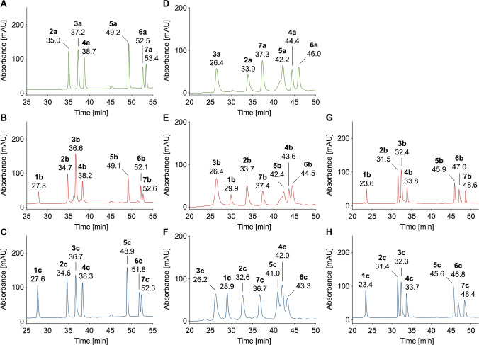
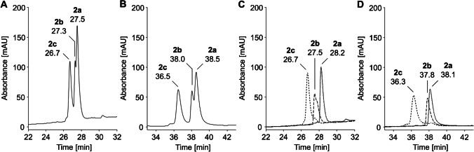
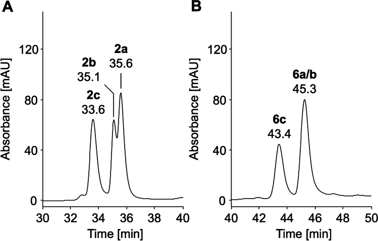
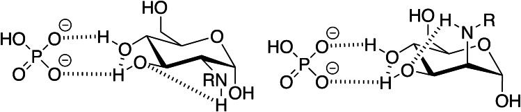
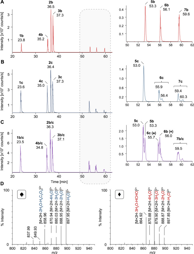
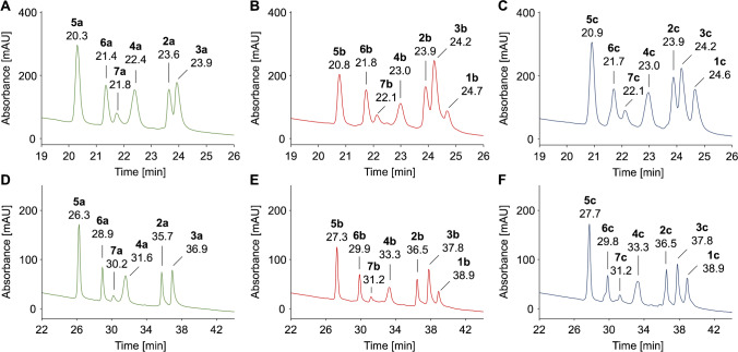
Amino groups in proteins can react with aldehyde groups in aldoses or keto groups in ketoses, e.g., D-glucose and D-fructose, yielding Schiff bases that rearrange to more stable Amadori and Heyns products, respectively. Analytical strategies to identify and quantify each glycation product in the presence of the corresponding isomer are challenged by similar physicochemical properties, impeding chromatographic separations, and by identical masses including very similar fragmentation patterns in tandem mass spectrometry. Thus, we studied the separation of seven peptide families, each consisting of unmodified, glucated, and fructated 15mer to 22mer peptides using reversed-phase (RP) and hydrophilic interaction chromatography (HILIC). In RP-HPLC using acidic acetonitrile gradients, unglycated peptides eluted ~ 0.1 to 0.8 min after the corresponding glycated peptides with four of seven peptides being baseline separated. Isomeric glucated and fructated peptides typically coeluted, although two late-eluting peptides were partially separated. Neutral eluents (pH 7.2) improved the chromatographic resolution (Rs), especially in the presence of phosphate, providing good and often even baseline separations for six of the seven isomeric glycated peptide pairs with fructated peptides eluting earlier (Rs = 0.7 to 1.5). Some glucated and unmodified peptides coeluted, but they can be distinguished by mass spectrometry. HILIC separated glycated and unmodified peptides well, whereas glucated and fructated peptides typically coeluted. In conclusion, HILIC efficiently separated unmodified and the corresponding glycated peptides, while isomeric Amadori and Heyns peptides were best separated by RP-HPLC using phosphate buffered eluents.

Keywords: Amadori and Heyns peptides; Fructation; Glucation; Hydrophilic interaction chromatography (HILIC); Reversed-phase high-performance liquid chromatography (RP-HPLC).


Conflict of interest statement

The authors declare no competing interests.

Figures

Fig. 1
Fig. 1
RP chromatograms of unmodified (green traces, n = 6), glucated (red traces, n = 7), and fructated (blue traces, n = 7) peptides (500 pmol each) showing the part of the chromatogram where the peptides eluted. Peptides were separated on a Jupiter C18 column (AC, GH) or Synergi Fusion RP column (DF) at 60 °C using eluent systems A (AC), B (DF), or C (GH) and gradients with a slope of 0.6% acetonitrile per minute. Absorbance was recorded at 214 nm. Full chromatograms are provided in the Supplement (Fig. S2, S12, S14). Peptide sequences and modification sites are provided in Table 1
Fig. 2
Fig. 2
RP chromatograms of unmodified (8a), glucated (8b), and fructated (8c) peptides (500 pmol each) of peptide family #2 (panels A and B) and an overlay of the RP chromatograms of individually injected peptides (panels C and D) displayed from 22 to 32 min and 33 to 43 min, respectively. Separations were performed on an Aqua C18 column at a column temperature of 60 °C using eluent system B and gradients with a slope of 1.8% (panels A and C) or 0.6% acetonitrile per minute (panels B and D). Absorbance was recorded at 214 nm. The full chromatograms are provided in the Supplement (Fig. S5, S6). Peptide sequences and modification sites are provided in Table 1
Fig. 3
Fig. 3
RP chromatograms of unmodified (a), glucated (b), and fructated (c) peptides (500 pmol each) of peptide family #2 (panel A) and #6 (panel B) displayed from 30 to 40 min and 40 to 50 min, respectively. Peptides were separated on a Synergi Fusion RP column (60 °C) using a linear 65-min gradient from 3 to 42% aqueous acetonitrile containing sodium phosphate (10 mmol/L, pH 7.2). Absorbance was recorded at 214 nm. The full chromatograms are provided in the Supplement (Fig. S7). Peptide sequences and modification sites are provided in Table 1
Fig. 4
Fig. 4
Possible hydrogen bonding interactions of 2-amino-2-deoxyglucosyl- and 2-amino-2-deoxymannosyllysine modification (HRP) with hydrogen phosphate
Fig. 5
Fig. 5
Extracted ion chromatograms (XICs) of triply (peptides #6, #2, #5, #4, and #7) and quadruply (peptides #3 and #1) protonated precursor ions of glucated peptides (b; panel A), fructated peptides (c; panel B), and a mixture of glycated peptides (panel C; 500 fmol each). Peptides were separated on a nanoRP-UPLC-ESI-Orbitrap-MS using an Acquity UPLC BEH C18 column (30 °C) and a linear 87-min gradient from 3 to 40% aqueous acetonitrile containing 0.1% formic acid. The inserts display the XICs from 50 to 62 min. Tandem mass spectra (panel D) recorded for the doubly protonated precursor ions at m/z 906.98 eluting at 55.7 min (6c) and 56.0 min (6b) when separating the glycated peptide mixture (panel C) confirming the structures of Amadori and Heyns peptides, respectively. Peptide sequences and modification sites are provided in Table 1
Fig. 6
Fig. 6
HILIC chromatograms of unmodified (panel A, D), glucated (panel B, E), and fructated peptides (panel C, F) (500 pmol each) displayed from 19 to 26 min or from 22 to 43 min. Peptides were separated on a Luna-HILIC column at room temperature using eluent system E and gradients with a slope of 1.8% (panels A–C) or 0.6% water per minute (panels D–F). Absorbance was recorded at 214 nm. Full chromatograms are provided in the Supplement (Fig. S16/S18). Peptide sequences and modification sites are provided in Table 1

Similar articles

Cited by

References

    1. Lustig RH. Ultraprocessed food: addictive, toxic, and ready for regulation. Nutr. 2020;2020(12):3401. doi: 10.3390/NU12113401. - DOI - PMC - PubMed
    1. Rondeau P, Bourdon E. The glycation of albumin: structural and functional impacts. Biochimie. 2011;93:645–658. doi: 10.1016/j.biochi.2010.12.003. - DOI - PubMed
    1. Singh VP, Bali A, Singh N, Jaggi AS. Advanced glycation end products and diabetic complications. Korean J Physiol Pharmacol. 2014;18:1–14. doi: 10.4196/kjpp.2014.18.1.1. - DOI - PMC - PubMed
    1. Spiller S, Frolov A, Hoffmann R. Quantification of specific glycation sites in human serum albumin as prospective type 2 diabetes mellitus biomarkers. Protein Pept Lett. 2017;24:1–1. doi: 10.2174/0929866524666170202124120. - DOI - PubMed
    1. Gugliucci A. Formation of fructose-mediated advanced glycation end products and their roles in metabolic and inflammatory diseases. Adv Nutr. 2017;8:54–62. doi: 10.3945/an.116.013912.54. - DOI - PMC - PubMed

LinkOut - more resources