. 2022 Aug 3;28(1):88.
doi: 10.1186/s10020-022-00494-5.

Oxalate-induced apoptosis through ERS-ROS-NF-κB signalling pathway in renal tubular epithelial cell

Affiliations

Oxalate-induced apoptosis through ERS-ROS-NF-κB signalling pathway in renal tubular epithelial cell

Shaoxiong Ming et al. Mol Med. .

Abstract

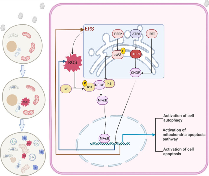
Background: Kidney stones are composed of approximately 70-80% calcium oxalate. However, the exact mechanism of formation of calcium oxalate kidney stones remains unclear. In this study, we investigated the roles of endoplasmic reticulum stress (ERS), reactive oxygen species (ROS), and the NF-κB signalling pathway in the pathogenesis of oxalate-induced renal tubular epithelial cell injury and its possible molecular mechanisms.

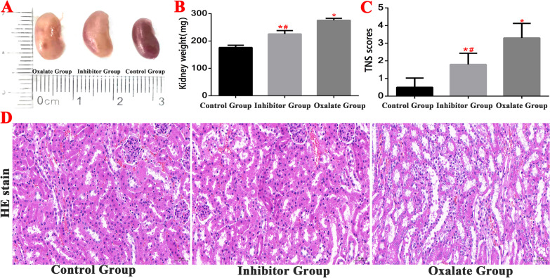
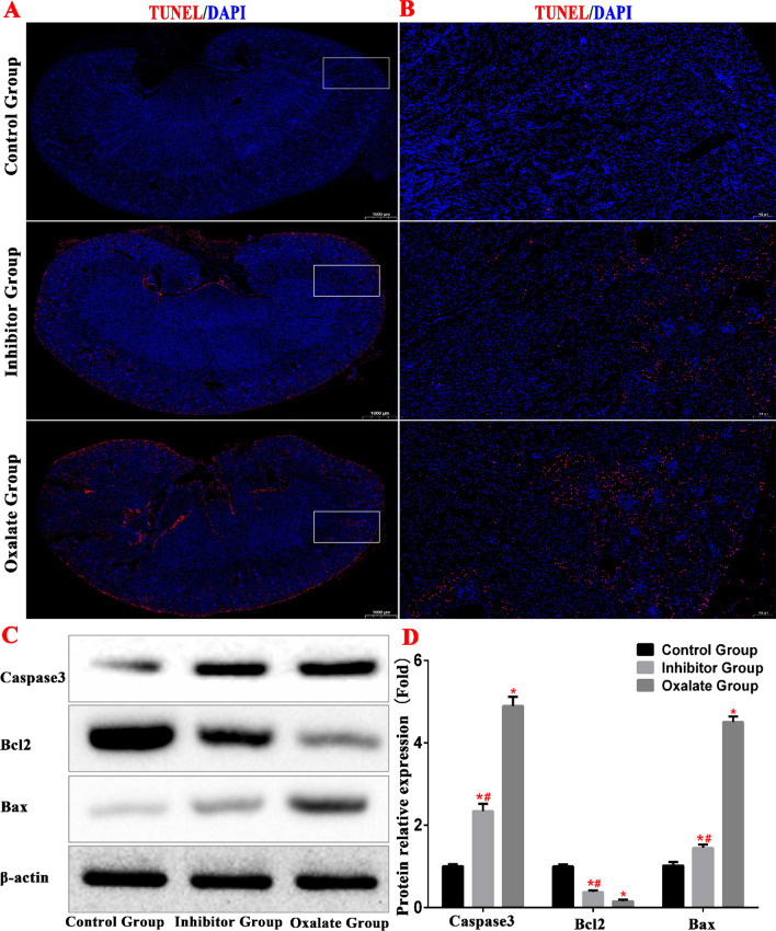
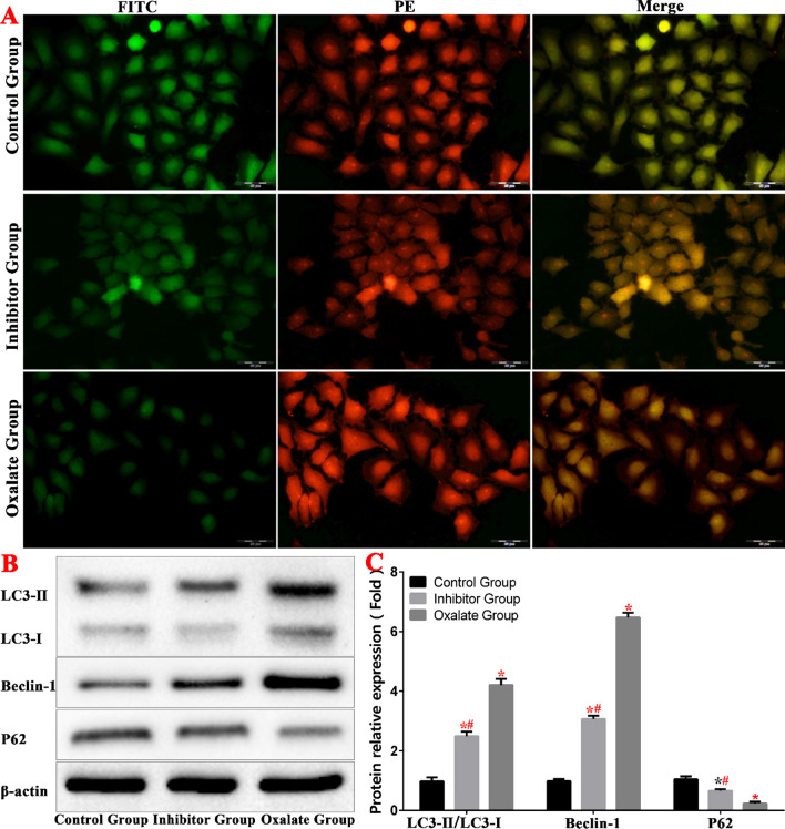
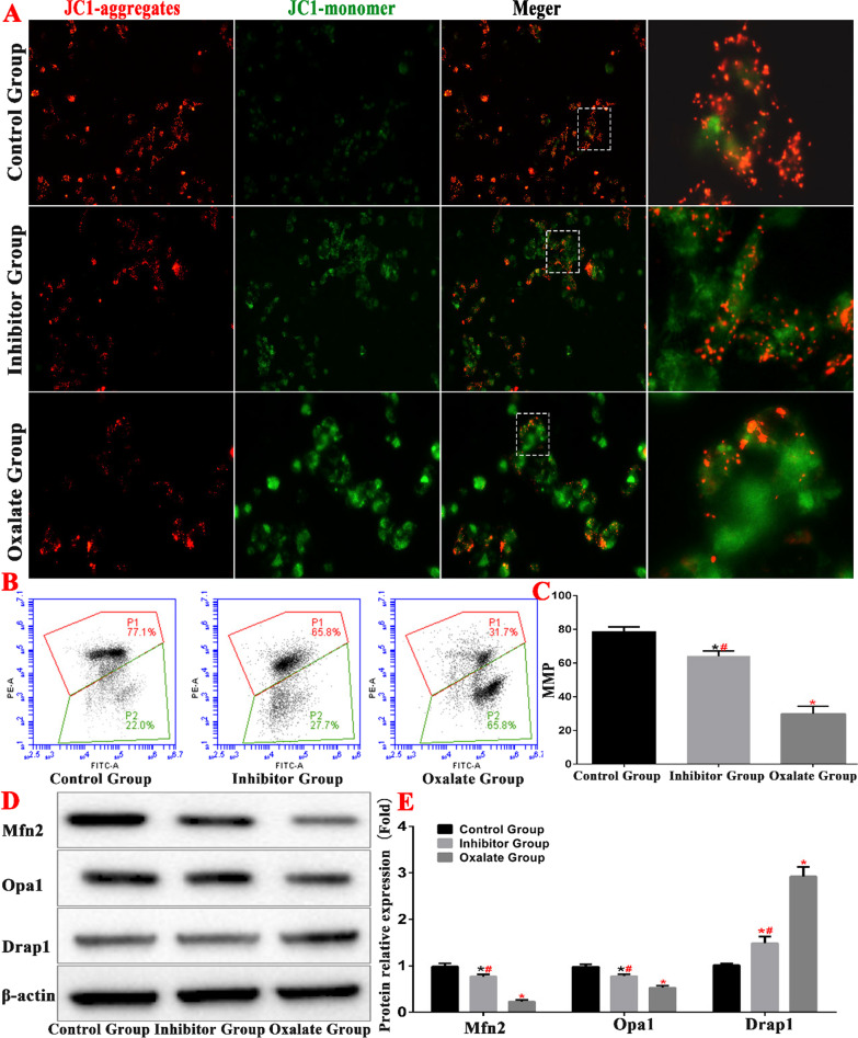
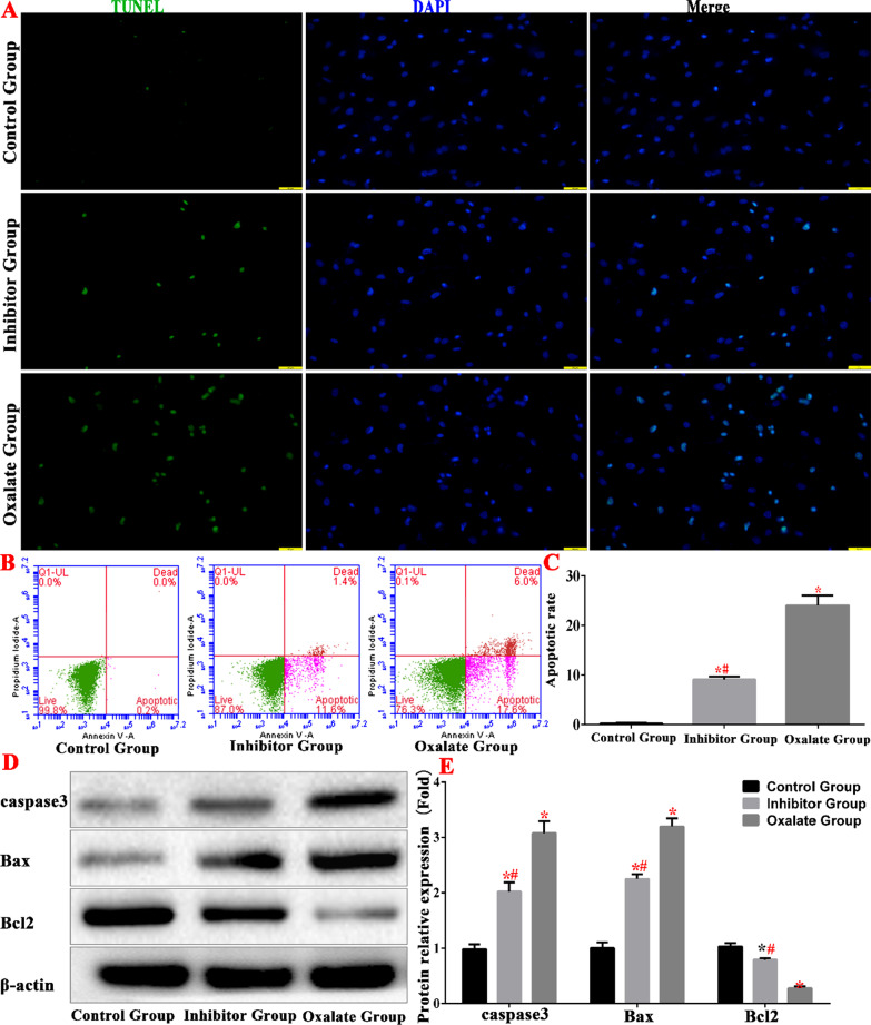
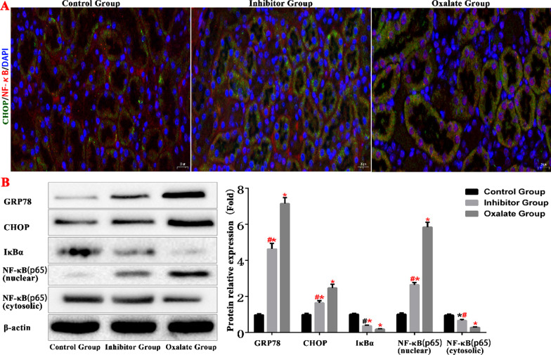
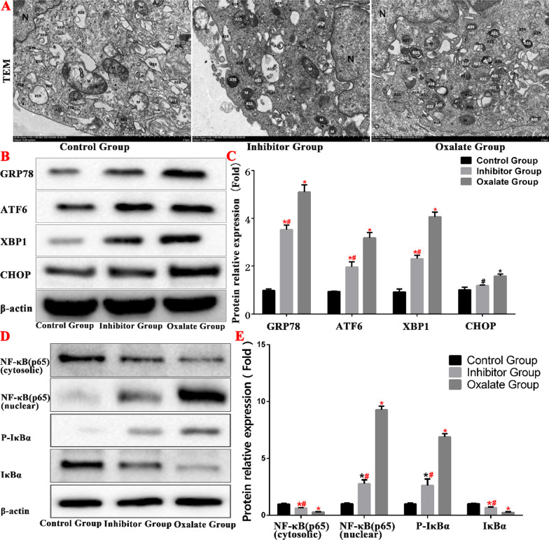
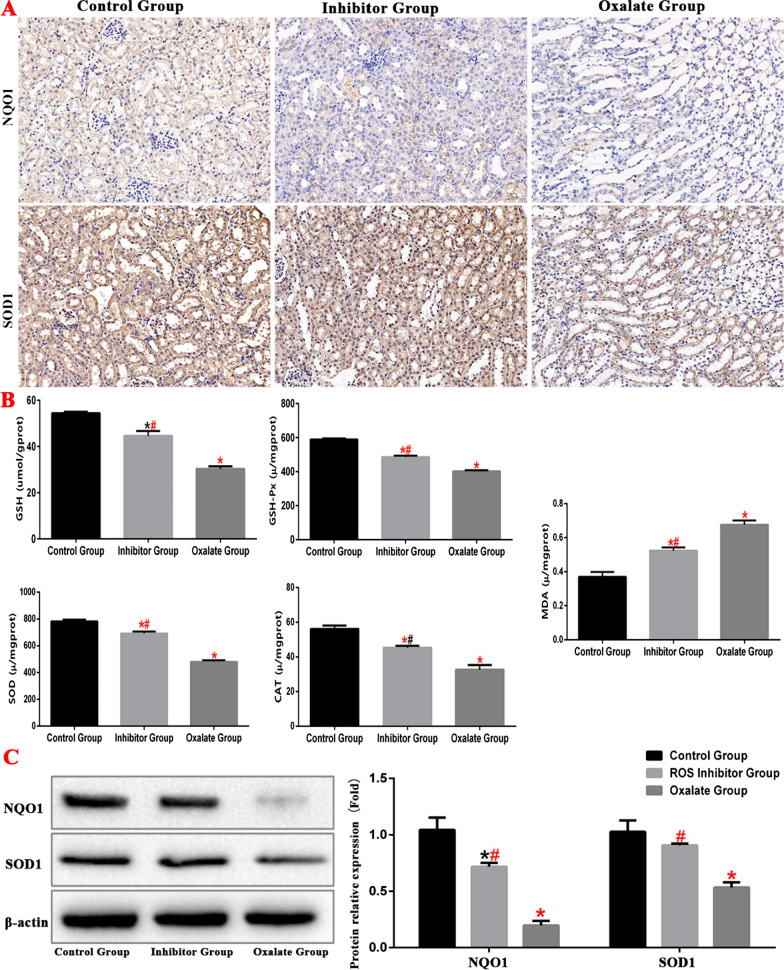
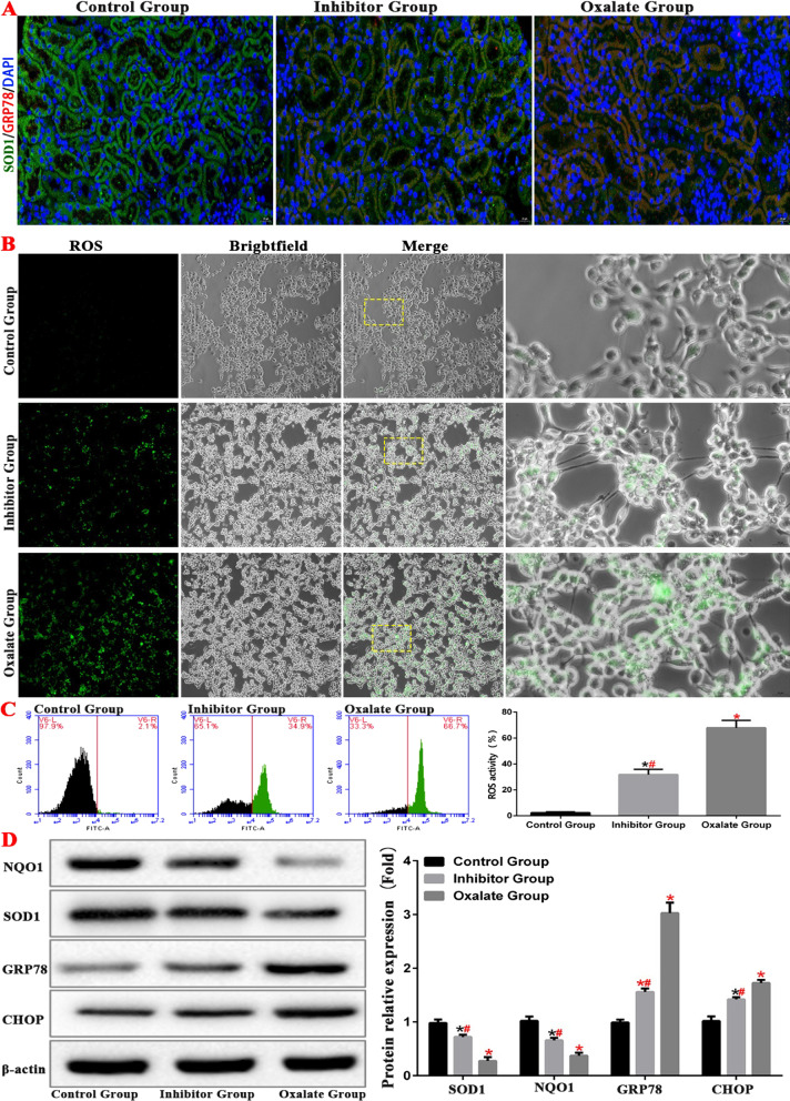
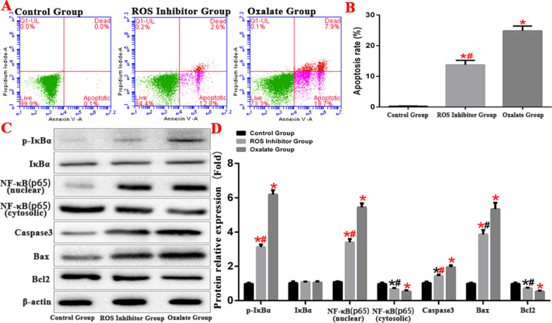
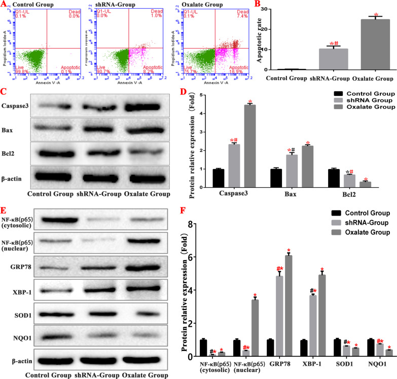
Methods: We established a model to evaluate the formation of kidney stones by intraperitoneal injection of glyoxylic acid solution into mice and assessed cell morphology, apoptosis, and the expression levels of ERS, ROS, and NF-κB signalling pathway-related proteins in mouse renal tissues. Next, we treated HK-2 cells with potassium oxalate to construct a renal tubular epithelial cell injury model. We detected the changes in autophagy, apoptosis, and mitochondrial membrane potential and investigated the ultrastructure of the cells by transmission electron microscopy. Western blotting revealed the expression levels of apoptosis and autophagy proteins; mitochondrial structural and functional proteins; and ERS, ROS, and NF-κB (p65) proteins. Lastly, we studied the downregulation of NF-κB activity in HK-2 cells by lentivirus interference and confirmed the interaction between the NF-κB signalling and ERS/ROS pathways.

Results: We observed swelling of renal tissues, increased apoptosis of renal tubular epithelial cells, and activation of the ERS, ROS, and NF-κB signalling pathways in the oxalate group. We found that oxalate induced autophagy, apoptosis, and mitochondrial damage in HK-2 cells and activated the ERS/ROS/NF-κB pathways. Interestingly, when the NF-κB signalling pathway was inhibited, the ERS/ROS pathway was also inhibited.

Conclusion: Oxalate induces HK-2 cell injury through the interaction between the NF-κB signalling and ERS/ROS pathways.

Keywords: Endoplasmic reticulum stress (ERS); Kidney stones; NF-κB signalling pathway; Oxalate; Reactive oxygen species (ROS).


Conflict of interest statement

The authors declare that they have no competing interests.

Figures

Fig.1
Fig.1
Oxalate induces renal tubular epithelial cell injury by activating ERS/ROS/NF-κB signaling pathway
Fig. 2
Fig. 2
Detection of kidney tissue damage in Kidney stone model. A, B Comparison of kidney weight detection of mice in each group. C, D HE staining was used to observe the kidney structure and compare the renal tubular injury scores of mice in each group. (*(Red)Compared with control group, P < 0.01. #(Red)Compared with oxalate group, P < 0.01)
Fig. 3
Fig. 3
A, B TUNEL staining was used to observe the renal apoptosis of mice in each group. C, D Western blot was used to detect the expression of apoptosis-related proteins. (*(Red)Compared with control group, P < 0.01. #(Red)Compared with oxalate group, P < 0.01)
Fig. 4
Fig. 4
Oxalate-induced autophagy in HK-2 cells. A Autophagy level of each group of autophagy lentivirus. B, C Western blot was used to detect the expression of autophagy-related protein in each group.(*compared with normal Control group, P < 0.05.*(Red)Compared with control group, P < 0.01. #(Red)Compared with oxalate group, P < 0.01)
Fig. 5
Fig. 5
Oxalate-induced mitochondrial damage in HK-2 cells. A MMP detection in JC-I fluorescence groups. B, C Comparison of MMP detection in each group by flow cytometry. D, E Western blot was used to detect the expression of mitochondrial related protein in each group (*compared with normal control group, P < 0.05.*(Red)Compared with control group, P < 0.01. #(Red)Compared with oxalate group, P < 0.01)
Fig. 6
Fig. 6
Oxalate-induced apoptosis of HK-2 cells. A detection of apoptosis in each groups. B, C Comparison of apoptosis detection in each group by flow cytometry. D, E Western blot was used to detect the expression of apoptosis-related proteins in each group. (*compared with normal control group, P < 0.05.*(Red)Compared with control group, P < 0.01. #(Red)Compared with oxalate group, P < 0.01)
Fig. 7
Fig. 7
ERS and NF-κB protein expression level in kidney tissue of mice (*compared with normal Control group, P < 0.05.*(Red)Compared with Control group, P < 0.01. #Compared with oxalate group, P < 0.05. #(Red) Compared with oxalate group, P < 0.01)
Fig. 8
Fig. 8
Oxalate activates the NF-κB pathway by stimulating ERS in HK-2 cells. A Ultrastructure observation of each group under transmission electron microscope. B, C Western blot was used to detect the expression of ERS-related proteins in each group. D, E Western blot was used to detect the expression of NF-κB pathway proteins in each group. (*compared with normal Control group, P < 0.05.*(Red)Compared with Control group, P < 0.01. #Compared with oxalate group, P < 0.05. #(Red)Compared with oxalate group, P < 0.01)
Fig. 9
Fig. 9
ERS and ROS protein expression level in kidney tissue of mice in each group. (*compared with normal control group, P < 0.05.*(Red)Compared with control group, P < 0.01. #Compared with oxalate group, P < 0.05. #(Red)Compared with oxalate group, P < 0.01)
Fig. 10
Fig. 10
ERS inhibits ROS levels in kidney tissue of mice and HK-2 cell. A The expressions of GPR78 and SOD1 in the kidney of model mice were detected by immunofluorescence. B, C The level of ROS in HK-2 cell model was detected by fluorescence microscope and flow cytometry. D Western blot was used to detect the expression of ERS and ROS related proteins protein in HK-2 cell model. (*compared with normal control group, P < 0.05.*(Red)Compared with control group, P < 0.01. #Compared with oxalate group, P < 0.05. #(Red)Compared with oxalate group, P < 0.01)
Fig. 11
Fig. 11
ROS induces apoptosis of HK-2 cells by activating NF-κB signaling pathway. A, B Comparison of apoptosis detection in each group by flow cytometry. C, D Western blot was used to detect the expression of apoptosis-related proteins and NF-κB protein in each group. (*compared with normal Control group, P < 0.05.*(Red)Compared with Control group, P < 0.01. #Compared with oxalate group, P < 0.05. #(Red)Compared with oxalate group, P < 0.01)
Fig. 12
Fig. 12
Inhibition of NF-κB signaling pathway can inhibit ERS and ROS levels in cells. A, B Comparison of apoptosis detection in each group by flow cytometry. C, D Western blot was used to detect the expression of apoptosis-related proteins and NF-κB protein in each group. E, F Western blot was used to detect the expression of ERS and ROS related proteins and NF-κB protein in each group. (*compared with normal Control group, P < 0.05.*(Red)Compared with control group, P < 0.01. #Compared with oxalate group, P < 0.05. #(Red)Compared with oxalate group, P < 0.01)

Similar articles

Cited by

References

    1. Cai Y, Arikkath J, Yang L, Guo ML, Periyasamy P, Buch S. Interplay of endoplasmic reticulum stress and autophagy in neurodegenerative disorders. Autophagy. 2016;12(2):225–244. doi: 10.1080/15548627.2015.1121360. - DOI - PMC - PubMed
    1. Cybulsky AV. Endoplasmic reticulum stress, the unfolded protein response and autophagy in kidney diseases. Nat Rev Nephrol. 2017;13(11):681–696. doi: 10.1038/nrneph.2017.129. - DOI - PubMed
    1. Delgado M, Tesfaigzi Y. BH3-only proteins, Bmf and Bim, in autophagy. Cell Cycle. 2013;12(22):3453–3454. doi: 10.4161/cc.26696. - DOI - PMC - PubMed
    1. El-Husseiny T, Buchholz N. The role of open stone surgery. Arab J Urol. 2012;10(3):284–288. doi: 10.1016/j.aju.2012.03.004. - DOI - PMC - PubMed
    1. Frank S, Gaume B, Bergmann-Leitner ES, Leitner WW, Robert EG, Catez F, et al. The role of dynamin-related protein 1, a mediator of mitochondrial fission, in apoptosis. Dev Cell. 2001;1(4):515–525. doi: 10.1016/s1534-5807(01)00055-7. - DOI - PubMed

Publication types

LinkOut - more resources