. 2022 Aug 4;24(1):46.
doi: 10.1186/s12968-022-00877-x.

Le Cœur en Sabot: shape associations with adverse events in repaired tetralogy of Fallot

Affiliations

Le Cœur en Sabot: shape associations with adverse events in repaired tetralogy of Fallot

Anna Mîra et al. J Cardiovasc Magn Reson. .

Abstract

Background: Maladaptive remodelling mechanisms occur in patients with repaired tetralogy of Fallot (rToF) resulting in a cycle of metabolic and structural changes. Biventricular shape analysis may indicate mechanisms associated with adverse events independent of pulmonary regurgitant volume index (PRVI). We aimed to determine novel remodelling patterns associated with adverse events in patients with rToF using shape and function analysis.

Methods: Biventricular shape and function were studied in 192 patients with rToF (median time from TOF repair to baseline evaluation 13.5 years). Linear discriminant analysis (LDA) and principal component analysis (PCA) were used to identify shape differences between patients with and without adverse events. Adverse events included death, arrhythmias, and cardiac arrest with median follow-up of 10 years.

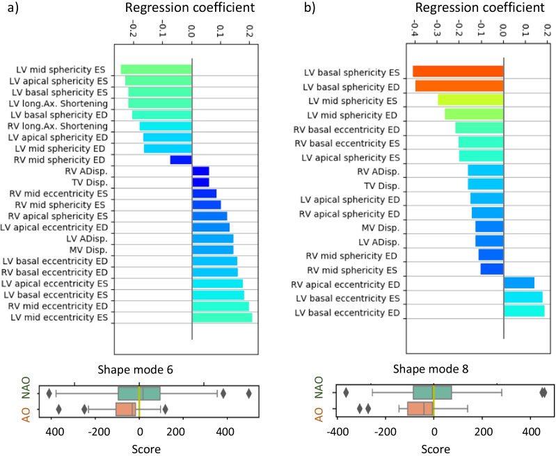
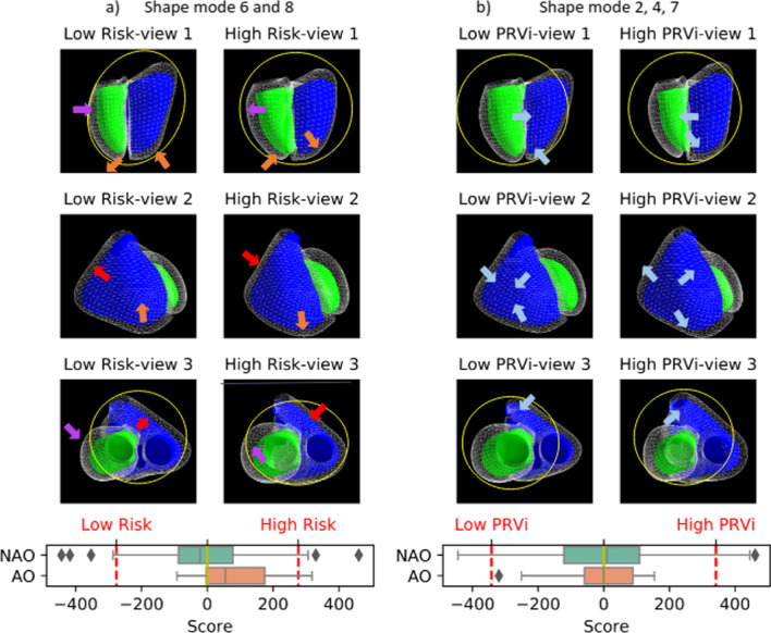
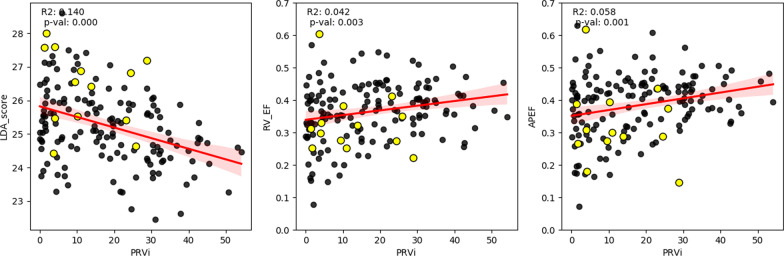
Results: LDA and PCA showed that shape characteristics pertaining to adverse events included a more circular left ventricle (LV) (decreased eccentricity), dilated (increased sphericity) LV base, increased right ventricular (RV) apical sphericity, and decreased RV basal sphericity. Multivariate LDA showed that the optimal discriminative model included only RV apical ejection fraction and one PCA mode associated with a more circular and dilated LV base (AUC = 0.77). PRVI did not add value, and shape changes associated with increased PRVI were not predictive of adverse outcomes.

Conclusion: Pathological remodelling patterns in patients with rToF are significantly associated with adverse events, independent of PRVI. Mechanisms related to incident events include LV basal dilation with a reduced RV apical ejection fraction.

Keywords: Biomarker; Biventricular shape; Magnetic resonance imaging; Tetralogy of Fallot.


Conflict of interest statement

A.D.M. and J.H.O. are co-founders of and have an equity interest in Insilicomed, and A.D.M. has an equity interest in Vektor Medical. A.D.M. and J.H.O. serve on the scientific advisory board of Insilicomed, and A.D.M. as scientific advisor to both companies. Some of their research grants have been identified for conflict of interest management based on the overall scope of the project and its potential benefit to these companies. The authors are required to disclose this relationship in publications acknowledging the grant support; however, the research subject and findings reported in this study did not involve the companies in any way and have no specific relationship with the business activities or scientific interests of either company. The terms of this arrangement have been reviewed and approved by the University of California San Diego in accordance with its conflict of interest policies.

Figures

Fig. 1
Fig. 1
Patient-specific biventricular shape modelling. Left—short axis contours, middle—long axis contours, right: biventricular shape. Contours: green—left ventricular (LV) epicardium, red—LV endocardium, yellow—right ventricle (RV) endocardium, orange—left atrium, cyan—right atrium. Surfaces: (right) green—RV, purple—LV
Fig. 2
Fig. 2
Volume parcellation. The right ventricle (RV) mesh is cropped according to the landmarks’ closest distance (PV—pulmonary valve, TV—tricuspid valve, A—RV apex). Cyan—inlet volume, green—outlet volume, and purple—apical volume
Fig. 3
Fig. 3
Definition of calliper distances. Yellow—left ventricle (LV). Blue—right ventricle (RV). HRV—the distance between tricuspid valve (TV) centroid and RV apex. HLV—the distance between the mitral valve (MV) centroid and RV apex. WRV—the distance between the RV free wall and the septum measured at mid RV height (r*HRV, r = ½) on the axis perpendicular to the septum. WLV—the distance between the LV endocardium and the septum measured at mid-LV height (r*HLV, r = ½) along the axis perpendicular to the septum. LRV—the distance between the two most distant points on the RV free wall at mid RV height (½*HLV) along the axis parallel to the septum
Fig. 4
Fig. 4
TOP: Association between geometrical features and the shape modes (SM): a SM6, b SM8. BOTTOM: Box-plots—score distribution of SM6 (a), SM8 (b) across the studied population for adverse outcomes (AO) and no adverse outcomes (NAO). The box denotes Q1 and Q3, whiskers Q1-1.5*IQR and Q3 + 1.5*IQR and diamonds are outliers. ED end-diastole, ES end-systole, LV left ventricle, RV right ventricle
Fig. 5
Fig. 5
Shape pattern associated with adverse events a and pulmonary regurgitation (b). Low risk: shape associated with no adverse outcomes. High risk: shape associated with adverse outcomes. Low pulmonary regurgitation: shape associated with reduced pulmonary regurgitation. High P: shape associated with high pulmonary regurgitation. Green and blue surfaces show LV and RV (respectively) at ES. Wireframe: LV and RV at ED. Red arrow: RV basal remodelling; purple—LV basal remodelling; orange—RV apical remodelling and LV apical displacement; blue—RV apical dilatation. Yellow circles illustrate the contrast between circular shape versus elliptical shape. Box plots: a Distribution of the risk score for adverse outcomes and no adverse outcomes, b) distribution of PR score for adverse outcomes and no adverse outcomes. The box denotes Q1 and Q3, whiskers Q1-1.5*IQR and Q3 + 1.5*IQR and diamonds are outliers. Red lines: + 2std and −2std representing the score of the plotted shapes
Fig. 6
Fig. 6
Association between pulmonary regurgitation and LDA risk score (SM6 + APEF), RVEF, and apical ejection fraction (APEF). Subjects with adverse events are marked in yellow circles

References

    1. Ferguson EC, Krishnamurthy R, Oldham SA. Classic imaging signs of congenital cardiovascular abnormalities. Radiographics. 2007;27(5):1323–1334. doi: 10.1148/rg.275065148. - DOI - PubMed
    1. Cohn JN, Ferrari R, Sharpe N. Cardiac remodeling–concepts and clinical implications: a consensus paper from an international forum on cardiac remodeling. Behalf of an International Forum on Cardiac Remodeling. J Am Coll Cardiol. 2000;35(3):569–582. doi: 10.1016/S0735-1097(99)00630-0. - DOI - PubMed
    1. Geva T, Sandweiss BM, Gauvreau K, Lock JE, Powell AJ. Factors associated with impaired clinical status in long-term survivors of tetralogy of Fallot repair evaluated by magnetic resonance imaging. J Am Coll Cardiol. 2004;43(6):1068–1074. doi: 10.1016/j.jacc.2003.10.045. - DOI - PubMed
    1. Valente AM, Gauvreau K, Assenza GE, Babu-Narayan SV, Schreier J, Gatzoulis MA, Groenink M, Inuzuka R, Kilner PJ, Koyak Z, et al. Contemporary predictors of death and sustained ventricular tachycardia in patients with repaired tetralogy of Fallot enrolled in the INDICATOR cohort. Heart. 2014;100(3):247–253. doi: 10.1136/heartjnl-2013-304958. - DOI - PMC - PubMed
    1. Pushparajah K, Duong P, Mathur S, Babu-Narayan S. Educational series in congenital heart disease: cardiovascular MRI and CT in congenital heart disease. Echo Res Pract. 2019 doi: 10.1530/ERP-19-0048. - DOI - PMC - PubMed

Publication types

MeSH terms