. 2022 Aug 3;19(1):22.
doi: 10.1186/s12983-022-00468-7.

Neurogenesis of the scallop Azumapecten farreri: from the first larval sensory neurons to the definitive nervous system of juveniles

Affiliations

Neurogenesis of the scallop Azumapecten farreri: from the first larval sensory neurons to the definitive nervous system of juveniles

Marina Kniazkina et al. Front Zool. .

Abstract

Background: Scallops are among the best-studied bivalve mollusks. However, adult nervous system and neurogenesis studies of scallops are limited. Here, we studied the localization of neurotransmitters (serotonin/5-HT, FMRFamide, catecholamines) in adult ganglia and larvae of Azumapecten farreri using histochemical and immunohistochemical methods.

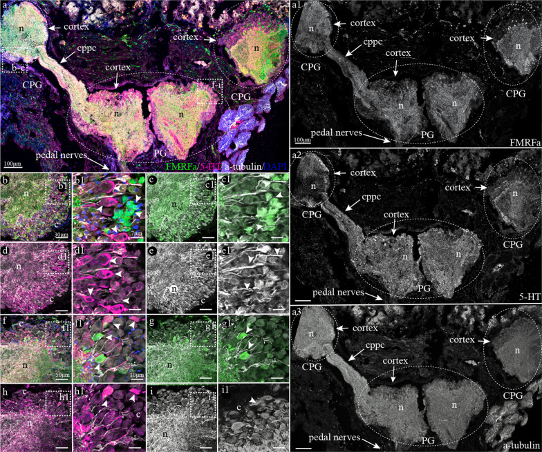
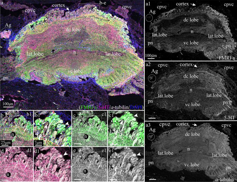
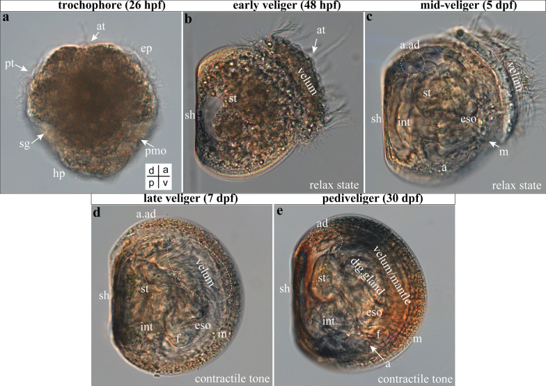
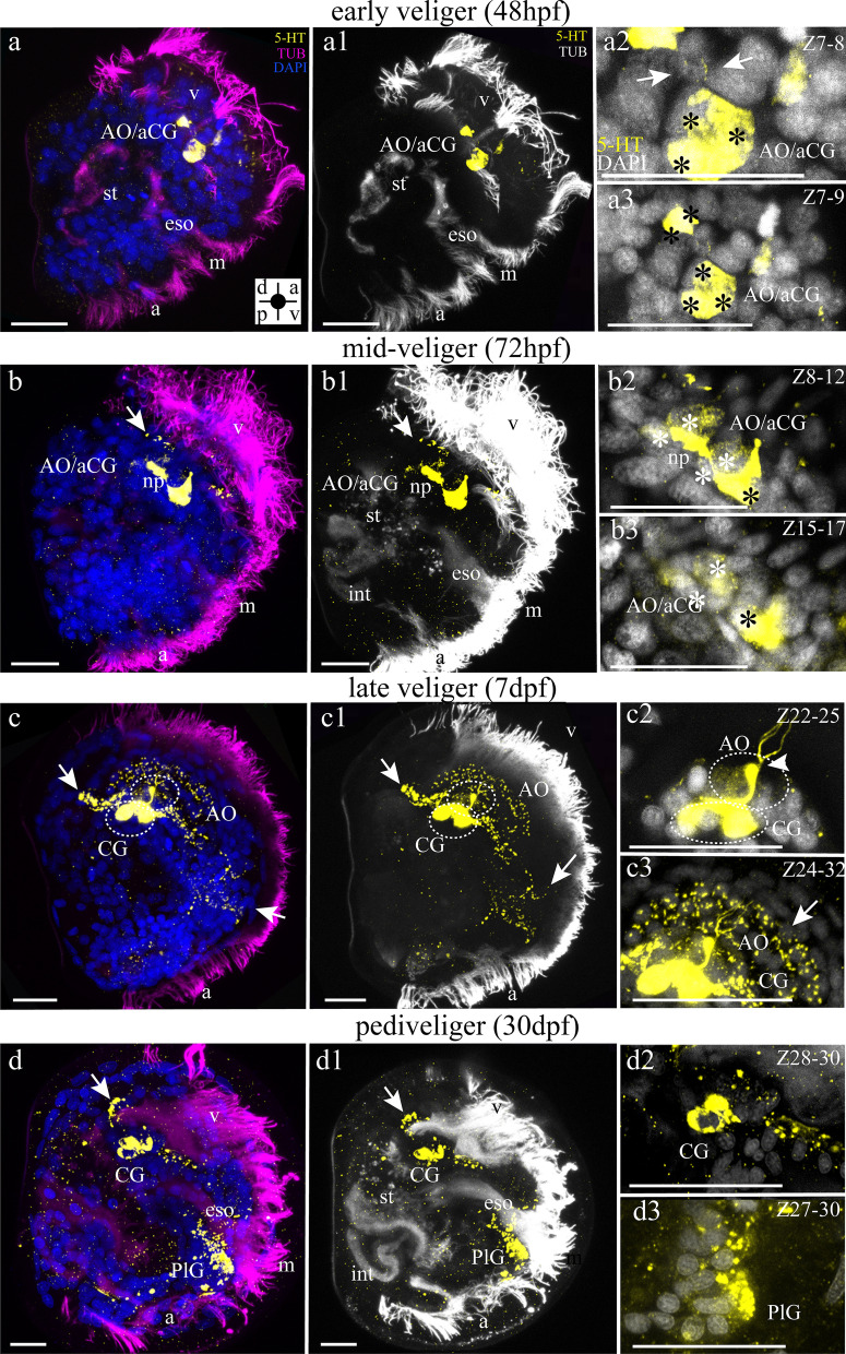
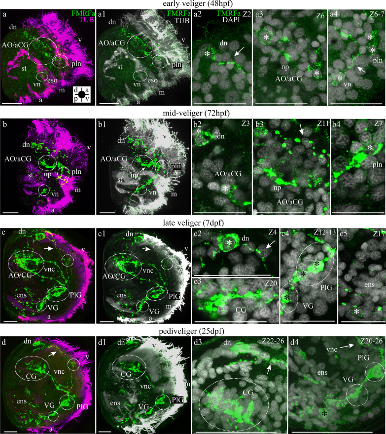
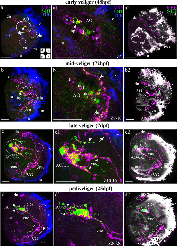
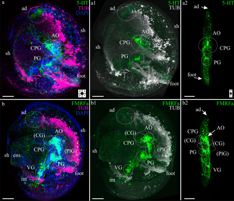
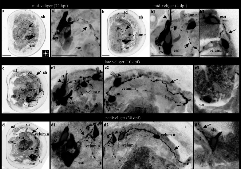
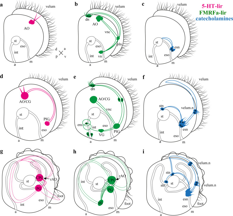
Results: We found peptide FMRFamide in all adult scallop ganglia, whereas 5-HT-like immunoreactive (lir) somata were exclusively detected in the cerebropleural, pedal, and accessory ganglia. Scallop larval neurogenesis starts with the emergence of the 5-HT-lir neurons, which are part of the apical organ (AO) at the early veliger stage. Near the AO, paired anlagen of cerebral ganglion (CG) developed. 5-HT-lir neurites of the CG innervate the velum, ventral, and dorsal parts of the larva at the late veliger stage. Scallop pediveligers possess 5-HT-lir CG, pleural ganglia, and immunopositive signals in the developing enteric nervous system. FMRFamide-lir is first detected in dorsal, ventral, and AO cells of early veligers. Later, FMRFamide-lir extends to the visceral nervous cord, all ganglia, as well as in the enteric nervous system in pediveligers. Catecholaminergic neurons are detected near the larval mouth, in the vellum, and in the stomach in veligers.

Conclusions: We described the distribution of neurotransmitters of the ganglia in adult scallops and the larval neurodevelopment in A. farreri. Immunostaining of neurotransmitters showed that the gross anatomy of adult scallop ganglia, in general, is similar to that in other bivalves, but complicated by the complexity of the structure of the ganglia and the appearance of additional ganglia not described in other molluscs. A comparison of larval neuromorphology suggests that 5-HT-lir structures are more conservative than FMRF-lir structures in Bivalvia. Notably, the latter are much more distributed in scallop A. farreri larvae than in other studied bivalves.

Keywords: Bivalve; Catecholamines; FMRFamide; Ganglia; Larvae; Neurogenesis; Neuromorphology; Serotonin.


Conflict of interest statement

The authors declare that they have no competing interests.

Figures

Fig. 1
Fig. 1
Cryo-section of the cerebropleural (CPG) and pedal (PG) ganglia of the adult scallop Azumapecten farreri immunostained with FMRFamide (green), serotonin (magenta), and acetylated α-tubulin (white) antibodies with DAPI (blue). a Tissue section view of the CPG connected with PG by cerebropleuro-pedal connective (cppc). The external part of the ganglia includes the neuronal somata (cortex) of ganglia; the inner part of ganglia is intertwined with neurites forming neuropil. Inserts: be1 Magnified parts of the CPG. fi1 The neuropil and cortex of the PG. Scale bars: a = 100 μm; b, c, d, e = 30 μm; b1, c1, d1, e1 = 7 μm; f, h, g, i = 50 μm; f1, h1, g1, i1 = 18 μm
Fig. 2
Fig. 2
Immunostaining of the visceral ganglion by FMRFamide (green), serotonin (magenta), and acetylated α-tubulin (white) antibodies with DAPI (blue) of the adult scallop Azumapecten farreri. a Tissue section of the visceral ganglion (VG) with accessory ganglion (Ag). The VG has a dorsal central lobe (dc lobe) ventral central lobe (vc lobe) and two lateral lobes (lat. lobe). Pallial nerves (pn) extending from the lateral lobes. On the upper side from the dorsal central lobe extend cerebropleuro-visceral connective (cpvc). The external part of the ganglia comprising of neuronal cells surrounds the central part of ganglia including the intertwined neurites (neuropil). Inserts: be1 Magnified parts of the VG. Scale bars: a = 150 μm; b, c, d, e = 50 μm; b1, c1, d1, e1 = 13 μm
Fig. 3
Fig. 3
Some stages of development of the scallop Azumapecten farreri. Light microscopy images. a trochophore, b early veliger, c middle veliger, d late veliger, e pediveliger stage. Abbreviations: ad—anterior adductor, at—apical tuf, ep—episphere, eso—esophagus, hp—hyposphere, int—intestine, m—mouth, pmo—presumptive mouth opening, pt—prototroch, sg—shell gland, sh—shell, st—stomach, v—velum. The orientation a—anterior, d—dorsal, p—posterior, v—ventral. Scale bars = 20 μm
Fig. 4
Fig. 4
Serotonin immunoreactivity (5-HT-lir) in the veliger larvae of Azumapecten farreri. Yellow—5-HT-lir; mangeta (ad) or white (a1d1)—cilia, α-tubulin immunoreactivity; blue (ad) or white (a2d2)—cell nuclei, DAPI. a The early veliger at 48 hpf. The apical organ (AO) comprises five 5-HT-lir cells with thin neurites running to the velum (v). Inserts: a1 Five cells with thin neurites (arrows). a2 Five cells with thin neurites (arrow) extending to the velum. b The middle veliger stage at 72 hpf. Cells of the AO are surrounded by neuropil and extend neurites (arrows) to the velum. Inserts: b1 Four paired cells of the AO (asterisks) and neurites extend to velum (arrow). b2 The neuropil of the AO with extending neurites (arrow). c The late veliger stage at 7 dpf. The AO is transformed into a cerebral ganglion (CG), which has thin neurites extending to the velum. From CG, neurites extend to the dorsal (arrow) and ventral parts of the larvae (arrow). Likewise on the ventral side of the larvae, neurites form a net of an enteric nervous system (ens). Inserts: c1 5-HT-lir neurites extend from CG to the dorsal side of the larvae (arrow) and to the velum (arrow) and c2 to the ventral side of the larvae and enteric nervous system (ens). d The pediveliger stage is 30 dpf. CG 5-HT-lir neurites extend to the dorsal part of larvae and to the ventral part of the larvae and enteric nervous system (ens). The two neurons located in the ventral part of the larvae are likely components of the prospective pleural ganglion (PlG). Inserts: d1 5-HT-lir innervation extends from CG to the dorsal side of the larva’ s body. d2 5-HT-lir cells like part of PlG. Additional abbreviations: a—anus, m—mouth, st—stomach. The orientation a- anterior, d—dorsal, p—posterior, v—ventral. The orientation a- anterior, d—dorsal, p—posterior, v—ventral. Scale bars = 20 μm
Fig. 5
Fig. 5
FMRFamide-like immunoreactivity (FMRFamide-lir) and acetylated α-tubulin staining and DAPI in the stages of the larvae of Azumapecten farreri. Green—FMRFamide, magenta—acetylated α-tubulin immunoreactivity (ad) or white (a1d1); white—nuclei, DAPI (a2, a3, b2, b3, c2c4, d3, d4). The apical pole is always upward, and the ventral side is on the right. a) FMRFamide-lir components in the 48-hpf early veliger stage with apical organ (AO), dorsal neurons (dn) and neurites extends to the ventral part of the larvae (arrows). Inserts: a1 Two paired cells of the apical organ (AO). a2 Dorsal neurons (dn) with cilia (arrowhead) and extended to ventral side of the larvae FMRFamide-lir neurites (arrow). b The larva of the middle veliger stage on 72 hpf, dorsal neurons and the nerve cord extends from AO to the ventral side of the larvae–visceral nerve cord (vnc). Inserts: b1 Number of cells in AO increased to eight (asterisks) and two paired dorsal neurons (asterisks, dn). b2 The connection of the visceral nerve cord and nerve cord extending from the dorsal neurons on the ventral side of the larva with appeared FMRFamide-lir ventral neurons (vn). b3 FMRFamide-lir neurites (arrow) in the ventral part of the larva and neurons of enteric innervation (asterisks). c Larvae at the veliger stage (7 dpf) demonstrate the presence of FMRFamide-lir elements in the CG, visceral ganglion (VG), pleural neurons (pln), dorsal neurons (dn) and enteric nervous system (ens). Inserts: c1 Net of neurites (arrow) between dorsal neurons (asterisks), CG (asterisks). c2, c3 Paired visceral nerve cords (vnc) connecting CG and pleural neurons (pln), which connect neurites (arrow) with VG and follows enteric innervation (arrows, asterisks, ens). d The 25-dpf pediveliger stage with the CG, paired visceral cords (vnc), pedal (PG), and visceral ganglia (VG) located along the visceraneural cord. Inserts: d1 Dorsal neurons (asterisks, dn) with cilia (arrowhead), neurites (arrow) and cerebral ganglion CG. d2 Visceral nerve cord (vnc) on the ventral side of the larva connected with pleural ganglion (PlG) and visceral ganglion (VG). d3 FMFRa-lir neuron of the gut (asterisks, ens). Additional abbreviations: a—anus, m—mouth, st—stomach v—velum. The orientation a—anterior, d—dorsal, p—posterior, v—ventral. Scale bars = 20 μm
Fig. 6
Fig. 6
Mutual distribution of FMRFamide-lir, 5-HT-lir and acetylated α-tubulin in the stages of the larvae of Azumapecten farreri. Green: 5-HT-lir, magenta: FMRFamide-lir, blue (ad, a1d1 or white (a2d2)): acetylated α-tubulin-lir. a FMRFamide-lir and 5-HT-lir components of the larval nervous system in the 48-hpf early veliger stage with apical organ (AO), dorsal neuron (dn) and neurites extending to the ventral part of the larvae. Inserts: a1 Two paired 5-HT-lir cells (asterisks) lie between two FMRFamide-lir cells (asterisk) of the apical organ (AO) near the apical tuft of cilia (at). In the central part of the AO, neurites form the neuropil. b The larva of the middle veliger stage is 72 hpf. AO includes 5-HT-lir cells (asterisk) and FMRFamidel-lir cells (asterisk). From FMRFamide-lir cells extending to the ventral side of the larva, there appear pleural neurons (pln) and down ventral neurons (vn). the dorsal part of the larva lie dorsal neurons (dn). The nerve cord extends from AO to the ventral side of the larvae—visceral nerve cord (vnc). Inserts: b1 The AO including 5-HT-lir cells (asterisk) and FMRFamide-lir cells (asterisk). c Larva at the veliger stage (7 dpf) demonstrate the presence of FMRFamide-lir elements in the CG (transformed from AO) including 5-HT-lir cells and FMRFamide-lir cells, visceral ganglion (VG) include only FMRFamide-lir elements, pleural ganglion (PlG) include only FMRFamide-lir elements, FMRFamide-lir dorsal neurons (dn) and interstitial innervation (ems). Inserts: c1 Transformed CG extending from four paired visceral cords (two 5-HT-lir cords and two FMRFamide-lir cords). d The 25-dpf pediveliger stage with the CG (including 5-HT-lir cells and FMRFamide-lir cells), four paired visceral cords (two 5-HT-lir cords and two FMRFamide-lir cords) (vnc), pedal ganglion (PG) including only FMRFamide-lir elements, and visceral ganglion (VG) including only FMRFamide-lir elements located along the visceral cords (vnc). Inserts: d1 Cerebral ganglion CG and visceral cord. From CG to the dorsal part of the larvae extends 5-HT-lir neurites and FMRFamide-lir neurites. Additional abbreviations: a—anus, m—mouth, st—stomach v—velum. The orientation a—anterior, d—dorsal, p—posterior, v—ventral. Scale bars = 20 μm
Fig. 7
Fig. 7
Neuromorphology of Azumapecten farreri pediveligers. a General view of 5-HT-lir (green) with the cerebropleural (CPG) and pedal (PG) ganglion together with acetylated α-tubulin (magenta) and DAPI (blue). a1a2 5-HT-lir (green) innervation of the foot is visible. Acetylated α-tubulin (pseudo-color, white) reveals foot and resorbing velum. Peripheral innervations of the mantle, anterior adductor muscles (ad) and enteric nervous system (ens) are visible. b General view of FMRFamide-lir (green) with the cerebropleural ganglion (CPG), pedal (PG), and visceral (VG) ganglia, acetylated α-tubulin (magenta) and DAPI (blue). b1b2 The FMRFamide-lir (green) innervations of the mantle, anterior adductor muscles (ad), and enteric nervous system (ens). Acetylated α-tubulin (pseudo-color, white) reveals foot and resorbing velum. Additional abbreviations: int—intestine, st—stomach, sh—shell. The orientation a—anterior, d—dorsal, p—posterior, v—ventral. Scale bars = 50 μm
Fig. 8
Fig. 8
FaGlu fluorescence of the catecholaminergic nervous system of A. farreri larvae. a Larvae in the middle veliger stage in 72 hpf have two catecholaminergic paired esophageal neurons (asterisks, esn) in a pharyngeal zone with two paired neurites along the esophagus (arrow). Inserts: a1 Magnified view of the two esophageal neurons (asterisks, esn) with neurites (arrow). b Middle veliger stage (4 dpf) has not only neurons of the esophagus (esophageal neurons, esn) and neurites but also innervation in the stomach of the larva like a net of neurites extending from esophageal neurons (arrows) and two appearing neurons in velum (velum.n). Inserts: b1 Esophageal neurons (asterisks, esn) have many cilia (arrowheads) near two paired neurons of the velum (asterisks, velum.n) connected with esophageal neurons by neurites (arrow). b2 The connection of the esophageal neurons with each other to neurites (arrows). c The 7 dpf-late veliger stage larva has esophageal neurons (esn, asterisk), velum.n with visible neurites along the velum (arrows), and more spread net of neurites with appeared catecholaminergic neuron (asterisks, stn) in the stomach zone. c1 Increased number of esophageal neurons from two to four cells (asterisks, esn) with cilia (arrowheads) and velum’s neurons (velum, n) connected to each other and to esophageal neurons by catecholamine-positive neurites (arrow). c2 Two neurons of the velum with extending catecholamine-positive nerve cord along the velum (arrow). c3 Catecholamine-positive neuron in the stomach of the larva (asterisks, stn). d Pediveliger stage on 30 dpf has esophageal neurons (esn) with connecting neurites similar to that in previous stages, stomach neuron (stn) with a net-like innervation in the stomach zone, velum’ s neurons (velum, n) with a neuronal cord along the velum with appearing additional neurons in velum (asterisk, velum, n). Inserts: d1 Six esophageal neurons of the pharyngeal zone (asterisks, esn), neurons of velum (asterisks, velum, n) and neurites (arrows). d2 Nerve cord of the velum (arrows) with two pairs of neurons (asterisks, velum, n). d3 Stomach innervation by two unpaired cells (asterisks, stn) with neurites (arrows). Additional abbreviations: v—velum. The orientation a—anterior, d—dorsal, p—posterior, v—ventral. Scale bars = 20 μm
Fig. 9
Fig. 9
Schematic presentation of the neurogenesis of A. farreri larvae. Serotonin-immunoreactive (5-HT-lir) elements are magenta; FMRFamide-lir elements are green; and catecholaminergic elements are blue. Black outlines provide a schematic overview of larval morphology and organs. d/v and a/p—orientation of larvae on the scheme. Three developmental stages are shown: ac early veliger, df late veliger, gi pediveliger. Somata are shown as asterisks. Prospective ganglia are shown as colored circles with the corresponding signatures of the ganglia or AO. The colored lines represent neurites connecting neurons or ganglia. Dotted lines represent the innervation of the pediveliger organs. Abbreviations: a—anus, AO—apical organ, CG—cerebral ganglia, CPG—cerebropleural ganglion, dn—dorsal neurons, eso—esophagus, esn—esophagus neurons, int—intestine, m—mouth, PG—pedal ganglia, PlG—pleural ganglion, st—stomach, stn -stomach neurons, velum, n—neurons of the velum, VG—visceral ganglia, vn—ventral neurons, vnc—visceral nerve cords. The orientation a—anterior, d—dorsal, p—posterior, v—ventral

Similar articles

Cited by

References

    1. Wilkens LA, Ache BW. Visual responses in the central nervous system of the scallop. Pecten ziczac. Experientia. 1977; 33:1338–1340 https://books.google.de/books?id=vhGdBAAAQBAJ&dq=wilkens+ache+1977&sourc...
    1. Odintsova N, Dyachuk V, Kiselev K, Shelud’ko N. Expression of thick filament proteins during ontogenesis of the mussel Mytilus trossulus (Mollusca: Bivalvia) Comput. Biochem. Physiol. B Biochem. Mol. Biol. 2006;144:238–244. doi: 10.1016/j.cbpb.2006.03.002. - DOI - PubMed
    1. Odintsova N, Dyachuk V, Nezlin L. Muscle and neuronal differentiation in primary cell culture of larval Mytilus trossulus (Mollusca: Bivalvia) Cell Tissue Res. 2010;339:625–637. doi: 10.1007/s00441-009-0918-3. - DOI - PubMed
    1. Dyachuk V, Wanninger A, Voronezhskaya EE. Innervation of bivalve larval catch muscles by serotonergic and FMRFamidergic neurons. Acta Biol Hung. 2012;63:221–229. doi: 10.1556/ABiol.63.2012.Suppl.2.30. - DOI - PubMed
    1. Leprêtre M, Almunia C, Armengaud J, Salvador A, Geffard A, Palos-Ladeiro MJ. The immune system of the freshwater zebra mussel, Dreissena polymorpha, decrypted by proteogenomics of hemocytes and plasma compartments. Proteomics. 2019;202:103366. doi: 10.1016/j.jprot.2019.04.016. - DOI - PubMed

LinkOut - more resources