. 2022 Aug 3;19(1):53.
doi: 10.1186/s12986-022-00686-0.

Salvianolic acid B activates chondrocytes autophagy and reduces chondrocyte apoptosis in obese mice via the KCNQ1OT1/miR-128-3p/SIRT1 signaling pathways

Affiliations

Salvianolic acid B activates chondrocytes autophagy and reduces chondrocyte apoptosis in obese mice via the KCNQ1OT1/miR-128-3p/SIRT1 signaling pathways

Tianwen Sun et al. Nutr Metab (Lond). .

Abstract

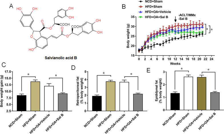
Purpose: Salvianolic acid B (Sal B) possesses strong anti-inflammatory and antioxidant activity. This study aims to explore the underlying mechanism of Sal B to improve the obesity-related osteoarthritis (OA).

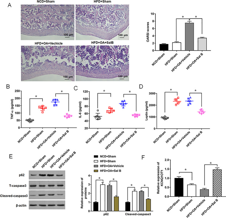
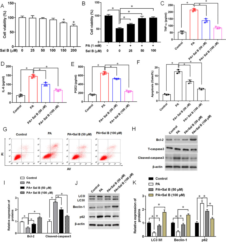
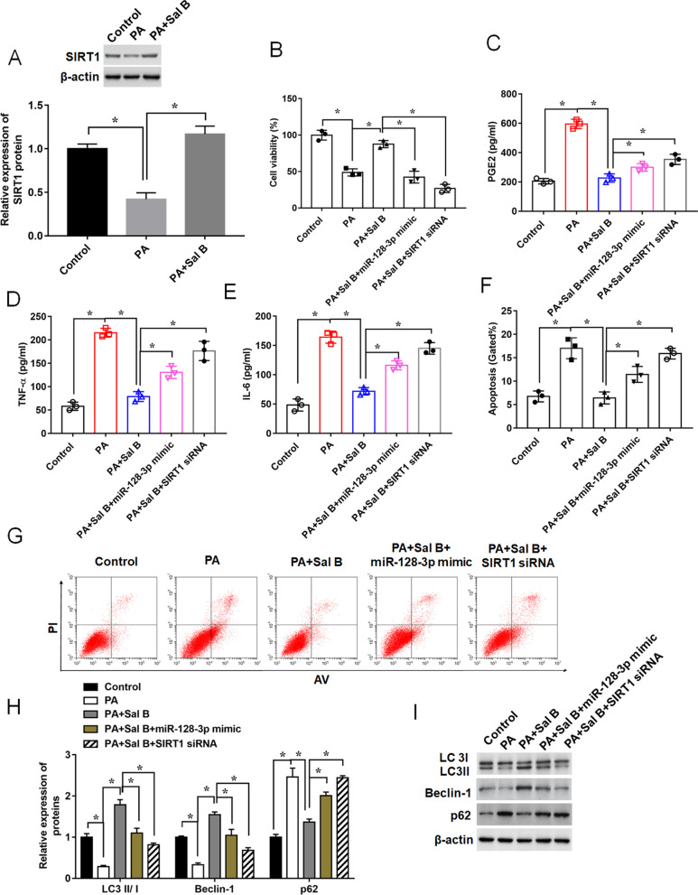
Methods: C57BL/6 J male mice were fed with a normal control diet (NCD), a high fat diet (HFD), or HFD with Sal B (25 mg/kg), and mouse body weights and osteoarticular inflammatory factor levels were examined. Mouse chondrogenic cell line ATDC5 were transfected with lncRNA KCNQ1 overlapping transcript 1 small hairpin RNA (KCNQ1OT1 shRNA), miR-128-3p mimic or Sirtuin-1 small interfering RNA (SIRT1 siRNA), then stimulated with Palmitic acid (PA) followed by the treatment of Sal B. Then, inflammatory response, apoptosis, and autophagy of ATDC5 cells in different groups were detected.

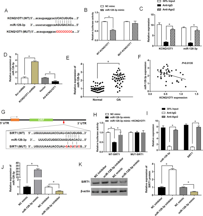
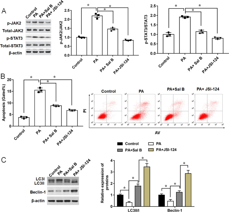
Results: Sal B reduced the body weight, decreased the levels of inflammatory markers, and improved cartilage damage in OA mice fed with HFD. KCNQ1OT1 was downregulated in OA mice fed with HFD, and PA-stimulated ATDC5 cells. Sal B protected ATDC5 cells against PA-mediated inflammation, apoptosis, and the inhibition of autophagy, while knockdown of KCNQ1OT1 reversed these results. KCNQ1OT1 was found to be functioned as a ceRNA to bind and downregulate the expression of miR-128-3p that was upregulated in PA-induced cells. Furthermore, SIRT1 was verified as a target of miR-128-3p. MiR-128-3p overexpression reversed the effects of Sal B on inflammatory response, apoptosis, and autophagy in PA-stimulated cells, and knockdown of SIRT1 displayed the similar results.

Conclusion: Sal B exerted a chondroprotective effect by upregulating KCNQ1OT1, which indicates Sal B can used for a therapeutic agent in obesity-related OA.

Keywords: Autophagy; KCNQ1OT1; Obesity; Osteoarthritis; Salvianolic acid B.


Conflict of interest statement

There is no conflict of interest to be declared by the authors.

Figures

Fig. 1
Fig. 1
Effects of Salvianolic acid B (Sal B) on body weight and fat pad weight. A Chemical skeleton structure of Sal B. B Body weights were measured weekly for 21 weeks. C Body weight gain obtained from the difference between the body weights during the 21 weeks. Retroperorenal fat (D) and Epididymal fat (E) were measured after 21 weeks. Male C57BL/6 mice were divided into four groups (n = 6 per group). Data are expressed as means ± SEM. Student’s t test was used for the comparison between 2 groups in this study. *P < 0.05
Fig. 2
Fig. 2
Sal B inhibits histological changes of knee joints and inflammatory response in mice fed with HFD. A Two knee joints per mouse with OA were collected after 21 weeks and stained with H & E (original magnification, 100 ×), Scale bars = 100 μm. BD Serum levels of TNF-α, IL-6 and leptin were measured by ELISA. E Relative expression levels of p62 and Cleaved-caspase3 were detected by Western blot analysis. F Expression of KCNQ1OT1 was determined by RT-qPCR. n = 6. Data are expressed as means ± SEM. Student’s t test was used for the comparison between 2 groups in this study. *P < 0.05
Fig. 3
Fig. 3
Sal B promotes chondrocyte autophagy, and reduces PA-induced apoptosis and inflammation. A Viability of ATDC5 cells was detected by MTT assay following the treatment of different concentrations of Sal B for 24 h. B Cell viability was measured in cells treated with Sal B (25 μM, 50 μM or 100 μM) followed by the treatment of PA (500 μM, 24 h). CE Concentrations of pro-inflammatory cytokines in the culture supernatant, F and G Apoptotic ratio, H and I expression levels of apoptosis-related proteins, and J and K expression levels of autophagy-related proteins were assayed, after ATDC5 cells were in turn treated with Sal B (50 μM or 100 μM) for 24 h, and 500 μM PA for another 24 h. n = 3. Data are expressed as means ± SEM. One-way ANOVA was used for the comparison in this study. *P < 0.05
Fig. 4
Fig. 4
Effects of KCNQ1OT1 knockdown on the protective functions of Sal B in PA-induced ATDC5 cells. A Relative expression of KCNQ1OT1 in cartilage tissues obtained from normal group and OA group was determined by RT-qPCR. B Expression of KCNQ1OT1, C Cell viability, DF Concentrations of pro-inflammatory cytokines in the culture supernatant, G and H Apoptotic ratio, and I and J expression levels of autophagy-related proteins were determined, after ATDC5 cells were in turn transfected with KCNQ1OT1 shRNA or Scr-shRNA, treated with 100 μM Sal B for 24 h, and treated with 500 μM PA for another 24 h. n = 3. Data are expressed as means ± SEM. Student’s t test and one-way ANOVA were used for the comparison in this study. *P < 0.05
Fig. 5
Fig. 5
KCNQ1OT1 is a ceRNA of miR-128-3p in the regulation of SIRT1 expression. A The binding sites of KCNQ1OT1 and miR-128-3p were predicted by bioinformatics analysis. B Dual luciferase reporter assay was performed to detect the luciferase activity of HEK-293 T cells co-transfected with miR-128-3p mimic and luciferase reporter vectors containing WT- or MUT- KCNQ1OT1 3’UTR. C The enrichment of KCNQ1OT1 and miR-128-3p in IgG or Ago2 immunoprecipitates detected by RIP and RT-qPCR. Anti-IgG served as a negative control, and 30% Input (cells extracts) served as a positive control. D Relative expression levels of miR-128-3p in ATDC5 cells transfected with KCNQ1OT1 shRNA, Ad-KCNQ1OT1 or their negative controls. E Expression of miR-128-3p in cartilage tissues obtained from normal group and OA group was determined. F The correlation analysis between KCNQ1OT1 and miR-128-3p was determined by spearman analysis. G The putative binding sites between miR-128-3p and SIRT1. H and I Dual-luciferase reporter gene assay and RIP analysis were performed to verify the correlation of miR-128-3p and SIRT1. Anti-IgG served as a negative control, and 30% Input (cells extracts) served as a positive control. J and K Expression levels of miR-128-3p and SIRT1 protein were detected in ATDC5 cells transfected with miR-128-3p mimic, miR-128-3p inhibitor and their negative control. Data are expressed as means ± SEM. Student’s t test was used for the comparison between 2 groups in this study. *P < 0.05
Fig. 6
Fig. 6
Influences of miR-128-3p overexpression or SIRT1 silence on the effects of Sal B on PA-mediated ATDC5 cells. ATDC5 cells were transfected with miR-128-3p mimic or SIRT1 siRNA, and treated with Sal B (100 μM, 24 h) followed by the treatment of PA (500 μM, 24 h). A Relative expression of SIRT1 was determined by Western blot assay. B Cell viability, CE Concentrations of pro-inflammatory cytokines in the culture supernatant, F and G Apoptotic ratio, and H and I expression levels of autophagy-related proteins were measured by MTT assay, ELISA assay, Flow cytometry analysis and Western blot, respectively. n = 3. Data are expressed as means ± SEM. Student’s t test was used for the comparison between 2 groups in this study. *P < 0.05
Fig. 7
Fig. 7
JAK2/STAT3 pathway inhibition weakens the effects of Sal B on PA-mediated ATDC5 cell autophagy and apoptosis. ATDC5 cells were treated with Sal B (100 μM, 24 h), followed by the treatment of PA or JSI-124 (10 μM, 24 h). A The protein levels of p-JAK2, JAK2, pSTAT3, STAT3 were determined by Western blot. B Apoptotic ratio was detected by Flow cytometry analysis. C Relative expression levels of autophagy-related proteins were measured by Western blot. n = 3. Data are expressed as means ± SEM. One-way ANOVA was used for the comparison between 2 groups in this study. *P < 0.05

References

    1. Abramoff B, Caldera FE. Osteoarthritis: pathology, diagnosis, and treatment options. Med Clin North Am. 2020;104:293–311. doi: 10.1016/j.mcna.2019.10.007. - DOI - PubMed
    1. Aili D, Wu T, Gu Y, Chen Z, Wang W. Knockdown of long non-coding RNA KCNQ1OT1 suppresses the progression of osteoarthritis by mediating the miR-211-5p/TCF4 axis in vitro. Exp Ther Med. 2021;21:455. doi: 10.3892/etm.2021.9886. - DOI - PMC - PubMed
    1. Almeida M, Porter RM. Sirtuins and FoxOs in osteoporosis and osteoarthritis. Bone. 2019;121:284–292. doi: 10.1016/j.bone.2019.01.018. - DOI - PMC - PubMed
    1. An T, Zhang J, Lv B, Liu Y, Huang J, Lian J, Wu Y, Gao S, Jiang G. Salvianolic acid B plays an anti-obesity role in high fat diet-induced obese mice by regulating the expression of mRNA, circRNA, and lncRNA. PeerJ. 2019;7:e6506. doi: 10.7717/peerj.6506. - DOI - PMC - PubMed
    1. Caramés B, Hasegawa A, Taniguchi N, Miyaki S, Blanco FJ, Lotz M. Autophagy activation by rapamycin reduces severity of experimental osteoarthritis. Ann Rheum Dis. 2012;71:575–581. doi: 10.1136/annrheumdis-2011-200557. - DOI - PMC - PubMed

LinkOut - more resources