. 2022 Aug 4;10(1):119.
doi: 10.1186/s40168-022-01317-9.

Gardnerella vaginalis alters cervicovaginal epithelial cell function through microbe-specific immune responses

Affiliations

Gardnerella vaginalis alters cervicovaginal epithelial cell function through microbe-specific immune responses

Lauren Anton et al. Microbiome. .

Abstract

Background: The cervicovaginal (CV) microbiome is highly associated with vaginal health and disease in both pregnant and nonpregnant individuals. An overabundance of Gardnerella vaginalis (G. vaginalis) in the CV space is commonly associated with adverse reproductive outcomes including bacterial vaginosis (BV), sexually transmitted diseases, and preterm birth, while the presence of Lactobacillus spp. is often associated with reproductive health. While host-microbial interactions are hypothesized to contribute to CV health and disease, the mechanisms by which these interactions regulate CV epithelial function remain largely unknown.

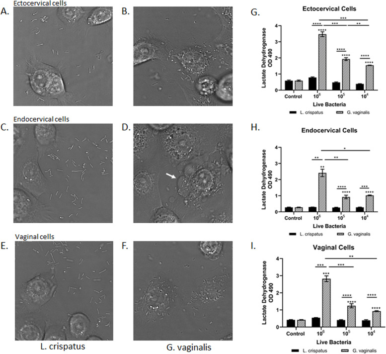
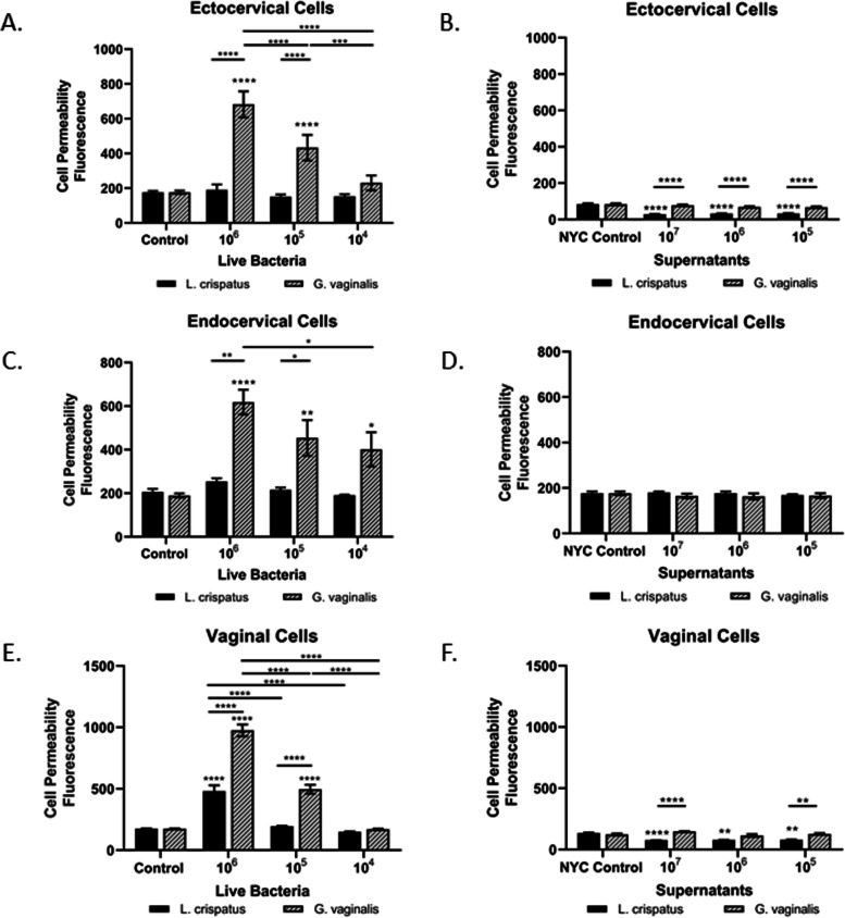
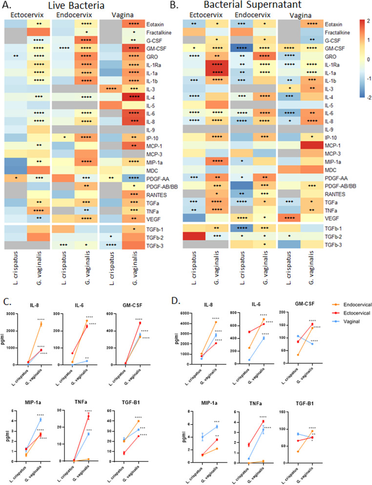
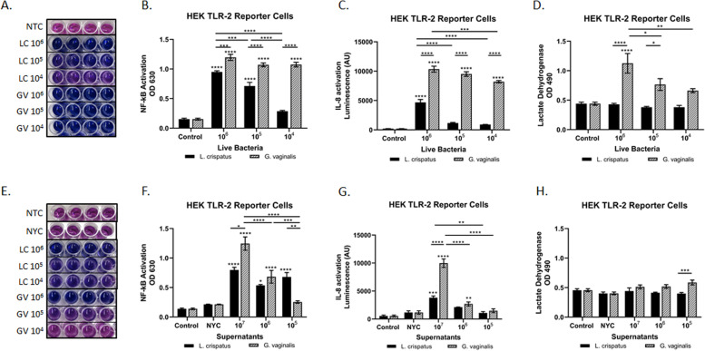
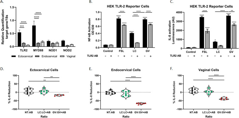
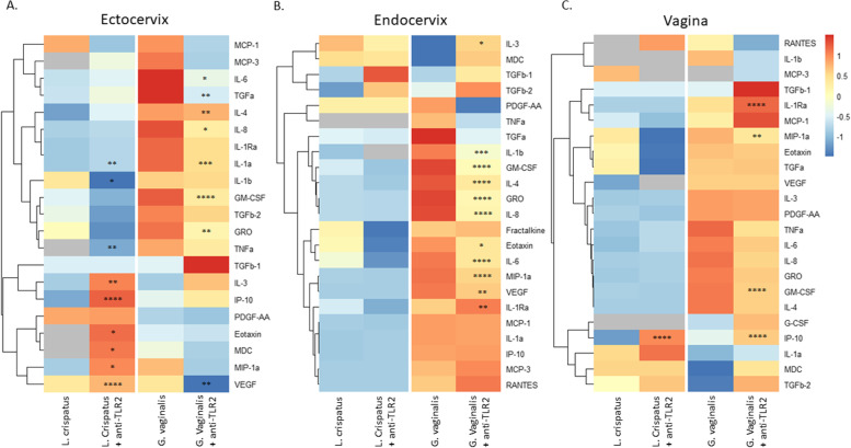
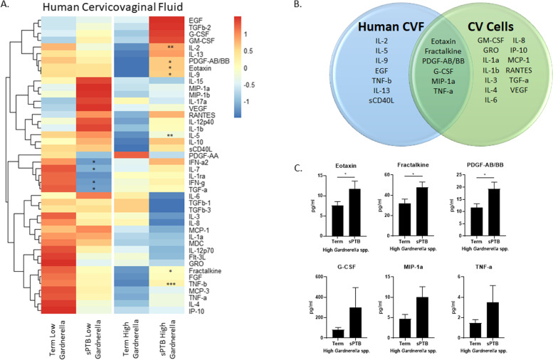
Results: Using an in vitro co-culture model, we assessed the effects of Lactobacillus crispatus (L. crispatus) and G. vaginalis on the CV epithelial barrier, the immune mediators that could be contributing to decreased barrier integrity and the immune signaling pathways regulating the immune response. G. vaginalis, but not L. crispatus, significantly increased epithelial cell death and decreased epithelial barrier integrity in an epithelial cell-specific manner. A G. vaginalis-mediated epithelial immune response including NF-κB activation and proinflammatory cytokine release was initiated partially through TLR2-dependent signaling pathways. Additionally, investigation of the cytokine immune profile in human CV fluid showed distinctive clustering of cytokines by Gardnerella spp. abundance and birth outcome.

Conclusions: The results of this study show microbe-specific effects on CV epithelial function. Altered epithelial barrier function through cell death and immune-mediated mechanisms by G. vaginalis, but not L. crispatus, indicates that host epithelial cells respond to bacteria-associated signals, resulting in altered epithelial function and ultimately CV disease. Additionally, distinct immune signatures associated with Gardnerella spp. or birth outcome provide further evidence that host-microbial interactions may contribute significantly to the biological mechanisms regulating reproductive outcomes. Video Abstract.

Keywords: Cervix; Epithelial barrier; Gardnerella vaginalis; Inflammation; Lactobacillus crispatus; Preterm birth; TLR2.


Conflict of interest statement

MAE receives salary support from NIH (NIAID, NINR, and NICHD). She is a consultant for MIRVIE. ESF has consulted for Astarte Medical Partners and Enzymetrics Bioscience, Inc. The other authors declare that they have no competing interests.

Figures

Fig. 1
Fig. 1
Co-localization of cervicovaginal epithelial cells with live G. vaginalis but not L. crispatus results in increased cell death. An in vitro live bacteria and host cervicovaginal co-culture model were created to study host microbial interactions in the CV space. Representative images of ectocervical (A, B), endocervical (C, D), and vaginal (E, F) epithelial cells interacting with L. crispatus (A, C, E) or G. vaginalis (B, D, F) are shown. Exposure of ectocervical (G), endocervical (H), and vaginal (I) cells to G. vaginalis but not L. crispatus results in dose-dependent cell death after 24 h. Values are mean ± SEM. Asterisks over the individual bars represent comparisons with control; asterisks over solid lines represent comparisons between treatment groups. *p < 0.05, **p < 0.01, ***p < 0.001, ****p < 0.0001
Fig. 2
Fig. 2
Live G. vaginalis increases epithelial barrier permeability. Cell permeability was measured in ectocervical, endocervical, and vaginal epithelial cells after 24-h exposure to live bacteria (A, C, E) or bacteria-free supernatants (B, D, F) of L. crispatus or G. vaginalis. Bacterial growth media alone acted as a negative control for the bacteria-free supernatants tested. Cell permeability is expressed as fluorescence OD measurements from a fluorescent plate reader and is indicative of the movement of FITC-dextran from the top to the bottom insert of a transwell chamber system. Values are mean ± SEM. Asterisks over the individual bars represent comparisons with control; asterisks over solid lines represent comparisons between treatment groups. *p < 0.05, **p < 0.01, ***p < 0.001, ****p < 0.0001
Fig. 3
Fig. 3
Live G. vaginalis activates the host-epithelial immune response, while bacteria-free supernatants alter immune activation in a nonbacterial-specific manner. Immune cytokines/chemokines released from ectocervical, endocervical, and vaginal cells after exposure to live L. crispatus and G. vaginalis (A) or their bacteria-free supernatants (B) for 24 h were measured by Luminex. Heat map depicts fold change (vs NTC for live bacteria or vs NYC for bacteria-free supernatants) by color and p-value by asterisks. p-value is based on pg/ml values. Representative graphs of cytokines (pg/mL) showing significant differences between L. crispatus and G. vaginalis exposure for both live bacteria (C) and bacteria-free supernatants (D) show epithelial cell-specific responses. Values are mean ± SEM. *p < 0.05, **p < 0.01, ***p < 0.001, ****p < 0.0001
Fig. 4
Fig. 4
L. crispatus and G. vaginalis activate NF-κB signaling through TLR2; however, only G. vaginalis results in increased IL-8 levels. The HEK TLR2 reporter cell line was used to determine if either live or bacteria-free supernatants from L. crispatus and G. vaginalis activated TLR2-mediated cell signaling. Representative images (A, E) and the corresponding quantification (B, F) of the QUANTI-Blue NF-κB detection assay, IL-8 activation (SEAP quantification) (C, G), and cytotoxicity (D, H) were all altered after exposure to live bacteria and bacteria-free supernatants from L. crispatus and G. vaginalis. For A and E, darker blue/purple indicates higher NF-kB. Values are mean ± SEM. Asterisks over the individual bars represent comparisons with control; asterisks over solid lines represent comparisons between treatment groups. *p < 0.05, **p < 0.01, ***p < 0.001, ****p < 0.0001
Fig. 5
Fig. 5
Blocking the TLR2 receptor significantly reduces L. crispatus and G. vaginalis-induced NF-κB and IL-8 activation. TLR2, MYD88, NOD1, and NOD2 are expressed in cervicovaginal epithelial cells, but expression varies by epithelial cell type (A). Blocking the TLR2 receptor in the HEK TLR2 reporter cells significantly reduced L. crispatus and G. vaginalis-induced NF-κB activation (B) and only G. vaginalis-induced IL-8 (C). The TLR2 agonist FSL was included as a positive control. Blocking the TLR2 receptor in cervicovaginal epithelial cells resulted in a reduction in G. vaginalis-induced IL-8, but no effect was seen in L. crispatus treated cells (D, E, F). Data is expressed as a percent reduction of the ratio of treatment alone to treatment plus anti-TLR antibody (A, B). Values are mean ± SEM. Asterisks over the individual bars represent comparisons to control; asterisks over solid lines represent comparisons between treatment groups. *p < 0.05, **p < 0.01, ***p < 0.001, ****p < 0.0001
Fig. 6
Fig. 6
Immune profile of cervicovaginal epithelial cells identifies cytokines mediated by activation of TLR2. Blocking the TLR2 receptor alters the immune cytokines/chemokines released from ectocervical (A), endocervical (B), and vaginal cells (C) after exposure to live L. crispatus and G. vaginalis. Heat map depicts fold change by color and p-value by asterisks. Fold change was calculated between the non-treated (NTC) control and live bacteria alone or live bacteria plus anti-TLR2 antibody. p-value is based on pg/ml values. *p < 0.05, **p < 0.01, ***p < 0.001, ****p < 0.0001
Fig. 7
Fig. 7
Blocking the TLR2 receptor does not mitigate G. vaginalis-induced increases in cell permeability. Cell permeability was measured in ectocervical (A), endocervical (B), and vaginal (C) epithelial cells after pretreatment with the anti-TLR2 antibody and subsequent exposure to live bacteria. G. vaginalis-induced cell permeability was unchanged after blocking the TLR2 receptor. Values are mean ± SEM
Fig. 8
Fig. 8
Immune profile from human cervicovaginal fluid is altered by abundance of Gardnerella spp. and delivery outcome. Cervicovaginal swabs collected from CST IV pregnant individuals at 16–20 weeks of gestation with either term or sPTB deliveries were used for Luminex assays to determine immune profiles of cytokines/chemokines. Heat map shows immune profiles vary by both Gardnerella spp. abundance as well as delivery outcome (A). Ven diagram shows overlapping cytokines (p < 0.10) induced in CVF from individuals with high Gardnerella spp. abundance and sPTB versus cervicovaginal epithelial cells exposed to live G. vaginalis in culture (B). Graphs of overlapping cytokines (C). Heat map (A) values are based on pg/mL and expressed on a relative scale. Values are mean ± SEM. *p < 0.05 and a trend for significance = p < 0.10

Similar articles

Cited by

References

    1. Kogut MH, Lee A, Santin E. Microbiome and pathogen interaction with the immune system. Poult Sci. 2020;99:1906–1913. doi: 10.1016/j.psj.2019.12.011. - DOI - PMC - PubMed
    1. Okumura R, Takeda K. Maintenance of intestinal homeostasis by mucosal barriers. Inflamm Regen. 2018;38:5. doi: 10.1186/s41232-018-0063-z. - DOI - PMC - PubMed
    1. Allaire JM, Crowley SM, Law HT, Chang SY, Ko HJ, Vallance BA. The intestinal epithelium: central coordinator of mucosal immunity. Trends Immunol. 2018;39:677–696. doi: 10.1016/j.it.2018.04.002. - DOI - PubMed
    1. Frank DN, St Amand AL, Feldman RA, Boedeker EC, Harpaz N, Pace NR. Molecular-phylogenetic characterization of microbial community imbalances in human inflammatory bowel diseases. Proc Natl Acad Sci U S A. 2007;104:13780–13785. doi: 10.1073/pnas.0706625104. - DOI - PMC - PubMed
    1. Kiely CJ, Pavli P, O’Brien CL. The role of inflammation in temporal shifts in the inflammatory bowel disease mucosal microbiome. Gut Microbes. 2018;9:477–485. - PMC - PubMed

Publication types

LinkOut - more resources