. 2022 Aug 3;20(1):172.
doi: 10.1186/s12915-022-01365-5.

Evolutionary conserved relocation of chromatin remodeling complexes to the mitotic apparatus

Affiliations

Evolutionary conserved relocation of chromatin remodeling complexes to the mitotic apparatus

Giovanni Messina et al. BMC Biol. .

Abstract

Background: ATP-dependent chromatin remodeling complexes are multi-protein machines highly conserved across eukaryotic genomes. They control sliding and displacing of the nucleosomes, modulating histone-DNA interactions and making nucleosomal DNA more accessible to specific binding proteins during replication, transcription, and DNA repair, which are processes involved in cell division. The SRCAP and p400/Tip60 chromatin remodeling complexes in humans and the related Drosophila Tip60 complex belong to the evolutionary conserved INO80 family, whose main function is promoting the exchange of canonical histone H2A with the histone variant H2A in different eukaryotic species. Some subunits of these complexes were additionally shown to relocate to the mitotic apparatus and proposed to play direct roles in cell division in human cells. However, whether this phenomenon reflects a more general function of remodeling complex components and its evolutionary conservation remains unexplored.

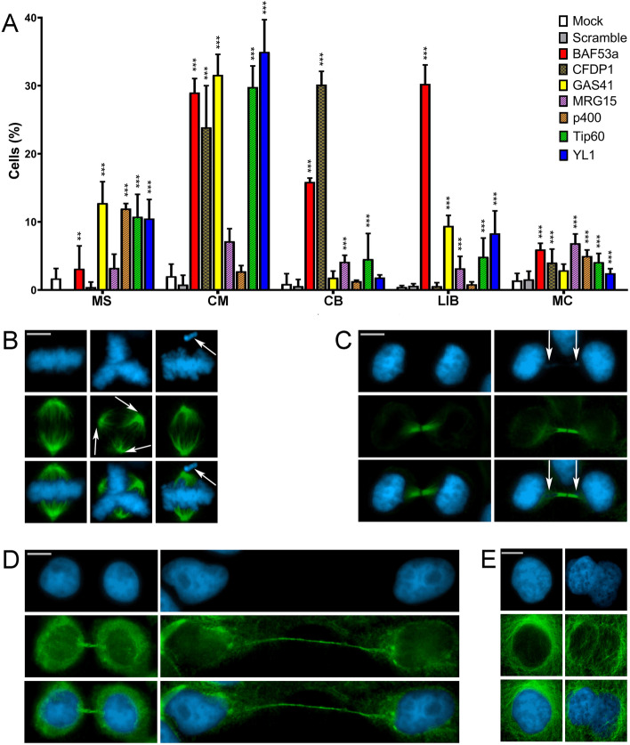
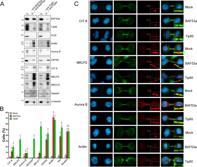
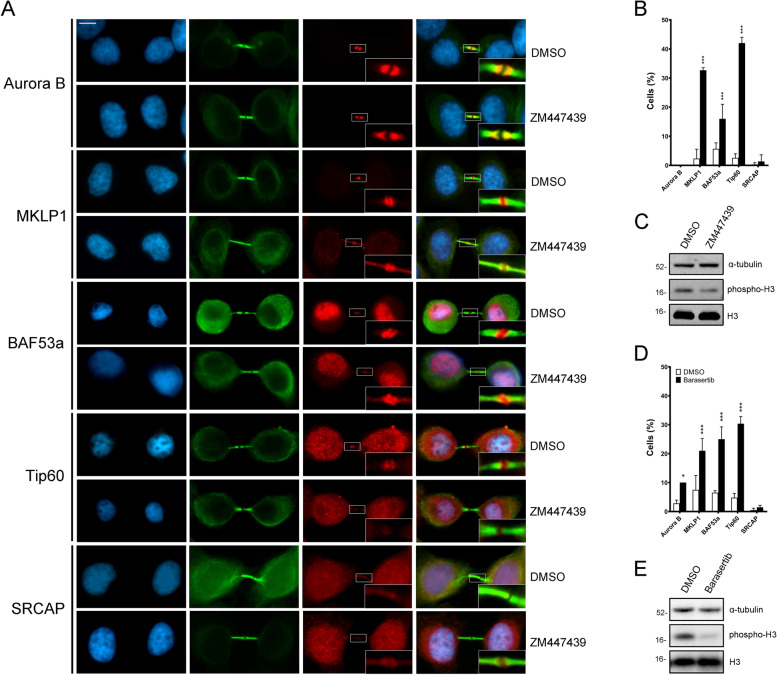
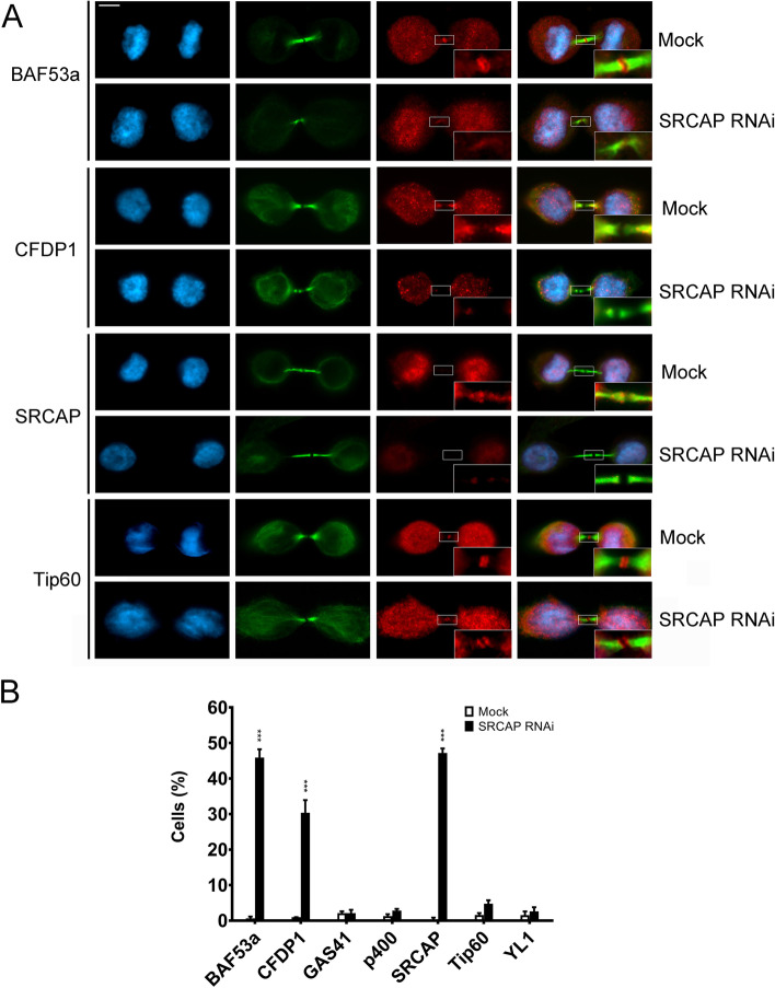
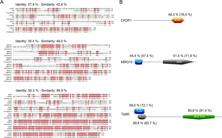
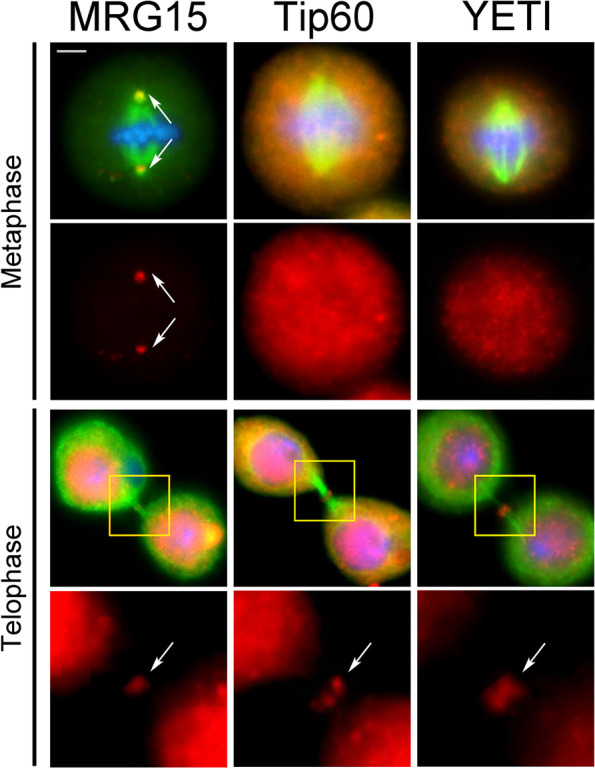
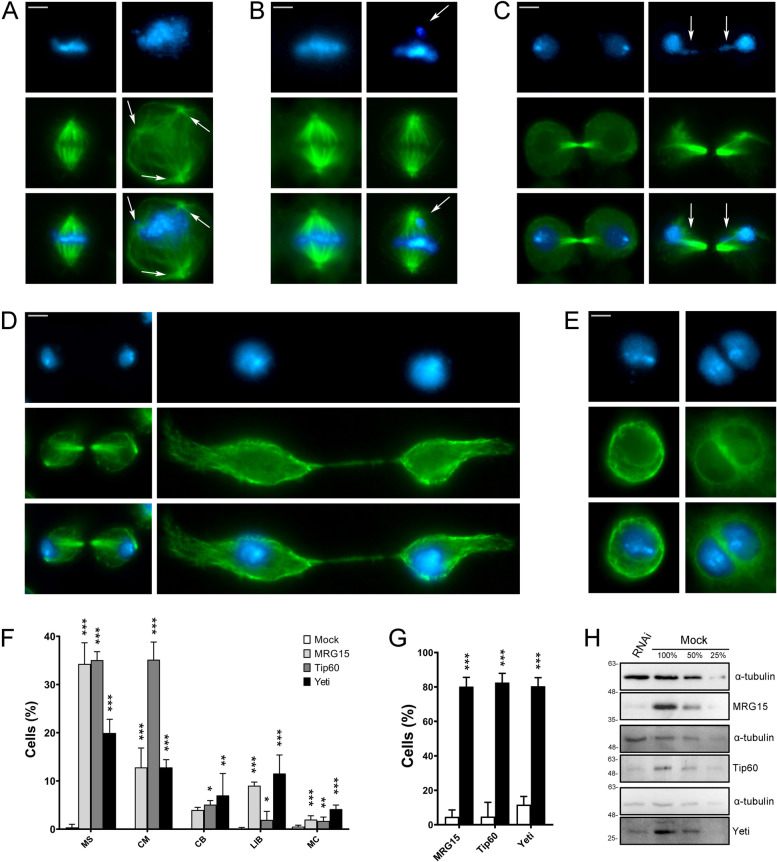
Results: We have combined cell biology, reverse genetics, and biochemical approaches to study the subcellular distribution of a number of subunits belonging to the SRCAP and p400/Tip60 complexes and assess their involvement during cell division progression in HeLa cells. Interestingly, beyond their canonical chromatin localization, the subunits under investigation accumulate at different sites of the mitotic apparatus (centrosomes, spindle, and midbody), with their depletion yielding an array of aberrant outcomes of mitosis and cytokinesis, thus causing genomic instability. Importantly, this behavior was conserved by the Drosophila melanogaster orthologs tested, despite the evolutionary divergence between fly and humans has been estimated at approximately 780 million years ago.

Conclusions: Overall, our results support the existence of evolutionarily conserved diverse roles of chromatin remodeling complexes, whereby subunits of the SRCAP and p400/Tip60 complexes relocate from the interphase chromatin to the mitotic apparatus, playing moonlighting functions required for proper execution of cell division.

Keywords: Cell division; Chromatin remodeling; Cytokinesis; Midbody; Moonlighting proteins.


Conflict of interest statement

The authors declare that they have no competing interests.

Figures

Fig. 1
Fig. 1
Schematic representation of midbody and subunit composition of SRCAP and p400/Tip60 chromatin remodeling complexes in human cells. A The midbody can be subdivided in three main regions [8, 35]: the midbody ring (red), containing anillin and citron kinase and other contractile ring components; the midbody central core (blue) marked by central spindle proteins such as the centralspindlin complex; and the midbody arms (green) where Aurora B and the other components of the chromosomal passenger complex (CPC) are recruited. B SRCAP (left) and p400/Tip60 (right) complexes share many subunits. The chromatin remodeling subunits (CRS) studied in this work are depicted in colours
Fig. 2
Fig. 2
Localization of SRCAP and p400/Tip60 chromatin remodeling subunits (CRS) to the centrosomes, spindle, and midbody in HeLa cells. Fixed HeLa cells stained with DAPI (blue), anti-CRS (red), and anti-α-tubulin (green). BAF53a, CFDP1, GAS41, and YL1 fluorescent signals were found at the spindle in both metaphase and/or anaphase; Tip60 localized at the centrosome and kinetochores (particularly evident at anaphase). In telophase, all the tested CRS were found at the midbody. Scale bar = 10 μm
Fig. 3
Fig. 3
Validation of antibodies against the CRS. A Total CRS amount assessed by Western blotting in RNAi-treated and mock-treated cells. B Histograms showing the fluorescence intensity of immunostaining in RNAi-treated and mock-treated cells. The colors of bars are referred to Fig. 1B. Bars with different dashed lines indicate SRCAP complex-specific subunits (CFDP1) or p400/Tip60-specific subunits (p400 and Tip60). Plain colored bars indicate subunits common to both complexes (BAF53a, GAS41, and YL1). The fluorescence intensity showed a decrease ranging from about 60 up to 75% in RNAi-treated HeLa cells compared to the mock-treated cells. Fluorescence intensity was assessed using the ImageJ software and statistical significance was verified by T- test. C Examples of IFM staining. The fluorescence intensity of BAF53a and Tip60 clearly decreased in RNAi-treated compared to mock-treated cells. Scale bar = 10 μm
Fig. 4
Fig. 4
Localization of CRS on isolated midbodies. A Immunolocalization. Fixed preparations of isolated midbodies were stained with DAPI (blue), antibodies against the protein of interest (red), and anti-α-tubulin (green). No DAPI staining was detected. The antibody staining for each tested protein clearly decorated the isolated midbodies. Scale bar = 5 μm. B Detection of CRS by Western blotting on midbody extracts from HeLa cells. Aurora B and α-tubulin were used as positive controls. ISWI was not detected (negative control)
Fig. 5
Fig. 5
RNAi mediated depletion of CRS affects cell division in HeLa cells. Cells were stained with DAPI (blue) and anti-α-tubulin (green). Scale bar = 10 μm. Five classes of defects were categorized. A Histograms showing the quantitative analysis of cell division defects. Colors are referred to Fig. 1B. Bars with two different dashed lines indicate SCRAP complex-specific subunits (CFDP1) or p400/Tip60-specific subunits (MRG15, p400, and Tip60). Plain colored bars indicate subunits common to both complexes (BAF53a, GAS41, and YL1). B Mock, left panel; multipolar spindles (MS), middle panel; chromosome misalignments (CM), right panel. C Mock, left panel; chromatin bridges (CB) right panel. D Mock, left panel; long intercellular bridges (LIB) right panel; no DAPI-stained trapped chromatin was observed. E Mock, left panel; multinucleated cells (MC), right panel. Three independent experiments were performed. The quantitative analysis of defects scored in RNAi-treated and control cells (Table 2) is based on the following numbers: at least 100 prometaphases and metaphases for MS, 70 metaphases for CM and ASM, 300 telophases for LIB and CB, and 5500 interphases for MC. Experimental groups were compared with control groups (mocks and scrambles) by Fisher’s exact test.*=P < 0.05; **=P < 0.005; and ***=P < 0.0005
Fig. 6
Fig. 6
Interactions between CRS and cytokinesis regulators. A Immunoprecipitation of protein extracts from cytoplasmic fraction of telophase synchronized HeLa cells (S2 fraction). IP sample immunoprecipitated with BAF53a or Tip60 antibodies (+anti-BAF53a; +anti-Tip60) were compared to negative controls (−anti-BAF53a or −anti-Tip60). Anillin, Aurora B, CEP55, CIT-K, MKLP2, MRG15, spastin, and α-tubulin were found in the IP samples immunoprecipitated with BAF53a or Tip60 antibodies, but not in the negative controls. Three independent IP experiments were performed. IN = input, IP = immunoprecipitation. Synchronization of the HeLa cells was performed according to Messina et al. [31]. B Histograms showing the quantitative analysis of mislocalizations of cytokinesis regulators at the midbody in mocks and BAF53a or Tip60 depleted cells. Three independent experiments were performed and at least 300 telophases were scored in both RNAi-treated and control cells. *=P<0.05,**=P<0.005, ***=P<0.0005. C Examples of mislocalization of cytokinesis regulators in mock and BAF53a or Tip60 depleted cells. From left to the right: DAPI (blue), anti-α-tubulin (green), cytokinesis regulators (red), and merge. Scale bar = 10 μm
Fig. 7
Fig. 7
Inhibition of Aurora B kinase activity with ZM447439 and Barasertib affected the localization of BAF53a and Tip60 to the midbody. A Treatment with ZM447439: from left to right: DAPI (blue), anti- α-tubulin (green), tested protein (red), and merge. Anti-MKLP1 and anti-Aurora B immunostaining were used as positive and negative controls, respectively. Scale bar = 10 μm. The localization pattern of BAF53a and Tip60, as well as that of MKLP1 were affected, while no effect was seen for SRCAP and Aurora B. B The effects found with ZM447439 treatment are summarized in the graph. At least 300 telophases were scored in three independent experiments for both treated cells and control HeLa cells. C To test the effectiveness of ZM447439 inhibition, phosphorylation of the histone H3 (target of Aurora B kinase) during mitosis was evaluated; α-tubulin was used as loading control. D The effects found with Barasertib treatment are summarized in the graph. At least 200 telophases were scored in two independent experiments for both treated cells and control HeLa cells. A slight effect on Aurora localization was seen at the limit of significance values. E To test the effectiveness of Barasertib inhibition, phosphorylation of the histone H3 (target of Aurora B kinase) during mitosis was evaluated; α-tubulin was used as loading control. *=P<0.05, **<=P<0.005, ***=P<0.0005
Fig. 8
Fig. 8
SRCAP-dependent localization of BAF53a and CFDP1 to the midbody. A Examples of mislocalization. DAPI (blue), anti-α-tubulin (green), tested CRS (red), and merge. Scale bar = 10 μm. B The graph summarized the results. At least of 300 telophases were scored in two independent experiments for both treated cells and control HeLa cells. ***=P<0.0005
Fig. 9
Fig. 9
Sequence conservation of Drosophila MRG15, dTip60, and YETI proteins with their human orthologs. A) BLAST alignments; B) High identity levels found in specific functional domains of CFDP1, MRG15 and Tip60
Fig. 10
Fig. 10
Immunostaining showing the localization of MRG15, Tip60, and YETI to the mitotic apparatus in D. melanogaster S2 cells. Cells were stained with DAPI (blue), anti-α-tubulin (green), and anti-MRG15, anti-Tip60, and anti-YETI (red). Scale bar = 5 μm. In addition to interphase nuclei, the anti-MRG15 decorated the centrosomes (metaphase) and midbody (telophase), while anti-Tip60 and anti-YETI staining was detected on the midbody
Fig. 11
Fig. 11
RNAi-mediated depletion of MRG15, Tip60, and YETI in S2 cells affects mitosis and cytokinesis. Examples of mitotic defects found in MRG15, Tip60, and YETI depleted S2 cells and their quantification. DAPI staining is shown in blue, α-tubulin in green. RNAi knockdown experiments were performed by transfecting S2 cells with specific siRNAs against each protein (see “Methods”). Scale bar = 5 μm. Five classes of defects were considered: A multipolar spindle (MS); B chromosome misalignments in metaphase (CM); C chromatin bridges (CB); D long intercellular bridges (LIB); E multinucleated cells (MC); F Quantitative analysis of defects scored in RNAi-treated and control cells (Table 5) is based on the following numbers: at least 100 prometaphases and metaphases for MS, 70 metaphases for CM, 300 telophases for LIB and CB, 5500 interphases for MC. Three independent experiments were performed. G Percent of cells showing a decrease of fluorescence intensity at the midbody in MRG15, Tip60, or YETI depleted cells (black histograms) compared to the mocks (white histograms). The results are based on three independent experiments. H WB showing a decrease of total amount of MRG15, Tip60, and YETI proteins in RNAi-treated S2 cells compared to the mocks; a-tubulin is used as loading control

Similar articles

Cited by

References

    1. Cleveland DW, Mao Y, Sullivan KF. Centromeres and kinetochores: from epigenetics to mitotic checkpoint signaling. Cell. 2003;112(4):407–421. doi: 10.1016/S0092-8674(03)00115-6. - DOI - PubMed
    1. Pollard TD. Nine unanswered questions about cytokinesis. J Cell Biol. 2017;216(10):3007–3016. doi: 10.1083/jcb.201612068. - DOI - PMC - PubMed
    1. Flemming W. Zellsubstanz, Kern und Zelltheilung. 1882.
    1. Skop AR, Liu H, Yates J, 3rd, Meyer BJ, Heald R. Dissection of the mammalian midbody proteome reveals conserved cytokinesis mechanisms. Science. 2004;305(5680):61–66. doi: 10.1126/science.1097931. - DOI - PMC - PubMed
    1. Glotzer M. The molecular requirements for cytokinesis. Science. 2005;307(5716):1735–1739. doi: 10.1126/science.1096896. - DOI - PubMed

Publication types

LinkOut - more resources