. 2022 Aug 3;13(1):394.
doi: 10.1186/s13287-022-03100-x.

Mesenchymal stem cell-derived extracellular vesicles protect retina in a mouse model of retinitis pigmentosa by anti-inflammation through miR-146a-Nr4a3 axis

Affiliations

Mesenchymal stem cell-derived extracellular vesicles protect retina in a mouse model of retinitis pigmentosa by anti-inflammation through miR-146a-Nr4a3 axis

Jia Zhang et al. Stem Cell Res Ther. .

Abstract

Background: Retinitis pigmentosa is a rod-cone degenerative disease that induces irreversible vision loss. This study probed the protective capacity of mesenchymal stem cell-derived small EVs (MSC-EVs) on the retinas of rd10 mice and the underlying mechanism.

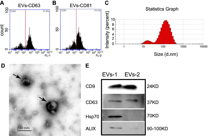
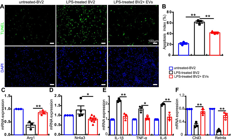
Methods: MSC-EVs were injected into the vitreous of rd10 mice at postnatal day 14 and P21; morphology and function were examined at P28. The mechanism of action was explored by using co-culture of photoreceptor cell line 661 W and microglia cell line BV2.

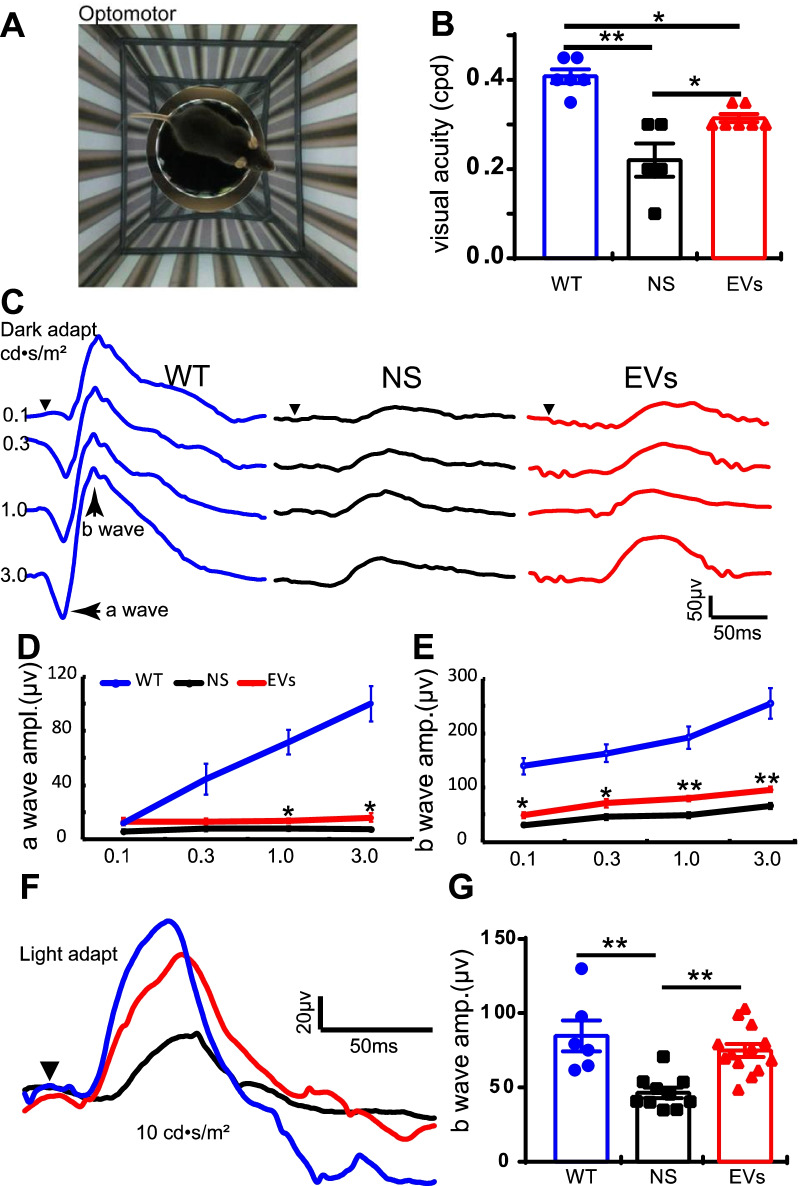
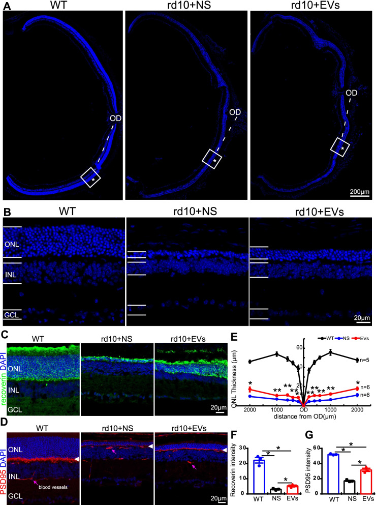
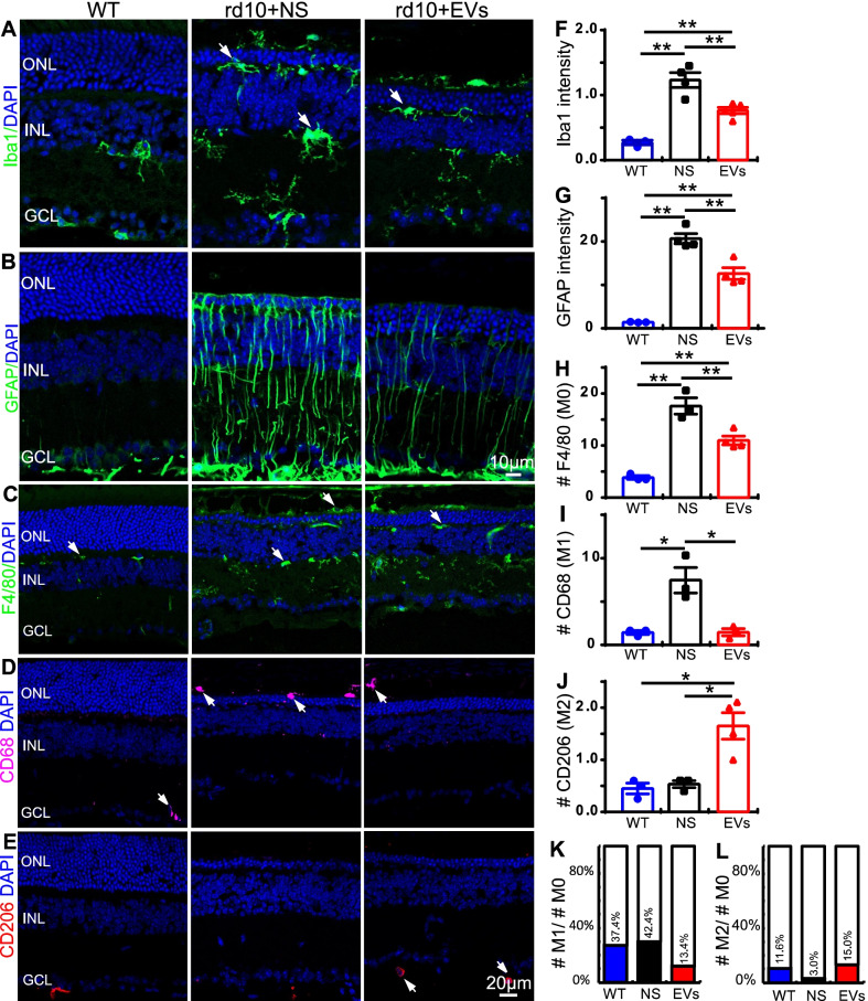
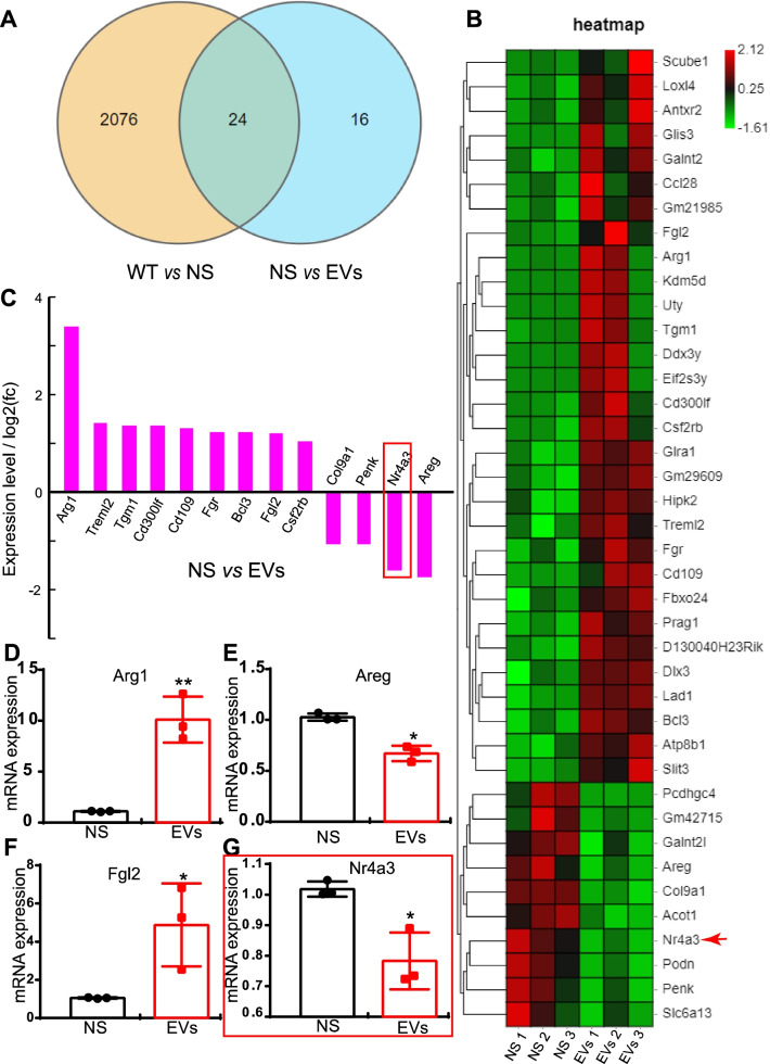
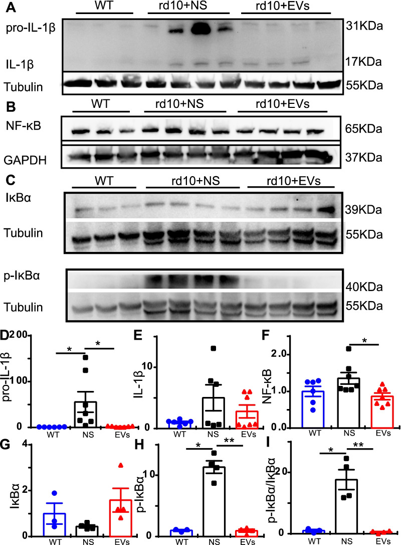
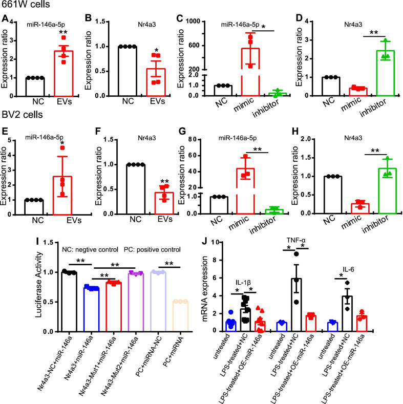
Results: Treatment with MSC-EVs increased the survival of photoreceptors and preserved their structure. Visual function, as reflected by optomotor and electroretinogram responses, was significantly enhanced in MSC-EVs-treated rd10 mice. Mechanistically, staining for Iba1, GFAP, F4/80, CD68 and CD206 showed that MSC-EVs suppressed the activation of microglial, Müller glial and macrophages. Furthermore, western blotting showed that the treatment inhibited the NF-κB pathway. RNA-seq and qPCR showed that MSC-EVs upregulated anti-inflammatory cytokines while downregulating pro-inflammatory cytokines. MSC-EVs application in vitro decreased the number of TUNEL-positive 661 W cells co-cultured with LPS-stimulated BV2, with similar impact on the cytokine expression as in vivo study. Genetic screening predicted miR-146a to be the downstream target of MSC-EVs, which was detected in MSC-EVs and upregulated in co-cultured 661 W cells and BV2 cells after MSC-EVs treatment. Upregulation of miR-146a by using its mimic decreased the expression of the transcription factor Nr4a3, and its downregulation inhibition promoted Nr4a3 expression in both 661 W and BV2 cells. Nr4a3 was further identified as the target gene of miR-146a by dual-luciferase assay. Furthermore, overexpressing miR-146a significantly decreased the expression of LPS-induced pro-inflammatory cytokines in BV2 cells.

Conclusions: MSC-EVs delays retinal degeneration in rd10 mice mainly by its anti-inflammatory effect via the miR-146a-Nr4a3axis. Hence, MSC-EVs may be used in the treatment of neurodegenerative diseases.

Keywords: Extracellular vesicles; Inflammation; Retinitis pigmentosa; miR-146a-Nr4a3 axis; rd10 mouse.


Conflict of interest statement

The authors declare that they have no competing interests.

Figures

Fig. 1
Fig. 1
Characterization of MSC-EVs. AB Flow cytometry results showed 73.4% and 74.5% of EVs were positive for CD63 A and CD81 (B). C Particle size analysis of the EVs showing the distribution of EVs with different sizes. Around 75% of the particles had the size of exosomes, i.e., 30–200 nm. D Morphologies of two typical EVs taken by transmission electron microscopy (indicated by arrows). E. Western blotting showing the expression of EVs’ markers (CD9, CD63, Hsp70 and ALIX) in two batches of exosomes, EVs-1 and EVs-2, with lower concentration
Fig. 2
Fig. 2
MSC-EVs improves visual function in rd10 mice. A Illustration of the optomotor system. B Quantification of the visual acuity in WT, NS-treated and MSC-EVs-treated rd10 mice. C Representative ERG traces under various light intensities ranging from 0.1 to 3.0 cd • s/m2 under dark adaptation in WT (blue), NS-treated rd10 mice (black) and MSC-EVs-treated rd10 mice (red) at P28. Arrowhead shows the starting time point of the flash. D, E Average peak amplitudes of dark-adapted a-wave (D) and b-wave (E). F Representative ERG traces at 10.0 cd • s/m2 flash under light adaptation with 20 cd • s/m2 background. G Average b-wave amplitudes of light-adapted condition in three groups. Data are represented as mean ± SEM. Student’s t test and one-way ANOVA test were applied. * P < 0.05, **, P < 0.01
Fig. 3
Fig. 3
MSC-EVs promote photoreceptor survival in rd10 mice at P28. A Images of eye cups with retinas stained with DAPI from three animal groups, with squared regions at 600 µm away from the center of the optic disk (OD) enlarged in B. ONL layer where photoreceptor somas are located (*) became thinner in rd10 mice, while MSC-EVs increased its thickness. C, D Images of immunofluorescence staining with an antibody against recoverin (green, C) or PSD95 (red, D) and DAPI (blue) of retinal slices in three animal groups. Arrowheads in D point to the photoreceptors presynaptic terminals that are labeled by anti-PSD95. Pink arrows point to blood vessels. E Quantification of the ONL thickness at 200 µm, 400 µm, 600 µm, 1000 µm and 2000 µm away from the OD at P28. F Quantification of the mean gray intensity in the three animal groups of recoverin (F) and PSD95 (G) in three animal groups. OD, optic disk; ONL, outer nuclear layer; INL, inner nuclear layer; and GCL, ganglion cell layer. Data are shown as mean ± SEM. * P < 0.05, **, P < 0.01. Student’s t test and one-way ANOVA were applied
Fig. 4
Fig. 4
MSC-EVs inhibit the reactive gliosis and immunoproliferation in the rd10 retina. A Retinal sections stained with DAPI (blue) and for Iba1 (green) in WT, NS-treated rd10 and MSC-EVs-treated rd10. In the rd10 retina, microglial cells presented as active state and distributed throughout the whole retina. MSC-EVs decreased its expression. B Retinal sections stained for GFAP (green). In the rd10 retina, GFAP was strongly expressed in Müller cells and extended from GCL to retinal OS; MSC-EVs decreased its expression. C-E Retinal sections stained for F4/80 (green, C), CD68 (pink, D) or CD206 (red, E). F, G Quantification of the Iba1-positive fluorescence intensity (F) and GFAP-positive fluorescence intensity (G). H-J Quantification of the number of F4/80 (M0 macrophage) (H)-, CD68 (M1 macrophage) (I)- and CD206 (M2 macrophage) (J)-positive cells. K, L Quantification of the ratio of M1/M0 number (K) and M2/M0 number (L). The MSC-EVs treatment reduced the fluorescence intensity of Iba1, the GFAP neurite intensity and the number of M0 and M1 macrophages in the rd10 retinas. White arrow indicated the cell somas of positive fluorescence. Data are shown as mean ± SEM. * P < 0.05, **, P < 0.01. One-way ANOVA
Fig. 5
Fig. 5
MSC-EVs regulate the gene expression of inflammatory cytokines in the rd10 retina. A A pie chart of RNA-seq results in WT, NS-treated and MSC-EVs-treated rd10 retinas showing the number of genes whose expression differs between the groups. B Heat map of RNA-seq results showing the 40 differentially expressed genes between NS-treated and MSC-EVs-treated rd10 retinas. C The expression level of 13 differentially expressed genes between NS-treated and MSC-EVs-treated rd10 retinas which are related to immunity and inflammation. D-G qPCR quantification of 4 differentially expressed genes between NS-treated rd10 and MSC-EVs-treated rd10 retinas. Data are represented as mean ± SEM. A Student’s t test was applied for qPCR data
Fig. 6
Fig. 6
MSC-EVs inhibit the activation of NF-κB signaling pathway in rd10 mice. A-C Western blotting results of IL-1β and its precursor (A), NF-κB (B), IκBα and p-IκBα (C) protein. D-I Quantitative analyses of the expression levels of IL-1β precursor, IL-1β, NF-κB, IκBα, p-IκBα and p-IκBα/ IκBα normalized to that of WT retina. Data are represented as mean ± SEM. * P < 0.05, **, P < 0.01, one-way ANOVA
Fig. 7
Fig. 7
MSC-EVs suppress inflammation in vitro. A TUNEL staining of 661W cells co-cultured with inactive-BV2, LPS-activated-BV2 or LPS-activated BV2 + EVs. 661W cells were stained with DAPI (blue, bottom panel) and apoptotic cells were labeled with TUNEL staining (green, top panel). B The percentage of TUNEL positive 661W cells over DAPI positive cells in the three groups. C-H mRNA expression of Arg1 (C), Nr4a3 (D), IL-1β, TNF-α, IL-6 (E), Chil3 and Retnla (F) in the treated group relative to that of the untreated group. Data are represented as mean ± S.E.M. One-way ANOVA and Student’s t-test was applied for statistics
Fig. 8
Fig. 8
MiR-146a directly regulates the expression of Nr4a3. A, B The expression of miR-146a-5p (A) and Nr4a3 (B) after MSC-EVs treatment for 24 h in 661 W cells. C, D The expression of miR-146a-5p (C) and Nr4a3 (D) after overexpressing miR-146a-5p by using the mimic or blocking it using inhibitor in 661 W cells. E, F The expression of miR-146a-5p(E) and Nr4a3 (F) after MSC-EVs treatment in BV2 cells. G, H The expression of miR-146a-5p (G) and Nr4a3 (H) after using the mimic or inhibitor in BV2 cells. The expression of Nr4a3 followed the changes of miR-146a-5p. I The luciferase activity of miR-146a-5p and Nr4a3 or with two forms mutated at the predicted binding sites of Nr4a3. Values were normalized to that of Nr4a3-negative control. J Overexpressing miR-146a in BV2 cells downregulated expression of the LPS-induced pro-inflammatory factors IL-1β, TNF-α and IL-6. Data are shown as mean ± SEM. One-way ANOVA and Student’s t test were applied for statistics. All experiments were repeated for 3 times

Similar articles

Cited by

References

    1. Hamel C. Retinitis pigmentosa. Orphanet J Rare Dis. 2006;1:40. doi: 10.1186/1750-1172-1-40. - DOI - PMC - PubMed
    1. Gagliardi G, Ben M'Barek K, Goureau O. Photoreceptor cell replacement in macular degeneration and retinitis pigmentosa: a pluripotent stem cell-based approach. Prog Retin Eye Res. 2019;71:1–25. doi: 10.1016/j.preteyeres.2019.03.001. - DOI - PubMed
    1. Genove G, Mollick T, Johansson K. Photoreceptor degeneration, structural remodeling and glial activation: a morphological study on a genetic mouse model for pericyte deficiency. Neuroscience. 2014;279:269–284. doi: 10.1016/j.neuroscience.2014.09.013. - DOI - PubMed
    1. Wang K, Xiao J, Peng B, Xing F, So KF, Tipoe GL, et al. Retinal structure and function preservation by polysaccharides of wolfberry in a mouse model of retinal degeneration. Sci Rep. 2014;4:7601. doi: 10.1038/srep07601. - DOI - PMC - PubMed
    1. Liu F, Zhang J, Xiang Z, Xu D, So KF, Vardi N, et al. Lycium barbarum polysaccharides protect retina in rd1 mice during photoreceptor degeneration. Invest Ophthalmol Vis Sci. 2018;59:597–611. doi: 10.1167/iovs.17-22881. - DOI - PMC - PubMed

Publication types

LinkOut - more resources