

Noninvasive hemoglobin sensing and imaging: optical tools for disease diagnosis
- PMID: 35922891
- PMCID: PMC9346606
- DOI: 10.1117/1.JBO.27.8.080901
Noninvasive hemoglobin sensing and imaging: optical tools for disease diagnosis
Abstract
Significance: Measurement and imaging of hemoglobin oxygenation are used extensively in the detection and diagnosis of disease; however, the applied instruments vary widely in their depth of imaging, spatiotemporal resolution, sensitivity, accuracy, complexity, physical size, and cost. The wide variation in available instrumentation can make it challenging for end users to select the appropriate tools for their application and to understand the relative limitations of different methods.
Aim: We aim to provide a systematic overview of the field of hemoglobin imaging and sensing.
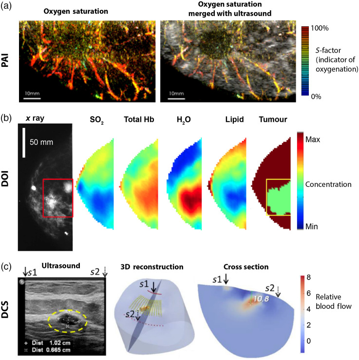
Approach: We reviewed the sensing and imaging methods used to analyze hemoglobin oxygenation, including pulse oximetry, spectral reflectance imaging, diffuse optical imaging, spectroscopic optical coherence tomography, photoacoustic imaging, and diffuse correlation spectroscopy.
Results: We compared and contrasted the ability of different methods to determine hemoglobin biomarkers such as oxygenation while considering factors that influence their practical application.
Conclusions: We highlight key limitations in the current state-of-the-art and make suggestions for routes to advance the clinical use and interpretation of hemoglobin oxygenation information.
Keywords: hemoglobin; imaging; sensing; spectroscopy.
Figures


Similar articles
-
Assessing the comparative effects of interventions in COPD: a tutorial on network meta-analysis for clinicians.Respir Res. 2024 Dec 21;25(1):438. doi: 10.1186/s12931-024-03056-x. Respir Res. 2024. PMID: 39709425 Free PMC article. Review.
-
Pulse oximetry screening for critical congenital heart defects.Cochrane Database Syst Rev. 2018 Mar 1;3(3):CD011912. doi: 10.1002/14651858.CD011912.pub2. Cochrane Database Syst Rev. 2018. PMID: 29494750 Free PMC article.
-
Contrast-enhanced ultrasound for the diagnosis of hepatocellular carcinoma in adults with chronic liver disease.Cochrane Database Syst Rev. 2022 Sep 2;9(9):CD013483. doi: 10.1002/14651858.CD013483.pub2. Cochrane Database Syst Rev. 2022. PMID: 36053210 Free PMC article.
-
Cost-effectiveness of using prognostic information to select women with breast cancer for adjuvant systemic therapy.Health Technol Assess. 2006 Sep;10(34):iii-iv, ix-xi, 1-204. doi: 10.3310/hta10340. Health Technol Assess. 2006. PMID: 16959170
-
Rapid molecular tests for tuberculosis and tuberculosis drug resistance: a qualitative evidence synthesis of recipient and provider views.Cochrane Database Syst Rev. 2022 Apr 26;4(4):CD014877. doi: 10.1002/14651858.CD014877.pub2. Cochrane Database Syst Rev. 2022. PMID: 35470432 Free PMC article.
Cited by
-
Multispectral imaging of nailfold capillaries using light-emitting diode illumination.J Biomed Opt. 2022 Dec;27(12):126002. doi: 10.1117/1.JBO.27.12.126002. Epub 2022 Dec 12. J Biomed Opt. 2022. PMID: 36519074 Free PMC article.
-
Carbon Monoxide Poisoning: Diagnosis, Prognostic Factors, Treatment Strategies, and Future Perspectives.Diagnostics (Basel). 2025 Feb 27;15(5):581. doi: 10.3390/diagnostics15050581. Diagnostics (Basel). 2025. PMID: 40075828 Free PMC article. Review.
-
Image-enhanced endoscopy in upper gastrointestinal disease: focusing on texture and color enhancement imaging and red dichromatic imaging.Clin Endosc. 2025 Mar;58(2):163-180. doi: 10.5946/ce.2024.159. Epub 2024 Nov 6. Clin Endosc. 2025. PMID: 39722144 Free PMC article. Review.
-
Distribution-informed and wavelength-flexible data-driven photoacoustic oximetry.J Biomed Opt. 2024 Jun;29(Suppl 3):S33303. doi: 10.1117/1.JBO.29.S3.S33303. Epub 2024 Jun 5. J Biomed Opt. 2024. PMID: 38841431 Free PMC article.
-
Investigation on the influence of the skin tone on hyperspectral imaging for free flap surgery.Sci Rep. 2024 Jun 17;14(1):13979. doi: 10.1038/s41598-024-64549-9. Sci Rep. 2024. PMID: 38886457 Free PMC article.
