[Preprint]. 2022 Jul 25:2022.07.25.22278025.
doi: 10.1101/2022.07.25.22278025.

Plasma proteomics of SARS-CoV-2 infection and severity reveals impact on Alzheimer and coronary disease pathways

Affiliations

Plasma proteomics of SARS-CoV-2 infection and severity reveals impact on Alzheimer and coronary disease pathways

Lihua Wang et al. medRxiv. .

Update in

Abstract

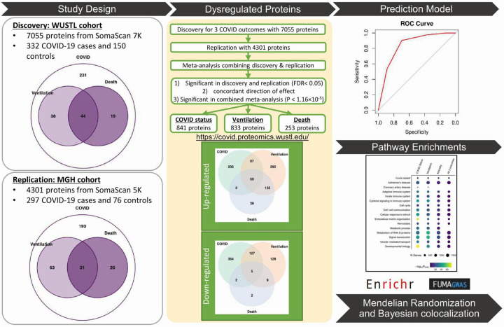
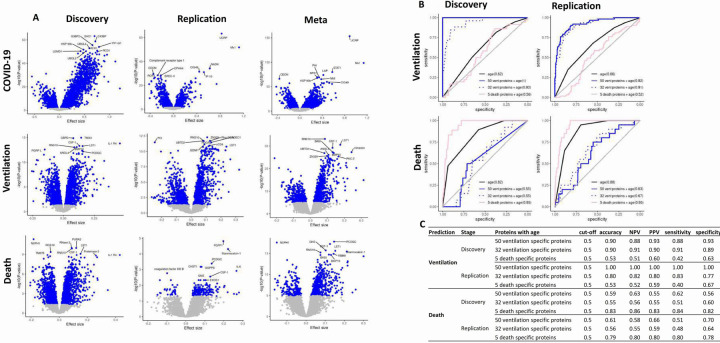
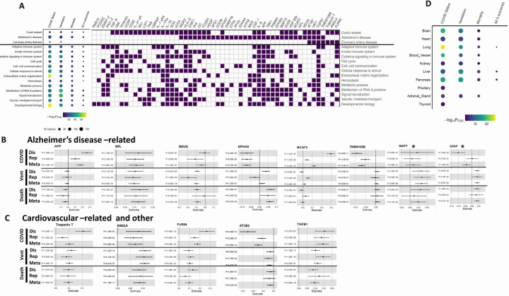
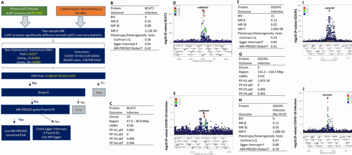
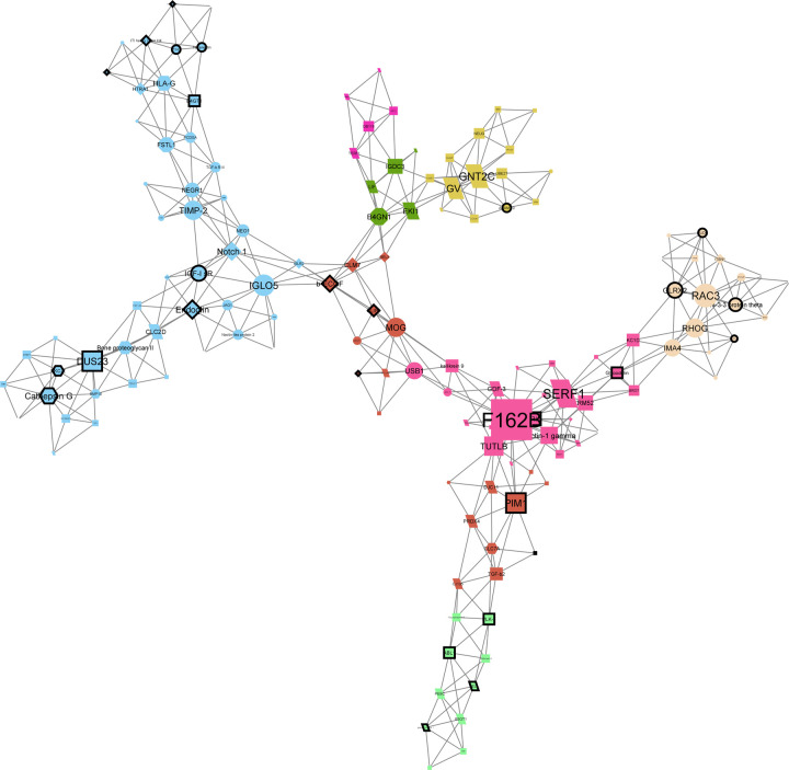
Identification of the plasma proteomic changes of Coronavirus disease 2019 (COVID-19) is essential to understanding the pathophysiology of the disease and developing predictive models and novel therapeutics. We performed plasma deep proteomic profiling from 332 COVID-19 patients and 150 controls and pursued replication in an independent cohort (297 cases and 76 controls) to find potential biomarkers and causal proteins for three COVID-19 outcomes (infection, ventilation, and death). We identified and replicated 1,449 proteins associated with any of the three outcomes (841 for infection, 833 for ventilation, and 253 for death) that can be query on a web portal ( https://covid.proteomics.wustl.edu/ ). Using those proteins and machine learning approached we created and validated specific prediction models for ventilation (AUC>0.91), death (AUC>0.95) and either outcome (AUC>0.80). These proteins were also enriched in specific biological processes, including immune and cytokine signaling (FDR ≤ 3.72×10 -14 ), Alzheimer's disease (FDR ≤ 5.46×10 -10 ) and coronary artery disease (FDR ≤ 4.64×10 -2 ). Mendelian randomization using pQTL as instrumental variants nominated BCAT2 and GOLM1 as a causal proteins for COVID-19. Causal gene network analyses identified 141 highly connected key proteins, of which 35 have known drug targets with FDA-approved compounds. Our findings provide distinctive prognostic biomarkers for two severe COVID-19 outcomes (ventilation and death), reveal their relationship to Alzheimer's disease and coronary artery disease, and identify potential therapeutic targets for COVID-19 outcomes.


Conflict of interest statement

Competing interests:

CC has received research support from: Biogen, EISAI, Alector and Parabon. The funders of the study had no role in the collection, analysis, or interpretation of data; in the writing of the report; or in the decision to submit the paper for publication. CC is a member of the advisory board of Vivid genetics, Halia Therapeutics and ADx Healthcare.

Figures

Figure 1:
Figure 1:. Study overview.
In the discovery stage, plasma protein level was measured using SomaScan v4.1 7K (7,055 proteins passed QC) on 332 COVID-19 cases, including 82 patients on ventilation and 63 patients died from SARS-CoV-2 infection, and 150 healthy controls recruited at Washington University in St Louis (WUSTL). Differential abundance analyses for infection, ventilation and death were performed. The publicly available Massachusetts General Hospital (MGH) COVID-19 cohort that includes 4,301 proteins for 297 cases and 76 controls were then used for replication. The replicated proteins were defined by the Benjamini-Hochberg false discovery rate (FDR) < 0.05 in discovery and replication stage, P < 1.16×10⁻⁵ in meta-analysis and the concordant direction of effect sizes. There were 841 proteins associated with infection, 833 proteins with ventilation and 253 with death, which were used to build and validate prediction models and perform pathway enrichment analyses. Mendelian randomization (MR) was performed using publicly available plasma protein quantitative trait loci (pQTL) of these differential abundant proteins and COVID-19 host genetics initiative (HGI) GWAS summary statistics to identify the causal proteins. For the MR nominated proteins, the genetic region harboring the shared causal variants that driving the causal relationship among them were further tested for Bayesian co-localization analyses. The proteomic data for the discovery cohort be interactively explored in our web portal (www.omics.wustl.edu/proteomics
Figure 2:
Figure 2:. Differentially abundant proteins for COVID-19 infection, ventilation and death and their prediction models.
A) Volcano plots of differential abundance analyses for COVID-19 infection, ventilation and death in the discovery, replication cohort and in the meta-analysis. Effect size is displayed in the x-axis and –log10(P-value) displayed in y-axis. The blue dots were proteins with the Benjamini-Hochberg FDR<0.05 in discovery and replication, P < 1.16×10⁻⁵ in meta-analysis. The protein names targeted by the top 10 aptamers are labeled for each of the 6 volcano plots. B) The ROC analyses with corresponding AUC were obtained from logistic regression model, which considered each outcome (ventilation and death) as response. The black solid curve included age alone as predictor. The blue solid curve included 50 ventilation specific proteins and age as predictors. The blue dotted curve included Lasso-selected 32 ventilation specific proteins and age as predictors. The pink solid curve included 5 death specific proteins and age as predictors. Y-axis represents sensitivity and x-axis represents specificity. C) Table with the performance measures for the 3 prediction models. It includes accuracy, negative predictive value (NPV), positive prediction value (PPV), sensitivity and specificity at 0.5 of Youden’s J statistic. This evaluation used the case-control balanced subsample by selecting age and gender matched or age matched controls in discovery and replication, respectively. For ventilation, the 2 models with 50 and 32 ventilation specific proteins outperformed, providing ~77–100% prediction values. For death, the model with 5 death specific proteins performed better, showing ~78–86% prediction values.
Figure 3.
Figure 3.. Pathway enrichment analyses using the robustly identified proteins.
A) Pathway enrichment analyses of proteins identified on the meta-analyses identified 16 different pathways. Size in the dot chart corresponds to the number of identified proteins and color corresponds to the FDR corrected significance. Several differentially abundant proteins (IL-6, IL-1b, IL-1Ra, and STAT1, among others) for the thre outcomes were enriched in COVID-19 related pathways. Proteins were significantly enriched for Alzheimer’s disease (AD) pathway (FDR=6.84×10⁻²² for COVID-19 infection; FDR=1.19×10⁻¹⁴ for ventilation; FDR=5.46×10⁻¹⁰ for death). Enrichment of COVID-19 infection (FDR=0.046) and ventilation (FDR=0.019) was observed for coronary artery disease. B) Forest plots of AD-related proteins: amyloid precursor protein (APP), Neurofilament light polypeptide (NFL), Neurogranin (NEUG), Ephrin type-A receptor 5 (EPHA5), Branched-chain-amino-acid aminotransferase, mitochondrial (BCAT2), Transmembrane protein 106B (TMEM106B), Microtubule-associated protein tau (MAPT*), Glial fibrillary acidic protein (GFAP*). *: showed a nominal significance (P<0.05). C) Forest plots of cardiovascular-related proteins: Troponin T (cardiac muscle), Angiopoietin-related protein 4 (ANGL4), FURIN, Sodium/potassium-transporting ATPase subunit beta-1 (AT1B1) and Transforming growth factor b1 (TGFB1). D) Tissue specificity enrichment analyses via FUMA for 30 tissues in GTEx v8 using the robustly identified proteins. In addition to enrichment for lung, we found enrichment for brain (P=2.03×10⁻¹⁹, 4.79×10⁻¹³, and 6.3×10⁻³ for COVID-19 infection, ventilation and death, respectively) and enrichment for heart (P=2.81×10⁻⁸, 6.02×10⁻⁸, and 3.03×10⁻³ for COVID-19 infection, ventilation and death, respectively).
Figure 4.
Figure 4.. Two-sample Mendelian randomization (MR) workflow and results nominating BCAT2 and GOLM1 as causal candidates for COVID-19 infection.
A) Two sample MR were performed to identify the causal plasma biomarkers for COVID-19 infection using pQTL summary statistics of each of 1,437 differential abundant proteins identified by our analyses and GWAS of COVID-19 infection from HGI as outcomes. The independent non-palindromic pQTL with P<1.5×10⁻⁶ were selected using clumping r2=0.001 and clumping kb=10000 as instruments. The inverse variance weighted method (IVW) was the primary methods of MR analyses. The criteria of significance for MR is P<3.48×10⁻⁵. If the number of instrument variables were larger than 3, MR pleiotropy residual sum and outlier (MR-PRESSO) was used to detect horizontal pleiotropy. If MR-PRESSO global P<0.05, we corrected horizontal pleiotropy via outlier removal. If MR-PRESSO global P>0.05 and Egger intercept P<0.05, P from MR-Egger method will be used for MR analyses. B) MR analyses identified BCAT2 as a causal plasma biomarker for COVID-19 infection. The MR effect estimate (β) is in the same direction as our differential abundant analyses. #IV: number of independent instrument variants: MR β: MR effect estimate; MR SE: MR standard error of estimated effect. C) Bayesian colocalization analysis found a presence of the functional variants who have driven this causal effect in chromosome 19. PP.H1.abf: Only BCAT2 has a genetic association in the region. PP.H2.abf: Only COVID-19 infection has a genetic association in the region. PP.H3.abf: Both BCAT2 and COVID-19 infection has a genetic association in the region, but with different causal variants. PP.H4.abf: BCAT2 and COVID-19 infection shared a single causal variant in the region. D) Locuszoom plot of rs4802507 in chromosome 19 for pQTL of BCAT2. E) Locuszoom plot of rs4802507 in chromosome 19 for GWAS of COVID-19 infection. F) MR analyses also identified GOLM1 as another causal plasma biomarker for COVID-19 infection. The MR β is in the same direction as our differential abundant analyses. G) Baysian colocalization analysis found a presence of the functional variants in chromosome 9. PP.H1.abf: Only GOLM1 has a genetic association in the region. PP.H2.abf: Only COVID-19 infection has a genetic association in the region. PP.H3.abf: Both GOLM1 and COVID-19 infection has a genetic association in the region, but with different causal variants. PP.H4.abf: GOLM1 and COVID-19 infection shared a single causal variant in the region. H) Sensitivity MR analyses by removal of variants at chromosome 9 ABO locus for GOLM1, MR P is still significant. I) Locuszoom plot of rs612169 in chromosome 9 for pQTL of GOLM1. J) Locuszoom plot of rs612169 in chromosome 9 for GWAS of COVID-19 infection.
Figure 5.
Figure 5.. A network plot containing 141 hub proteins identified from combined analysis of 1,449 differentially abundant proteins.
MEGENA network plot produced in Cytoscape using 141 hub proteins identified from MEGENA analysis of all differentially abundant proteins across the three COVID-19 oucomes. Colors represent clusters identified by MEGENA. Nodes with dark outlines represent proteins targeted by drugs according to DrugBank. Node shape corresponds to the analyses where that protein was significant. Protein nodes shaped as circles are significantly dysregulated only in COVID-19 infection (case vs control) status, triangles in infection and death (patients who died vs who did not), diamonds in infection and ventilation (patients on ventilation vs without ventilation), hexagons in all three, octagons in death, rectangles in ventilation, and parallelograms in ventilation and death.

Similar articles

References

    1. Rahman A. et al., Silent hypoxia in COVID-19: pathomechanism and possible management strategy. Mol Biol Rep 48, 3863–3869 (2021). - PMC - PubMed
    1. Wu Z., McGoogan J. M., Characteristics of and Important Lessons From the Coronavirus Disease 2019 (COVID-19) Outbreak in China: Summary of a Report of 72314 Cases From the Chinese Center for Disease Control and Prevention. JAMA 323, 1239–1242 (2020). - PubMed
    1. Davis H. E. et al., Characterizing long COVID in an international cohort: 7 months of symptoms and their impact. EClinicalMedicine 38, 101019 (2021). - PMC - PubMed
    1. Lee M. H. et al., Microvascular Injury in the Brains of Patients with Covid-19. N Engl J Med 384, 481–483 (2021). - PMC - PubMed
    1. Meinhardt J. et al., Olfactory transmucosal SARS-CoV-2 invasion as a port of central nervous system entry in individuals with COVID-19. Nat Neurosci 24, 168–175 (2021). - PubMed

Publication types