

Case Reports
doi: 10.1016/j.radcr.2022.07.019.
eCollection 2022 Oct.
Radiation-associated angiosarcoma of the breast with initial presentation as non-mass enhancement on MRI
Affiliations
- PMID: 35923341
- PMCID: PMC9340125
- DOI: 10.1016/j.radcr.2022.07.019
Item in Clipboard
Case Reports
Radiation-associated angiosarcoma of the breast with initial presentation as non-mass enhancement on MRI
Radiol Case Rep.
.
Abstract
Radiation-associated angiosarcoma of the breast (RAASB) is a rare and aggressive malignancy occurring after radiation therapy as part of breast cancer treatment. RAASB usually presents several years after prior radiation and typically involves the skin with or without involvement of the parenchyma. Most RAASB are detected as cutaneous changes on physical exam. Herein, we present a unique case of a clinically occult RAASB diagnosed as non-mass enhancement on annual surveillance breast MRI.
Keywords: Angiosarcoma; Breast imaging; MRI; Sarcoma; Ultrasound; Women's imaging.
© 2022 The Authors. Published by Elsevier Inc. on behalf of University of Washington.
Figures
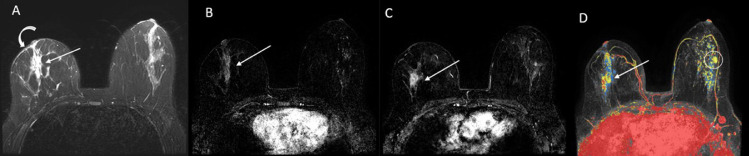
T2-weighted image demonstrates segmental increased signal in the right breast (straight arrow) and right breast skin thickening (curved arrow). (B and C) T1-weighted postcontrast subtraction images demonstrate segmental non-mass enhancement in the right breast (straight arrows) and mild background parenchymal enhancement in the left breast. (D) Maximum intensity projection (MIP) image of tissue contrast kinetics demonstrates segmental non-mass enhancement with washout kinetics (red color) in the right breast (straight arrow) as well as background parenchymal enhancement in the left breast. Incidentally noted fibroadenoma of the left breast (circle) was unchanged since initial MRI.
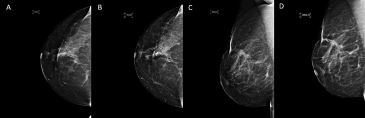
Right craniocaudal (RCC) and right mediolateral oblique (RMLO) views demonstrate benign calcifications and stable post surgical changes of the lumpectomy site 2 years prior (A and C) and at the time of diagnosis (B and D).
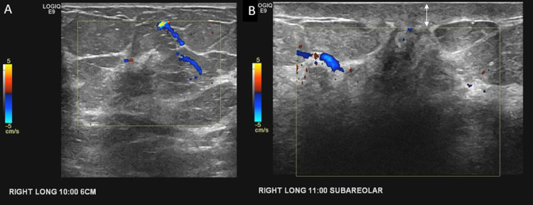
Gray-scale ultrasound images with Doppler demonstrate two irregular hypoechoic right breast masses with peripheral vascularity noted at the 10-o'clock (A) and 11-o'clock (B) positions. Note the mild skin thickening (double arrow).
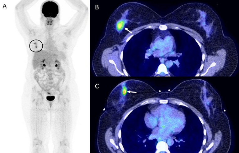
Maximum intensity projection (MIP) image from an FDG PET/CT exam demonstrates two FDG avid masses in the right breast (circle). (B and C) Axial fused FDG PET/CT images demonstrate a superior FDG avid mass with SUVmax of 4.4 (arrow), and a second, more inferior mass with SUVmax of 5.4 (arrow).

Gross pathologic image of the right breast simple mastectomy specimen. There is an angiosarcoma forming an 8.3 × 5.8 × 4.5 cm mass in the central portion of the breast. The tumor does not involve the skin (dermis or epidermis). The closest soft tissue margin is deep and negative by 2.2 cm. The closest peripheral skin margin is inferior and negative by 2.5 cm.
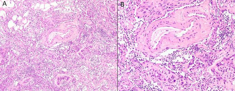
Postirradiation angiosarcoma of the mammary parenchyma, growing in a diffusely infiltrative fashion between non-neoplastic ducts, lobules, and thick-walled blood vessels (H+E, ×100). (B) Higher power view of high-grade mammary angiosarcoma, showing both small fascicles of hyperchromatic spindled endothelial cells and formation of irregular, slit-like vascular channels lined by protuberant, hyperchromatic endothelia (H+E, ×200).
Similar articles
-
Radiation-associated angiosarcoma of the breast: analysis of diagnostic tools in a registry-based population.Acta Radiol. 2022 Jan;63(1):22-27. doi: 10.1177/0284185120980142. Epub 2020 Dec 21. Acta Radiol. 2022. PMID: 33349000
-
Radiation-associated angiosarcoma of the breast: radical resection technique of the entire radiation field.Ther Adv Med Oncol. 2025 Feb 18;17:17588359251317842. doi: 10.1177/17588359251317842. eCollection 2025. Ther Adv Med Oncol. 2025. PMID: 39975512 Free PMC article.
-
Radiation-Associated Angiosarcoma of the Breast and Chest Wall Treated with Thermography-Controlled, Contactless wIRA-Hyperthermia and Hypofractionated Re-Irradiation.Cancers (Basel). 2021 Aug 3;13(15):3911. doi: 10.3390/cancers13153911. Cancers (Basel). 2021. PMID: 34359812 Free PMC article.
-
Cutaneous angiosarcoma and atypical vascular lesions of the skin and breast after radiation therapy for breast carcinoma.Am J Clin Pathol. 1994 Dec;102(6):757-63. doi: 10.1093/ajcp/102.6.757. Am J Clin Pathol. 1994. PMID: 7801888 Review.
-
Spontaneous regression of breast angiosarcoma after conservative treatment with radiotherapy: a case report and review of the literature.J Med Ultrason (2001). 2015 Jul;42(3):427-32. doi: 10.1007/s10396-014-0607-z. Epub 2014 Dec 27. J Med Ultrason (2001). 2015. PMID: 26576798 Review.
Cited by
-
Radiation-associated breast angiosarcoma after strut-adjusted volume implant brachytherapy.Radiol Case Rep. 2024 Jun 28;19(9):3888-3894. doi: 10.1016/j.radcr.2024.05.081. eCollection 2024 Sep. Radiol Case Rep. 2024. PMID: 39040825 Free PMC article.
References
Publication types
LinkOut - more resources
Full Text Sources
