. 2022 Jul 13;32(2):235-252.
doi: 10.1055/s-0042-1744142. eCollection 2022 Jun.

Solid Primary Retroperitoneal Masses in Adults: An Imaging Approach

Affiliations

Solid Primary Retroperitoneal Masses in Adults: An Imaging Approach

Vaibhav Gulati et al. Indian J Radiol Imaging. .

Abstract

Mass lesions in the retroperitoneal space may be primary or secondary. Primary retroperitoneal mass lesions are relatively uncommon as compared to pathology that arises secondarily from retroperitoneal organs. These may be solid or cystic lesions. The overlapping imaging features of various solid primary retroperitoneal tumors make the diagnosis difficult, and hence, histopathology remains the mainstay of diagnosis. This paper provides a brief review of the anatomy of the retroperitoneal space and provides an algorithmic approach based on cross-sectional imaging techniques to narrow down the differential diagnosis of solid primary retroperitoneal masses encountered in the adult population.

Keywords: Algorithmic approach; Anatomy; Imaging; Retroperitoneal space; Solid.


Conflict of interest statement

Conflicts of Interest None declared.

Figures

Fig. 1
Fig. 1
Cross-sectional diagram of the abdomen depicting the retroperitoneal compartments: APR, anterior pararenal space; PPR, posterior pararenal space; PR, perirenal space. Green—posterior renal fascia. Pink—anterior renal fascia. Yellow—transversalis fascia. Blue—lateroconal fascia.
Fig. 2
Fig. 2
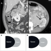
( A ) Coronal contrast-enhanced CT image of a 45-year-old male shows a heterogeneously enhancing mass lesion with positive beak/claw sign ( arrows ) along its contact surface with the right kidney (K) in a case of renal cell carcinoma. ( B ) Axial contrast-enhanced CT image in an 18-year-old female reveals heterogeneously enhancing mass that demonstrates negative beak sign ( arrows ) in relation to the left kidney. This indicates a primary retro-peritoneal origin of the mass. Schematic diagrams illustrating ( C ) the positive “beak” sign and ( D ) negative “beak” sign.
Fig. 3
Fig. 3
( A ) Post-contrast axial CT image of a 55-year-old female with colon carcinoma demonstrates a heterogeneously enhancing retroperitoneal mass involving the right colon which is embedded within the mass ( arrow ), illustrating the positive embedded organ sign. ( B ) Post-contrast axial CT image in a 50-year-old female with primary retroperitoneal sarcoma shows a right-sided heterogenous infiltrative mass in the retroperitoneum with involvement of the adjacent posterior abdominal wall muscles. The mass displaces and deforms the ascending colon ( arrow ) illustrating the negative embedded organ sign. Schematic diagrams illustrating the ( C ) positive “embedded organ” sign and ( D ) negative “embedded organ” sign.
Fig. 4
Fig. 4
( A ) Contrast-enhanced axial CT image of a 35-year-old female with retroperitoneal pheochromocytoma shows a large heterogeneous mass in the right suprarenal location immediately behind the IVC displacing it anteriorly, with an undetectable right adrenal gland illustrating the “phantom organ” sign. ( B ) Schematic diagram illustrating the “phantom organ” sign.
Fig. 5
Fig. 5
Retroperitoneal well-differentiated liposarcoma in a 35-year-old woman. ( A, B ) Axial contrast-enhanced CT images demonstrate a large, illdefined, fatty mass lesion ( asterisk ) involving the right anterior pararenal space surrounding and anteriorly displacing the ascending colon ( C ). There is medial displacement of small bowel loops and mesentery. Note the subtle soft-tissue septations and nodularity within the mass ( arrow ).
Fig. 6
Fig. 6
Retroperitoneal myxoid liposarcoma in a 60-year-old female with a palpable abdominal mass. ( A, B ) Axial and ( C ) coronal contrast-enhanced CT images show a large well-circumscribed heterogeneous left retroperitoneal mass displacing the left kidney (K) anteromedially, spleen superiorly, and bowel loops infero-medially. The lesion shows variable CT attenuation areas related to the presence of macroscopic fat (ROI 1), soft-tissue density component (ROI 2), and low attenuation myxoid (ROI 3) dominant component. Fat in the margin of the mass may simulate extraperitoneal fat and be missed.
Fig. 7
Fig. 7
Retroperitoneal liposarcoma in a 50-year-old man. ( A, B, C ) Axial and ( D ) coronal postcontrast CT images show a large heterogeneously enhancing left-sided retroperitoneal mass with a well-differentiated fatty component ( white asterisk ) superiorly in the perinephric region with linear septations within ( arrow ) and dedifferentiated mass-like areas of soft tissue ( black asterisk ) inferiorly. There is involvement of left psoas muscle (P) with mass-effect and displacement of the left kidney. Distinct, large, heterogeneously enhancing necrotic soft-tissue mass on the right, likely metastasis.
Fig. 8
Fig. 8
Retroperitoneal IVC leiomyosarcoma in a 28-year-old male with abdominal pain . ( A ) Contrast-enhanced axial and ( B ) coronal CT images demonstrate a relatively well-circumscribed heterogeneously enhancing mass in the retroperitoneum with extensive necrosis. There is expansion of the IVC (I) with both extraluminal and intraluminal components. There is involvement of both suprarenal and infrarenal segments of the IVC with cranial extension of tumor into the intrahepatic IVC ( large arrow ). The tumor thrombus extends into the right renal vein ( small arrow ) causing mild right-sided hydronephrosis.
Fig. 9
Fig. 9
Contrast-enhanced CT ( A, B, C ) axial and ( D ) coronal images in a 34-year-old male with a palpable mass show a large, circumscribed mass in left retroperitoneum encasing the left kidney (K) with negative beak sign along with anterior displacement of the spleen (S). The lesion is heterogeneous with areas of solid soft tissue ( black asterisk ) and cystic and myxoid components ( white asterisk ). In the absence of fat, myxoid variety of liposarcoma and undifferentiated pleomorphic sarcoma (MFH) appear similar and cannot be differentiated on imaging alone. This was undifferentiated pleomorphic sarcoma.
Fig. 10
Fig. 10
Retroperitoneal lymphadenopathy in a 48-year-old woman with Non-Hodgkin's lymphoma. ( A , B , C ) Axial post-contrast CT images demonstrate a homogeneously enhancing lymph node conglomerate in the retroperitoneum that anteriorly displaces the IVC (I) and surrounds abdominal aorta (A). The pancreas is also involved and is not visualized separately from the lesion. Note the lymphomatous deposit in the spleen ( small arrow ) and mild ascites ( asterisk ). ( D ) Coronal CT image shows encasement of the superior mesenteric artery by the lymph nodal mass with extensive mesenteric lymphadenopathy.
Fig. 11
Fig. 11
Retroperitoneal lymphadenopathy in a 37-year-old male with metastatic testicular carcinoma. ( A ) Axial and ( B ) coronal post-contrast CT images through the abdomen reveal multiple enlarged retroperitoneal lymph nodes in the paracaval and left paraaortic locations with heterogeneous peripheral enhancement and central necrosis.
Fig. 12
Fig. 12
Post-contrast CT and T2-weighted MRI ( A , B ) axial and ( C , D ) coronal images of a 17-year-old girl demonstrate a circumscribed homogenous retroperitoneal mass in the right paravertebral region. The mass displaces the right kidney (K) postero-inferiorly and IVC (I) anteriorly. High signal intensity areas ( asterisk ) correspond to areas of cystic/myxoid degeneration. Characteristic location, CT, and MRI appearance suggest a retroperitoneal neurogenic tumor, likely Schwannoma.
Fig. 13
Fig. 13
Retroperitoneal neurofibroma in a 39-year-old female. Contrast-enhanced axial T1- weighted MRI demonstrates a well-defined homogeneously enhancing mass lesion in the left paravertebral region, which displaces the left psoas muscle postero-laterally. There is a dumbbell-shaped intraspinal extension of the lesion with widening of the neural foramen ( white arrow ).
Fig. 14
Fig. 14
Malignant peripheral nerve sheath tumor in a 28-year-old female. Fat saturated T2-weighted ( A ) coronal and ( B ) post-contrast T1-weighted axial MRI show an ill-defined infiltrative heterogeneous mass lesion involving the left lumbosacral plexus with intraspinal component ( long white arrow ). The lesion appears heterogeneously hyperintense on T2-weighted images with heterogeneous post-contrast enhancement and areas of necrosis. The lesion is invading the left iliac bone and sacrum ( short white arrows ).
Fig. 15
Fig. 15
Retroperitoneal paraganglioma in a 41-year-old female with hypertension. ( A ) T1-weighted sagittal, ( B ) fat-saturated T2-weighted coronal , and ( C ) T2-weighted axial MRI show a circumscribed, retroperitoneal mass lesion in the left para-aortic prevertebral location with homogenous low to intermediate T1 signal intensity and high T2 signal intensity. A low signal intensity peripheral capsule can also be appreciated on T2-weighted images. ( D ) Fat suppressed, contrast-enhanced T1-weighted axial MRI demonstrates moderate to high, relatively homogenous enhancement in the lesion.
Fig. 16
Fig. 16
Retroperitoneal extramedullary hematopoiesis in a 20-year-old female with myelofibrosis. ( A ) Contrast-enhanced axial CT image shows a well-defined, round, minimally enhancing soft-tissue mass lesion ( white arrow ) in the left perinephric space of the retroperitoneum. The lesion is compressing the left pelvi-ureteric junction with mild hydronephrosis. Similar appearing multiple lobulated mass lesions also noted involving the right kidney ( black arrows ). ( B ) Coronal bone window CT image demonstrates diffusely increased density of the axial skeleton (osteosclerosis) due to myelofibrosis.
Fig. 17
Fig. 17
Idiopathic retroperitoneal fibrosis in a 38-year-old man. ( A , B ) Contrast-enhanced axial and ( C ) coronal CT images demonstrate a well-defined, retroperitoneal, soft-tissue mass ( asterisk ) surrounding the infrarenal abdominal aorta (A) and extending inferiorly to involve the common iliac arteries bilaterally. ( D ) Delayed excretory phase axial CT image shows partial encasement with narrowing of both ureters ( arrows ) with hydronephrosis ( not shown ). ( E ) Axial T1- and ( F ) T2-weighted MRI show intermediate T1 signal and low T2 signal intensity, indicative of mature fibrosis.
Fig. 18
Fig. 18
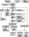
Diagnostic algorithm for primary retroperitoneal solid lesions.

Similar articles

Cited by

References

    1. Rajiah P, Sinha R, Cuevas C, Dubinsky T J, Bush W H, Jr, Kolokythas O. Imaging of uncommon retroperitoneal masses. Radiographics. 2011;31(04):949–976. - PubMed
    1. Brennan C, Kajal D, Khalili K, Ghai S. Solid malignant retroperitoneal masses—a pictorial review. Insights Imaging. 2014;5(01):53–65. - PMC - PubMed
    1. Osman S, Lehnert B E, Elojeimy S et al.A comprehensive review of the retroperitoneal anatomy, neoplasms, and pattern of disease spread. Curr Probl Diagn Radiol. 2013;42(05):191–208. - PubMed
    1. Ishikawa K, Nakao S, Nakamuro M, Huang T P, Nakano H. The retroperitoneal interfascial planes: current overview and future perspectives. Acute Med Surg. 2016;3(03):219–229. - PMC - PubMed
    1. Goenka A H, Shah S N, Remer E M.Imaging of the retroperitoneum Radiol Clin North Am 20125002333–355., vii - PubMed