Review
. 2022 Jul 5;32(2):213-223.
doi: 10.1055/s-0042-1744520. eCollection 2022 Jun.

Imaging Tips and Tricks in Management of Renal and Urothelial Malignancies

Affiliations
Review

Imaging Tips and Tricks in Management of Renal and Urothelial Malignancies

Shanti Ranjan Sanyal et al. Indian J Radiol Imaging. .

Abstract

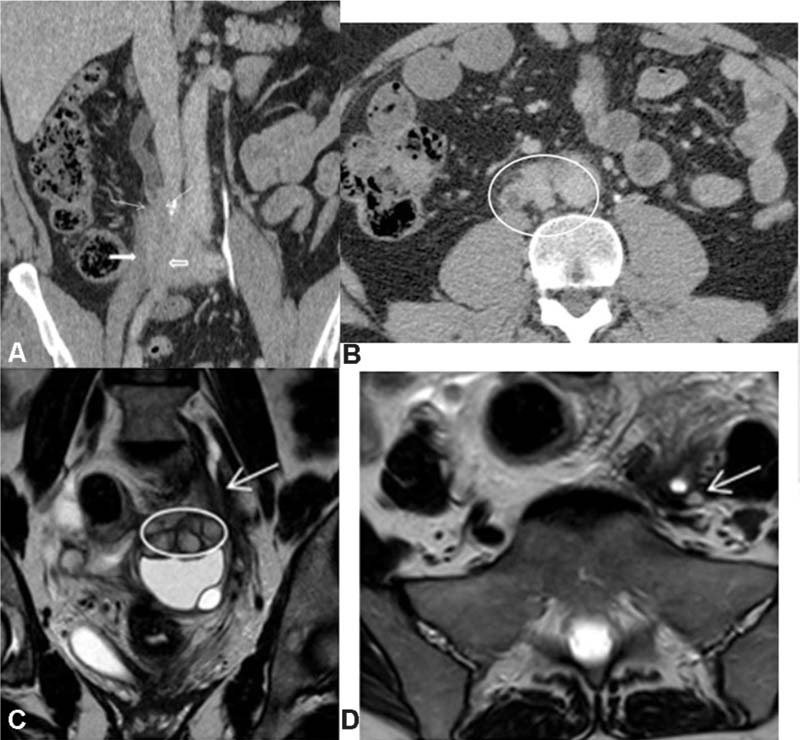
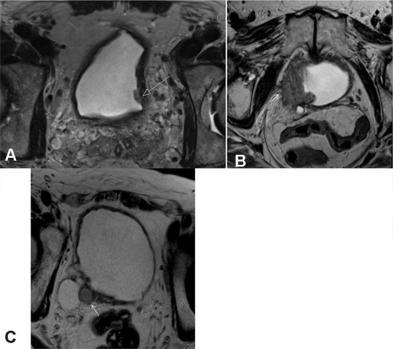
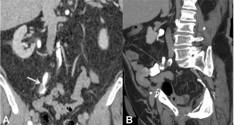
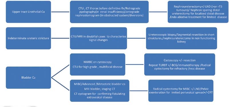
Management of urological malignancies has evolved significantly with continually changing guidelines and treatment options which demand more centralized involvement of radiology than ever before. Radiologists play a pivotal role in interpreting complex cancer scans and guiding clinical teams toward the best management options in the light of clinical profile. Management of complex uro-oncology cases is often discussed in multidisciplinary meetings which are essential checkpoints to evaluate an overall picture and formulate optimal treatment plans. The aim of this article is to provide a radiological perspective with practical guidance to fellow radiologists participating in uro-oncology multidisciplinary meetings based on commonly encountered case scenarios, updated guidelines, and cancer pathways. Crucial imaging tips with regards to renal and urinary tract cancers, upon which therapeutic decisions are made, have been condensed in this article after reviewing several complex cases from urology multidisciplinary meetings and European Association of Urology guidelines. Outline of various diagnostic and management strategies, key staging features, surveillance guidelines, and, above all, what the onco-urologists want to know from radiologists have been succinctly discussed in this article.

Keywords: imaging tips; management; renal masses; surveillance; urinary tract.


Conflict of interest statement

Conflicts of Interest/Competing Interests The authors declare that they have no financial or nonfinancial competing interests.

Figures

Fig. 1
Fig. 1
( A ) Axial-enhanced CT images demonstrated a right renal interpolar lesion being considered for partial nephrectomy, but close to renal hilum. The mass also shows calcification inside ( white arrow ), which favors more toward RCC with osteometaplasia. ( B ) Complex cystic mass in right kidney lower pole with MIP image demonstrating multiple accessory renal arteries ( black arrows ).
Fig. 2
Fig. 2
( A ) CT coronal reformatted images show a complex cystic mass ( solid white arrow ) with an apparently enhancing nodule ( dashed arrow ) in left kidney upper pole and another hypodense lesion more inferiorly ( circle ). ( B ) Contrast-enhanced MR images downstage the complex cyst in left kidney upper pole ( solid white arrow ) with thin septation; however, the mural nodule ( dashed arrow ) turned out to be a separate proteinaceous cyst without any obvious enhancement. The more inferior hypodense lesion is a solid enhancing mass more clearly demonstrated ( circle ). This information was vital to the surgeon who could spare the cyst and perform partial nephrectomy for the smaller solid lesion. CT, computed tomography; MR, magnetic resonance.
Fig. 3
Fig. 3
VHL patient with multiple recurrent tumor and postpartial nephrectomy-related complication. ( A ) Axial-enhanced CT shows a left renal mass ( arrow ) with pancreatic cysts ( ellipse ). ( B ) Postpartial nephrectomy noncontrast CT scan shows a hyperdense focus at the resection site. ( C ) CT angiogram confirms this to be a small intrarenal pseudoaneurysm ( arrow ). ( D ) Postsurveillance CT scan 1 year later depicts a new intrarenal lesion. CT, computed tomography; VHL, Von Hippel–Lindau disease.
Fig. 4
Fig. 4
( A ) Axial CT in a case of right renal RCC (*) with pancreatic ( arrow ) and small-bowel mesenteric ( circle ) metastases. ( B ) Not a candidate for surgical debulking.
Fig. 5
Fig. 5
( A ) Axial-enhanced CT images show some soft tissue ( arrow ) at the site of previous partial nephrectomy (PN) with mild thickening along right peritoneal reflection ( circle ). ( B ) On follow-up scan interval growth in size of soft tissue nodule at the site of PN in consistent with local recurrence.
Fig. 6
Fig. 6
Imaging algorithm and management strategies in accordance with type of renal mass on cross-sectional imaging.
Fig. 7
Fig. 7
( A ) Axial and reformatted coronal CT images: UTUC (pattern 1): circumferential smooth wall urothelial thickening ( arrow ) with no spiculation. ( B ) Axial CT images: UTUC (pattern 2): circumferential wall thickening of right distal ureter ( arrow ) with spiculation. ( C ) Axial CT urogram images: UTUC (pattern 3): small intraluminal soft tissue filling defect in left distal ureter ( arrow ) with smooth external surface and no spiculation. ( D ) Axial CT images reveal UTUC (pattern 4): asymmetric nodular wall thickening in left proximal ureter anteriorly ( arrow ) with spiculation. ( E ) Axial CT images: UTUC (pattern 5): intraluminal mass with smooth external surface. ( F ) Axial CT images: UTUC (pattern 6): intraluminal mass with irregular external surface and speculation ( arrow ) in left proximal ureter. CT, computed tomography; UTUC, upper tract urothelial cancer.
Fig. 8
Fig. 8
( A ) Axial CT images in a different case: central RCC ( arrows ), invading renal sinus, T3 tumor is a close differential for mass like TCC. ( B ) Mass like TCC with ureteric involvement—reniform shape of kidney maintained—important to differentiate from RCC. CT, computed tomography; RCC, renal cell cancer; TCC, transitional cell carcinoma.
Fig. 9
Fig. 9
( A ) Coronal reformatted CT images show soft tissue lesion involving right distal ureter ( thin arrows ) extending to involve right-sided iliac vessels ( block arrows ). ( B ) Axial CT images demonstrate soft tissue thickening ( circle ) encompassing ureters and retroperitoneal vessels. ( C ) T2W coronal MR images in a different case show left distal ureteric stricture ( arrow ) with endometriotic deposits ( oval ) seen on left ovary. ( D ) T2W axial MR—endometriosis with secondary ureteric stricture confirmed on laparoscopic biopsy. CT, computed tomography; T2W, T2 weighted.
Fig. 10
Fig. 10
Bladder tumor imaging subtypes: ( A ) T2W axial images demonstrate the superficial lesion in bladder, clearly demarcated from the hypointense mural layer. ( B ) Axial T2WMR imaging shows muscle invasive tumor with extravesical spread and infiltration to right obturator internus. ( C ) Axial T2W MR images demonstrate T2 intermediate signal tumor in bladder diverticulum ( arrow ). MR, magnetic resonance; T2W, T2 weighted.
Fig. 11
Fig. 11
Post TURBT CT urogram 2 weeks for superficial papillary tumor with preserved muscle on biopsy. ( A ) Axial image shows extensive bladder wall thickening, perivesical stranding and a suspicious right internal iliac lymph node ( arrow ). ( B ) On follow-up CTU after 3 months, although bladder inflammatory changes resolve, right internal iliac node ( arrow ) persists. ( C ) The node ( arrow ) demonstrates stable appearances with no significant uptake on a follow-up PET CT and no other evidence of disease elsewhere, hence considered a reactive node. CT, computed tomography; CTU, computed tomography urography; PET, positron emission tomography; TURBT, transurethral resection of a bladder tumor.
Fig. 12
Fig. 12
( A ) Postradical cystectomy coronal CTU images in a different patient: recurrent soft tissue ( arrow ) seen at ureteroileal anastomosis on postcystectomy surveillance scan. ( B ) Sagittal CTU maximum intensity projection images. CTU, computed tomography urography.
Fig. 13
Fig. 13
Imaging algorithm and management strategies for urothelial cancers.

Similar articles

References

    1. Wah T M. Image-guided ablation of renal cell carcinoma. Clin Radiol. 2017;72(08):636–644. - PubMed
    1. Israel G M, Bosniak M A. Pitfalls in renal mass evaluation and how to avoid them. Radiographics. 2008;28(05):1325–1338. - PubMed
    1. Prasad S R, Humphrey P A, Catena J Ret al.Common and uncommon histologic subtypes of renal cell carcinoma: imaging spectrum with pathologic correlation Radiographics 200626061795–1806., discussion 1806–1810 - PubMed
    1. Childs D D, Clingan M J, Zagoria R J et al.In-phase signal intensity loss in solid renal masses on dual-echo gradient-echo MRI: association with malignancy and pathologic classification. AJR Am J Roentgenol. 2014;203(04):W421–W428. - PubMed
    1. Uzzo R G, Novick A C. Nephron sparing surgery for renal tumors: indications, techniques and outcomes. J Urol. 2001;166(01):6–18. - PubMed