Upregulated Expression of IL2RB Causes Disorder of Immune Microenvironment in Patients with Kawasaki Disease
- PMID: 35924269
- PMCID: PMC9343205
- DOI: 10.1155/2022/2114699
Upregulated Expression of IL2RB Causes Disorder of Immune Microenvironment in Patients with Kawasaki Disease
Abstract
Aims: The clinical diagnosis of Kawasaki disease (KD) is not easy because of many atypical manifestations. This study is aimed at finding potential diagnostic markers and therapeutic targets for KD and analysing their correlation with immune cell infiltrations.
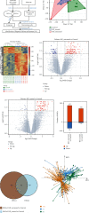
Methods: First, we downloaded the KD dataset from the Gene Expression Omnibus (GEO) database and used R software to identify differentially expressed genes (DEGs) and perform functional correlation analysis. Then, CIBERSORT algorithm was used to evaluate immune cell infiltrations in samples. Coexpression analysis between DEGs and infiltrating immune cells was performed to screen the main infiltrating immune cells. Subsequently, the least absolute shrinkage and selection operator (LASSO) logistic regression analysis was used to screen the core genes related to KD. Finally, correlation analysis between the core genes and the main infiltrating immune cells was performed.
Results: 327 DEGs were screened out in this study. Among them, 72 shared genes were the category of genes most likely to be disease-causing for they did not change before and after treatment. After analysis, it was found that expression level of IL2RB in KD tissues was significantly upregulated, the number of resting CD4+ memory T cells was decreased, and the decrease was significantly negatively correlated with the upregulated expression of IL2RB. Therefore, it was speculated that the upregulated expression of IL2RB disrupted Th1/Th2 cell differentiation balance, which led to a decrease of resting CD4+ memory T cells and finally caused disorder of immune microenvironment in patients with KD.
Conclusions: Upregulated expression of IL2RB leads to disorder of immune microenvironment in patients with KD and eventually causes the occurrence and development of KD. Therefore, IL2RB may serve as a diagnostic marker and potential therapeutic target for KD.
Copyright © 2022 Yunfei Liao et al.
Conflict of interest statement
The authors declare that there is no conflict of interest regarding the publication of this paper.
Figures


Similar articles
-
Interleukin 2 receptor subunit beta as a novel hub gene plays a potential role in the immune microenvironment of abdominal aortic aneurysms.Gene. 2022 Jun 15;827:146472. doi: 10.1016/j.gene.2022.146472. Epub 2022 Apr 4. Gene. 2022. PMID: 35381314
-
Bioinformatic analysis of underlying mechanisms of Kawasaki disease via Weighted Gene Correlation Network Analysis (WGCNA) and the Least Absolute Shrinkage and Selection Operator method (LASSO) regression model.BMC Pediatr. 2023 Feb 24;23(1):90. doi: 10.1186/s12887-023-03896-4. BMC Pediatr. 2023. PMID: 36829193 Free PMC article.
-
Similarity of immune-associated markers in COVID-19 and Kawasaki disease: analyses from bioinformatics and machine learning.BMC Pediatr. 2025 May 19;25(1):400. doi: 10.1186/s12887-025-05752-z. BMC Pediatr. 2025. PMID: 40383755 Free PMC article.
-
Prediction of Immune Infiltration Diagnostic Gene Biomarkers in Kawasaki Disease.J Immunol Res. 2022 Jun 17;2022:8739498. doi: 10.1155/2022/8739498. eCollection 2022. J Immunol Res. 2022. PMID: 35755167 Free PMC article.
-
Identify potential prognostic indicators and tumor-infiltrating immune cells in pancreatic adenocarcinoma.Biosci Rep. 2022 Feb 25;42(2):BSR20212523. doi: 10.1042/BSR20212523. Biosci Rep. 2022. PMID: 35083488 Free PMC article. Review.
Cited by
-
Inflammation and Neurodegeneration in Glaucoma: Isolated Eye Disease or a Part of a Systemic Disorder? - Serum Proteomic Analysis.J Inflamm Res. 2024 Feb 13;17:1021-1037. doi: 10.2147/JIR.S434989. eCollection 2024. J Inflamm Res. 2024. PMID: 38370463 Free PMC article.
-
Integration of scRNA-Seq and bulk RNA-Seq uncover perturbed immune cell types and pathways of Kawasaki disease.Front Immunol. 2023 Sep 28;14:1259353. doi: 10.3389/fimmu.2023.1259353. eCollection 2023. Front Immunol. 2023. PMID: 37841239 Free PMC article.
-
A comprehensive integration of data on the association of ITPKC polymorphisms with susceptibility to Kawasaki disease: a meta-analysis.BMC Med Genomics. 2025 Mar 20;18(1):56. doi: 10.1186/s12920-025-02121-8. BMC Med Genomics. 2025. PMID: 40114120 Free PMC article.
References
MeSH terms
Substances
LinkOut - more resources
Full Text Sources
Medical
Molecular Biology Databases
Research Materials

