. 2022 Aug 5;55(4):338-356.
doi: 10.5090/jcs.22.066.

Medical Complications of Lung Transplantation

Affiliations

Medical Complications of Lung Transplantation

Moo Suk Park. J Chest Surg. .

Abstract

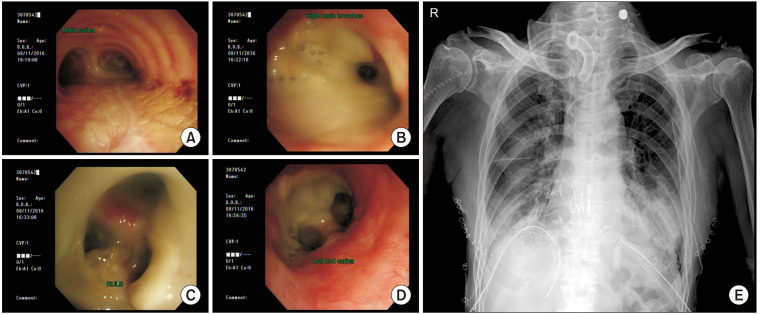
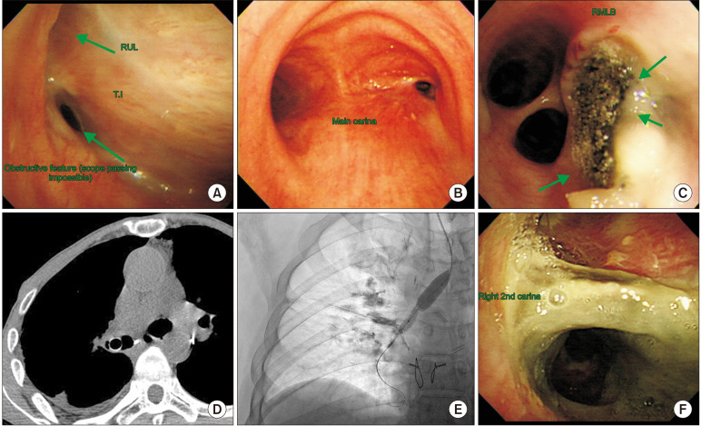
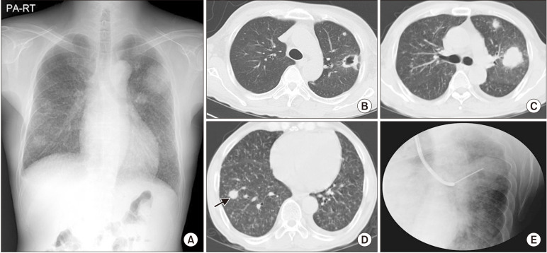
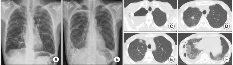
Lung transplantation (LT) is now considered as an effective treatment option for end-stage lung diseases that improves the short and long-term survival rates and quality of life. As increasingly many LT procedures are being performed, the medical complications of LT are also increasing in frequency and emerging as a very important issue for transplant clinicians. Although chronic lung allograft dysfunction and infection are major causes of death after LT, many medical complications, several of which result from immunosuppressive treatment, contribute to increased mortality and morbidity. This article reviews the most frequent and important medical complications of LT, accompanied by a review of the literature and studies from South Korea, including lung allograft rejection, infection, and non-allograft organ systemic complications.

Keywords: Complications; Graft rejection; Immunosuppression therapy; Infections; Lung transplantation; Organ.


Figures

Fig. 1
Fig. 1
A 65-year-old male patient with idiopathic pulmonary fibrosis with transmitted donor multidrug-resistant Acinetobacter baumannii and Klebsiella pneumoniae in pre-donation culture by bronchoscopy. (A–D) Bronchoscopic findings and (E) chest X-ray showed pneumonia in both lower lobes. RULB, right upper lobe bronchus.
Fig. 2
Fig. 2
(A–F) Multiple cases of airway stenosis after Aspergillus and bacterial tracheobronchitis infection (arrows). RUL, right upper lobe; RMLB, right middle lobe bronchus; T.I, truncus intermedius.
Fig. 3
Fig. 3
(A–E) Invasive pulmonary Aspergillus infection confirmed by transbronchial lung biopsy (arrow). PA, posterior-anterior; RT, right.
Fig. 4
Fig. 4
(A–F) A 49-year-old male patient who underwent lung transplantation due to graft-versus-host disease after hematopoietic stem cell transplantation. A 1-month post-lung transplantation sputum culture showed Mycobacterium tuberculosis from the donor. PA, posterior-anterior; RT, right.
Fig. 5
Fig. 5
(A–F) A 62-year-old male patient who underwent lung transplantation due to chronic bronchiectasis and cor pulmonale. Five years after lung transplantation, mixed-type chronic lung allograft dysfunction with airway obstructive bronchiolitis obliterans syndrome and restrictive allograft syndrome in both upper lobes developed, and sputum culture showed non-tuberculous mycobacteria (Mycobacterium intracellulare). PA, posterior-anterior; RT, right.
Fig. 6
Fig. 6
(A–H) A 64-year-old female patient who underwent single lung transplantation due to idiopathic pulmonary fibrosis. Four months post-lung transplantation, Nocardia farcina infection developed in the lung and brain through a leg wound (arrows). PA, posterior-anterior; RT, right.
Fig. 7
Fig. 7
A 34-year-old male patient who underwent bilateral lung transplantation due to idiopathic pulmonary fibrosis. (A) At 5 days post-transplant, fever, low prograft level, pulmonary edema, and hypoxemia developed, and bronchoalveolar lavage fluid analysis showed a total of 531 cells with 8% of polynuclear neutrophils and (B) improved acute rejection after methylprednisolone (500 mg) pulse infusion for 3 days and slow tapering from prednisolone.
Fig. 8
Fig. 8
(A–E) Severe gastroparesis and superior mesenteric artery syndrome requiring decompressive surgery (arrows).
Fig. 9
Fig. 9
(A, B) Two cases of suspected short telomere syndrome combined with pulmonary fibrosis, liver diseases, bone marrow suppression, and frequent infection; the diagnosis was confirmed by next-generation sequencing.
Fig. 10
Fig. 10
(A–G) A 58-year-old male patient who underwent lung transplantation due to idiopathic pulmonary fibrosis. At 10 days post-lung transplantation, a chest spiral computed tomography (CT) scan and leg extremity CT scan showed deep vein thrombosis and pulmonary thromboembolism (arrows).
Fig. 11
Fig. 11
A 59-year-old female patient who underwent lung transplantation due to destroyed lung and cor pulmonale. (A) At 1 month post-transplant, posterior reversible encephalopathy syndrome (PRES) first developed with seizures, hallucinations, and drowsiness (red circle). (B) After 3 months, PRES relapsed with seizures and high blood pressure.

Similar articles

Cited by

References

    1. Chambers DC, Cherikh WS, Goldfarb SB, et al. The International Thoracic Organ Transplant Registry of the International Society for Heart and Lung Transplantation: thirty-fifth adult lung and heart-lung transplant report-2018; focus theme: multiorgan transplantation. J Heart Lung Transplant. 2018;37:1169–83. doi: 10.1016/j.healun.2018.07.020. - DOI - PubMed
    1. Chambers DC, Yusen RD, Cherikh WS, et al. The Registry of the International Society for Heart and Lung Transplantation: thirty-fourth adult lung and heart-lung transplantation report-2017; focus theme: allograft ischemic time. J Heart Lung Transplant. 2017;36:1047–59. doi: 10.1016/j.healun.2017.07.016. - DOI - PubMed
    1. Lyu DM, Zamora MR. Medical complications of lung transplantation. Proc Am Thorac Soc. 2009;6:101–7. doi: 10.1513/pats.200808-077GO. - DOI - PubMed
    1. Thaniyavarn T, Grewal HS, Goldberg HJ, Arcasoy SM. Nonallograft complications of lung transplantation. Thorac Surg Clin. 2022;32:243–58. doi: 10.1016/j.thorsurg.2022.01.004. - DOI - PubMed
    1. Arcasoy SM, Wilt J. Medical complications after lung transplantation. Semin Respir Crit Care Med. 2006;27:508–20. doi: 10.1055/s-2006-954611. - DOI - PubMed