Review
. 2022 Aug 4;7(8):576-586.
doi: 10.1530/EOR-22-0009.

Comprehensive management of posterior shoulder instability: diagnosis, indications, and technique for arthroscopic bone block augmentation

Affiliations
Review

Comprehensive management of posterior shoulder instability: diagnosis, indications, and technique for arthroscopic bone block augmentation

Abdul-Ilah Hachem et al. EFORT Open Rev. .

Abstract

Recurrent posterior glenohumeral instability is an entity that demands a high clinical suspicion and a detailed study for a correct approach and treatment. Its classification must consider its biomechanics, whether it is due to functional muscular imbalance or to structural changes, volition, and intentionality. Due to its varied clinical presentations and different structural alterations, ranging from capsule-labral lesions and bone defects to glenoid dysplasia and retroversion, the different treatment alternatives available have historically had a high incidence of failure. A detailed radiographic assessment, with both CT and MRI, with a precise assessment of glenoid and humeral bone defects and of glenoid morphology, is mandatory. Physiotherapy focused on periscapular muscle reeducation and external rotator strengthening is always the first line of treatment. When conservative treatment fails, surgical treatment must be guided by the structural lesions present, ranging from soft tissue repair to posterior bone block techniques to restore or increase the articular surface. Bone block procedures are indicated in cases of recurrent posterior instability after the failure of conservative treatment or soft tissue techniques, as well as symptomatic demonstrable nonintentional instability, presence of a posterior glenoid defect >10%, increased glenoid retroversion between 10 and 25°, and posterior rim dysplasia. Bone block fixation techniques that avoid screws and metal allow for satisfactory initial clinical results in a safe and reproducible way. An algorithm for the approach and treatment of recurrent posterior glenohumeral instability is presented, as well as the author's preferred surgical technique for arthroscopic posterior bone block.

Keywords: bone block; bone defect; instability; posterior; shoulder.


Figures

Figure 1
Figure 1
Preoperative CT of a shoulder with posterior instability and bone defect. (A) Sagittal cut immediately medial to the humeral head showing the ‘best-fit circle’. (B) Gamma angle (γ) measurement in an axial cut. A circle matching the humeral articular surface is drawn (white circle), before measuring the angle formed from the center of the circle to the bicipital groove and to the medial border of the humeral defect (green lines). (C) Measurement of the glenoid retroversion in an axial cut using the Friedman method (α).
Figure 2
Figure 2
Treatment algorithm for patients with recurrent posterior glenohumeral instability.
Figure 3
Figure 3
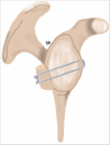
Graphic representation of the ‘Posterior Bone Block Cerclage’ technique. Using a system of ultra-high strength tapes without metal, the posterior bone defect is reconstructed with an iliac crest bone graft. Interconnection of the tapes over the graft’s cortical surface, associated to the use of a mechanical tensioner, achieves a strong and stable fixation parallel to the native glenoid surface.
Figure 4
Figure 4
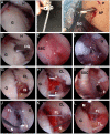
Glenoid preparation and tunnel drilling. (A, B, C, D, E, F, G, H, I, J, K, and L) Right shoulder, patient in lateral decubitus position, arthroscopic view from the anterosuperior portal. After release of the posterior capsulolabral complex from the glenoid rim, a drilling guide is placed parallel to the articular surface, using an accessory posterior portal when necessary. Two K-wires are then drilled into the glenoid using the guide, identifying their exit point through the anterior capsule and posterior to the subscapularis tendon. Two 2.4 mm canulated drills are then used to create the tunnels over the K-wires, before using a nitinol wire loop to replace each drill with a transport suture loop, in such a way that one loop faces anteriorly and the other posteriorly. +, inferior cannulated drill; ‡, superior cannulated drill; *, K-wires. AP, accessory posterior portal; CL, capsulolabral complex; DG, drill guide; FL, Fiberlink; G, glenoid; H, humeral head; IS, infraspinatus; M, arthroscopic measuring probe; N, nitinol wire loop; P, posterior portal; SSC, subscapularis; TL, Tigerlink.
Figure 5
Figure 5
Graft placement and capsulolabral complex repair. (A and B) Extra articular view showing graft preparation, cerclage fixation system and the interconnection of the pre-formed, racking hitch knots. (C, D, E, F, G, H, and I) Right shoulder, patient in lateral decubitus position, arthroscopic view from the anterosuperior portal. Using the transport sutures in the glenoid tunnels, two ultra-high-strength tape cerclage systems are transported through the graft and the glenoid, first into the graft from the cortical surface to the cancellous side, then from posterior to anterior in the first glenoid tunnel, returning from anterior to posterior in the second glenoid tunnel and finally through the second graft tunnel from the cancellous to the cortical side. The tape cerclage systems are interconnected with the pre-formed knots and the graft is placed into the joint, locking the system with additional manual knots. Finally, the capsulolabral complex is repaired to the native glenoid, leaving the graft in an extra-articular position. AP, accessory posterior portal; BB, iliac crest bone block; CL, capsulolabral complex; F, Fibertak; FTC, Fibertape Cerclage; H, humeral head; K, locking knot; TTC, Tigertape Cerclage.
Figure 6
Figure 6
Twelve-month follow-up CT of a patient treated with posterior bone block. (A) Sagittal cut with the preoperative ‘best-fit circle’ superimposed, showing adequate restoration of the glenoid surface and graft consolidation. (B) 3D reconstruction.

References

    1. Woodmass JM, Lee J, Wu IT, Desai VS, Camp CL, Dahm DL, Krych AJ. Incidence of posterior shoulder instability and trends in surgical reconstruction: a 22-year population-based study. Journal of Shoulder and Elbow Surgery 201928611–616. (10.1016/j.jse.2018.08.046) - DOI - PubMed
    1. Provencher MT, Leclere LE, King S, McDonald LS, Frank RM, Mologne TS, Ghodadra NS, Romeo AA. Posterior instability of the shoulder: diagnosis and management. American Journal of Sports Medicine 201139874–886. (10.1177/0363546510384232) - DOI - PubMed
    1. Moroder P, Scheibel M. ABC classification of posterior shoulder instability. Obere Extremitat 20171266–74. (10.1007/s11678-017-0404-6) - DOI - PMC - PubMed
    1. Chang ES, Greco NJ, McClincy MP, Bradley JP. Posterior shoulder instability in overhead athletes. Orthopedic Clinics of North America 201647179–187. (10.1016/j.ocl.2015.08.026) - DOI - PubMed
    1. Frank RM, Romeo AA, Provencher MT. Posterior glenohumeral instability: evidence-based treatment. Journal of the American Academy of Orthopaedic Surgeons 201725610–623. (10.5435/JAAOS-D-15-00631) - DOI - PubMed