Review
. 2022 Oct;24(10):1309-1325.
doi: 10.1007/s11886-022-01746-w. Epub 2022 Aug 4.

Diagnostic and Management Strategies in Patients with Late Recurrent Angina after Coronary Artery Bypass Grafting

Affiliations
Review

Diagnostic and Management Strategies in Patients with Late Recurrent Angina after Coronary Artery Bypass Grafting

Ruben W de Winter et al. Curr Cardiol Rep. 2022 Oct.

Abstract

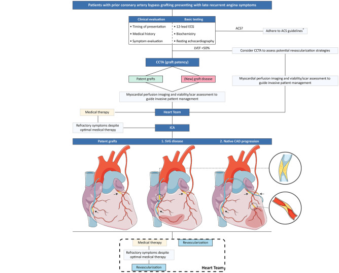
Purpose of review: This review will outline the current evidence on the anatomical, functional, and physiological tools that may be applied in the evaluation of patients with late recurrent angina after coronary artery bypass grafting (CABG). Furthermore, we discuss management strategies and propose an algorithm to guide decision-making for this complex patient population.

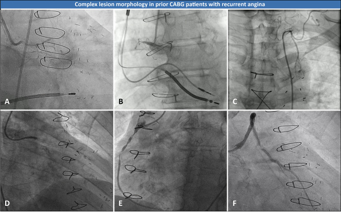
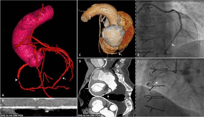
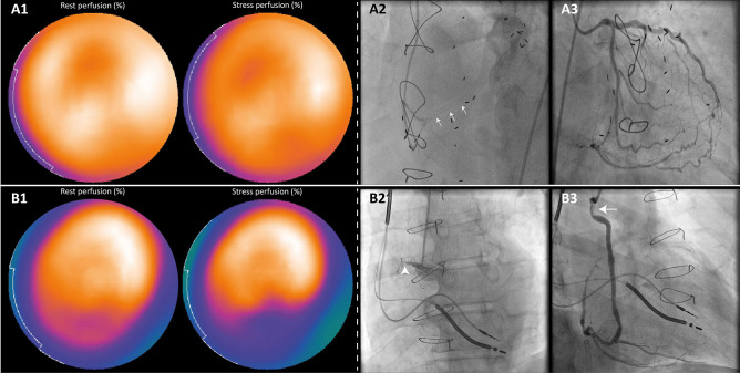
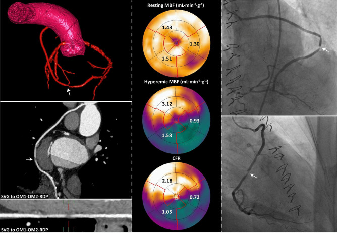
Recent findings: Patients with prior CABG often present with late recurrent angina as a result of bypass graft failure and progression of native coronary artery disease (CAD). These patients are generally older, have a higher prevalence of comorbidities, and more complex atherosclerotic lesion morphology compared to CABG-naïve patients. In addition, guideline recommendations are based on studies in which post-CABG patients have been largely excluded. Several invasive and non-invasive diagnostic tools are currently available to assess graft patency, the hemodynamic significance of native CAD progression, left ventricular function, and myocardial viability. Such tools, in particular the latest generation coronary computed tomography angiography, are part of a systematic diagnostic work-up to guide optimal repeat revascularization strategy in patients presenting with late recurrent angina after CABG.

Keywords: Diagnostic algorithm; Late recurrent angina; Prior coronary artery bypass grafting; Repeat revascularization.


Conflict of interest statement

None declared.

Figures

Fig. 1
Fig. 1
Advanced CAD and complex lesion morphology in patients presenting with late recurrent angina after CABG. Coronary angiography images showing advanced CAD and complex lesion morphology in prior CABG patients with recurrent angina symptoms. A Severely diseased LCA, B occluded RCA, C dysfunctional left internal mammary artery, D in-stent occlusion SVG on diagonal branch, E stenotic lesions SVG on PDA, F third time in-stent restenosis SVG on OM. CAD coronary artery disease, CABG coronary artery bypass grafting, LCA left coronary artery, OM obtuse marginal branch, PDA posterior descending artery, RCA right coronary artery, SVG saphenous vein graft
Fig. 2
Fig. 2
CCTA to evaluate bypass graft failure. CCTA and subsequent invasive coronary angiography images in a post-CABG patient with recurrent angina symptoms. AD CCTA reconstructed images showing a significant lesion in a saphenous vein jump graft with distal anastomoses on the intermediate branch, an OM branch and the PDA. The lesion is located between the anastomosis on the OM branch and the PDA (white arrows). In addition, a CTO was found in the RCA, depicted in the 3-dimensional reconstruction image in (A), indicated with the white arrowhead. E Subsequent angiography images confirmed the lesion in the venous jump graft (white arrow) (F) and in stent occlusion of the RCA (white arrowhead). CCTA coronary computed tomography angiography, CTO chronic total coronary occlusion; other abbreviations as in Fig. 1
Fig. 3
Fig. 3
SPECT perfusion imaging in patients with previous CABG. Rest and stress (intravenous adenosine 140 mg/kg/min) SPECT perfusion results acquired during a 2-day protocol using a weight-adjusted dose (370 to 550 MBq) of.99mTc tetrofosmin as a radiopharmaceutical, in conjunction with ICA images are illustrated. A1 SPECT-MPI in a patient presenting with dyspnea on exertion (RDS 2) and index bypass surgery 14 years ago showed mildly reduced tracer-uptake in the distal and mid anterior segments which persisted during hyperemia (differential diagnosis: a prior MI or an attenuation artefact). A2 Angiography showed a small caliber single SVG on the PDA with chronic in-stent occlusion of the distal native PDA indicated with the white arrows. A3 Extensive collateral circulation from the LCA supplied the occluded PDA (Rentrop grade 3, CC score 2). Despite the presence of well-developed collaterals, myocardial ischemia in the CTO territory is observed in over 90% of patients [115]. However, SPECT did not reveal ischemia in the inferior wall and may have been false negative. B1 Perfusion scintigraphy in a patient presenting with stable angina symptoms (CCS 4) 42 years after CABG revealed reduced tracer uptake in the inferior/inferolateral mid and basal segments which expanded during stress. B2, B3 ICA showed a CTO of the RCA (white arrowhead) and an obstructive stenosis in the proximal part of the SVG on the PDA (white arrow). SPECT-MPI results were therefore graded true positive. CC collateral connection, CCS Canadian Cardiovascular Society Angina Score, ICA invasive coronary angiography, MBq megabecquerel, MI myocardial infarction, MPI myocardial perfusion imaging, RDS Rose Dyspnea Scale, SPECT single-photon emission computed tomography; other abbreviations as in Figs. 1 and 2
Fig. 4
Fig. 4
Hybrid imaging to predict hemodynamically significant bypass graft lesions. The left panels show CCTA images of a post-CABG patient with recurrent angina and a saphenous vein jump graft on the first and second OM branches and the PDA. A lesion in the vein graft was observed located at the second anastomosis with the OM2 branch (white arrows). [15O]H2O PET perfusion imaging showed myocardial ischemia in the LCx (hyperemic MBF 0.93 mL min−1 g−1 and CFR 0.72) and RCA coronary vascular territories (hyperemic MBF 1.58 mL min−1 g.−1 and CFR 1.05). The combined anatomical and functional information can assist the Heart Team to decide to refer a patient to the catheterization laboratory or rather select a conservative strategy. This patient was referred for subsequent ICA, which confirmed a lesion in the SVG as the possible cause of ischemia. CFR coronary flow reserve, LCx left circumflex coronary artery, MBF myocardial blood flow, PET positron emission tomography; other abbreviations as in Figs. 1, 2, and 3
Fig. 5
Fig. 5
Management algorithm for patients with late recurrent angina after CABG. We propose an algorithm for the outpatient clinic to guide decision-making for patients presenting with late recurrent angina after CABG. A central role is depicted for CCTA, particularly to assess graft patency. Ideally, the Heart Team is provided with information on the timing of recurrent symptoms after initial CABG, cardiac history (including details on left ventricular function and valve abnormalities), CCTA images to establish protected and unprotected coronary territories, and stress testing results to evaluate myocardial ischemia and scar/viability if indicated, to guide revascularization decision-making. The use of the algorithm at the outpatient clinic can be illustrated by 2 example cases. Case 1. A patient with a history of CABG (2002) presented at the outpatient clinic with exertional dyspnea since 6 weeks. He had suffered frequent COPD exacerbations in the past but has been free of dyspnea symptoms for several years now. After basic assessment at the outpatient clinic, he was referred for CCTA, which showed a patent LIMA graft on the LAD. A significant lesion was observed in the single SVG on the RCA. SPECT-MPI did not show signs of myocardial ischemia and a normal LV function. After pulmonary assessment, the pulmonologist concluded a stable pulmonary function. However, dyspnea symptoms persisted despite optimal anti-ischemic therapy. Because myocardial ischemia was clinically expected, The Heart Team decided to refer the patient for ICA despite inconclusive stress testing results. Coronary angiography images confirmed a significant graft lesion and the patient was scheduled to undergo PCI of the native RCA. Case 2. A patient with late recurrent angina who underwent CABG 9 years previously, was referred for CCTA after a thorough evaluation of medical history, graft anatomy from the index surgery report, and basic testing. CCTA showed patent grafts (LIMA-LAD, Ao-OM1, Ao-RCA). The native coronary arteries were heavily calcified and obstructive three vessel disease was observed. Myocardial perfusion imaging with CMR showed moderate ischemia in the anterior wall without myocardial fibrosis. The Heart Team subsequently referred the patient for ICA and a significant lesion distal to the anastomosis of the LIMA with the LAD was found. The patient was rescheduled for discussion in the Heart Team and eventually underwent PCI of the distal LAD lesion. *ACS guidelines [116, 117]. (Parts of the figure were drawn by using pictures from Servier Medical Art. Servier Medical Art by Servier is licensed under a Creative Commons Attribution 3.0 Unported License [https://creativecommons.org/licenses/by/3.0/]). Ao ascending aorta, ACS acute coronary syndrome, CMR cardiac magnetic resonance imaging, COPD chronic obstructive pulmonary disease, LAD left anterior descending coronary artery, LIMA left internal mammary artery, LV left ventricle, PCI percutaneous coronary intervention; other abbreviations as in Figs. 1, 2, 3, and 4

Similar articles

Cited by

References

    1. • Neumann F-J, Sousa-Uva M, Ahlsson A, et al. 2018 ESC/EACTS guidelines on myocardial revascularization. Eur Heart J. 2018;40(2):87-165. Most recent myocardial revascularization guidelines. - PubMed
    1. • Lawton JS, Tamis-Holland JE, Bangalore S, et al. 2021 ACC/AHA/SCAI guideline for coronary artery revascularization: a report of the American College of Cardiology/American Heart Association Joint Committee on Clinical Practice Guidelines. J Am Coll Cardiol. 2021. Most recent myocardial revascularization guidelines. - PubMed
    1. Fearon WF, Zimmermann FM, De Bruyne B, et al. Fractional flow reserve-guided PCI as compared with coronary bypass surgery. N Engl J Med. 2021. - PubMed
    1. Morrison DA, Sethi G, Sacks J, et al. Percutaneous coronary intervention versus repeat bypass surgery for patients with medically refractory myocardial ischemia: AWESOME randomized trial and registry experience with post-CABG patients. J Am Coll Cardiol. 2002;40(11):1951–1954. doi: 10.1016/S0735-1097(02)02560-3. - DOI - PubMed
    1. Sabik JF, 3rd, Blackstone EH, Gillinov AM, et al. Occurrence and risk factors for reintervention after coronary artery bypass grafting. Circulation. 2006;114(1 Suppl):I454–I460. - PubMed

MeSH terms