Review
. 2022 Aug 4;79(8):464.
doi: 10.1007/s00018-022-04491-z.

Evolution and molecular interactions of major histocompatibility complex (MHC)-G, -E and -F genes

Affiliations
Review

Evolution and molecular interactions of major histocompatibility complex (MHC)-G, -E and -F genes

Antonio Arnaiz-Villena et al. Cell Mol Life Sci. .

Abstract

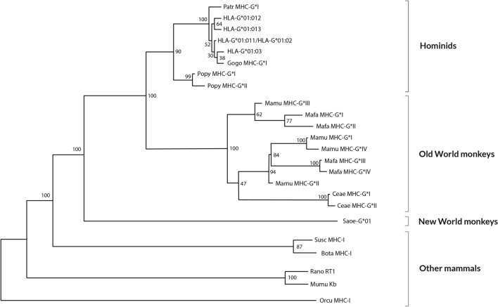
Classical HLA (Human Leukocyte Antigen) is the Major Histocompatibility Complex (MHC) in man. HLA genes and disease association has been studied at least since 1967 and no firm pathogenic mechanisms have been established yet. HLA-G immune modulation gene (and also -E and -F) are starting the same arduous way: statistics and allele association are the trending subjects with the same few results obtained by HLA classical genes, i.e., no pathogenesis may be discovered after many years of a great amount of researchers' effort. Thus, we believe that it is necessary to follow different research methodologies: (1) to approach this problem, based on how evolution has worked maintaining together a cluster of immune-related genes (the MHC) in a relatively short chromosome area since amniotes to human at least, i.e., immune regulatory genes (MHC-G, -E and -F), adaptive immune classical class I and II genes, non-adaptive immune genes like (C2, C4 and Bf) (2); in addition to using new in vitro models which explain pathogenetics of HLA and disease associations. In fact, this evolution may be quite reliably studied during about 40 million years by analyzing the evolution of MHC-G, -E, -F, and their receptors (KIR-killer-cell immunoglobulin-like receptor, NKG2-natural killer group 2-, or TCR-T-cell receptor-among others) in the primate evolutionary lineage, where orthology of these molecules is apparently established, although cladistic studies show that MHC-G and MHC-B genes are the ancestral class I genes, and that New World apes MHC-G is paralogous and not orthologous to all other apes and man MHC-G genes. In the present review, we outline past and possible future research topics: co-evolution of adaptive MHC classical (class I and II), non-adaptive (i.e., complement) and modulation (i.e., non-classical class I) immune genes may imply that the study of full or part of MHC haplotypes involving several loci/alleles instead of single alleles is important for uncovering HLA and disease pathogenesis. It would mainly apply to starting research on HLA-G extended haplotypes and disease association and not only using single HLA-G genetic markers.

Keywords: Apes; Complotypes; Disease; Evolution; HLA; HLA-E; HLA-F; HLA-G; Haplotypes; MHC; Monkeys.


Conflict of interest statement

The authors declare no relevant competing interests to disclose.

Figures

Fig. 1
Fig. 1
HLA gene complex is located in the short arm of human chromosome 6 (6p21.3). HLA-G, -E and -F mRNA transcription and translation scheme and HLA-G membrane and soluble isoforms are shown (see text). Exons (E) of each gene are shown in upper panels of the figure. A (*) symbol indicates a stop codon: it may be localized in E6 in HLA-E, -F and -G genes. HLA-G also presents stop codons in intron 2 or intron 4 depending on alternative splicing process which gives rise to different isoforms. Stop codon may be maintained in mature mRNA due to a reading-through mechanism in humans and primates which is described also in other HLA genes (i.e., HLA-DRB6). The presence of a selenocysteine insertion sequence (SECIS) at the 3 untranslated region leads to a selenocysteine incorporation at UGA (stop) codons [–18]; this may be the cause for stop codon maintenance in HLA-G, -E and -F translation. Beta-2 microglobulin (β2m) is represented bound to protein molecules in purple color. See also references [19, 20]
Fig. 2
Fig. 2
HLA-G protein alleles. Codon and aminoacidic changes among different alleles in exon 2, exon 3 and exon 4 are shown. The letter “N” at the end of some alleles shown in the table denotes null allele. These null alleles bear a stop codon due to single-base deletions or point mutation which give rise to an incomplete HLA-G protein translation. HLA-G*01:05N has a single cytosine deletion at codon 130 (CTG → TGC) which produces a reading frameshift change, causing a premature stop signal at codon 189 (GTG → TGA) [38, 39] and consequently a shorter protein with α1 functional domain at least [38, 40]. HLA-G*01:21N has a premature stop codon due to a punctual mutation in codon 226 (CAG → TAG) of coding sequence which leads to a non-complete translated protein [40]. The number of HLA-G protein alleles is rapidly growing; see IMGT-HLA database to be up to date on new alleles (https://www.ebi.ac.uk/ipd/imgt/hla; accessed September 2021) [41]
Fig. 3
Fig. 3
Relatedness Neighbor-Joining (NJ) dendrogram constructed with MHC-G exons1, 2, 3 and 4 sequences of man (HLA), chimpanzee (Patr), gorilla (Gogo), orangutan (Popy), rhesus monkey (Mamu), crab-eating macaque (Mafa), grivet (Ceae) and New World ape cotton-top tamarin (Saoe). It is shown that MHC-G of Saguinus oedipus diverges from all the other tested apes MHC-G [74]. Other mammals MHC-I sequences included in the analysis have been taken from GenBank: pig (Susc MHC-I; accession AF014002), cow (Bota MHC-I; accession X80936), mouse (MumuKb; accession U47328), rat (RanoRT1; accession X90376), and rabbit (Orcu MHC-I; accession K02441). Bootstrap values are shown
Fig. 4
Fig. 4
World map showing HLA-G*01:05N null allele frequencies in different populations. Populations are within white squares and HLA-G*01:05N frequencies are within blue squares. Note highest frequencies at Middle East (see text) [63]
Fig. 5
Fig. 5
(1) HLA-G*01:04 frequencies (red squares) are different over the World. Higher frequencies are found in Japanese, Iranians, and South Koreans; Europeans and Amerindians show lower frequencies. (2) HLA-G*01:01 frequencies (green squares) do not clearly differ among World populations [63]
Fig. 6
Fig. 6
A Neighbor-Joining dendrogram showing that HLA-E may be the most ancient MHC molecule in humans. HLA sequences have been taken from IMGT/HLA database [41] and Felis catus MHC-I (GenBank accession NM_001305029.1) has been taken as outgroup. Bootstrap values are shown

Similar articles

Cited by

References

    1. Klein J, Sato A. The HLA system. First of two parts. N Engl J Med. 2000;343:702–709. doi: 10.1056/NEJM200009073431006. - DOI - PubMed
    1. Hviid TV. HLA-G in human reproduction: aspects of genetics, function, and pregnancy complications. Hum Reprod Update. 2006;12:209–232. doi: 10.1093/humupd/dmi048. - DOI - PubMed
    1. Donadi EA, Castelli EC, Arnaiz-Villena A, Roger M, Rey D, Moreau P. Implications of the polymorphism of HLA-G on its function, regulation, evolution and disease association. Cell Mol Life Sci. 2011;68(3):369–395. doi: 10.1007/s00018-010-0580-7. - DOI - PMC - PubMed
    1. Carosella ED, Moreau P, LeMaoult J, Rouas-Freiss N. HLA-G: from biology to clinical benefits. Trends Immunol. 2008;29(3):125–132. doi: 10.1016/j.it.2007.11.005. - DOI - PubMed
    1. Carosella ED, Favier B, Rouas-Freiss N, Moreau P, LeMaoult J. Beyond the increasing complexity of the immunomodulatory HLA-G molecule. Blood. 2008;111(10):4862–4870. doi: 10.1182/blood-2007-12-127662. - DOI - PubMed

Substances