. 2022 Aug 2;63(9):5.
doi: 10.1167/iovs.63.9.5.

Phenotypic Expression of CFH Rare Variants in Age-Related Macular Degeneration Patients in the Coimbra Eye Study

Affiliations

Phenotypic Expression of CFH Rare Variants in Age-Related Macular Degeneration Patients in the Coimbra Eye Study

Cláudia Farinha et al. Invest Ophthalmol Vis Sci. .

Abstract

Purpose: To determine the association between rare genetic variants in complement factor H (CFH) and phenotypic features in age-related macular degeneration (AMD) patients from the Coimbra Eye Study (CES).

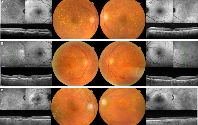
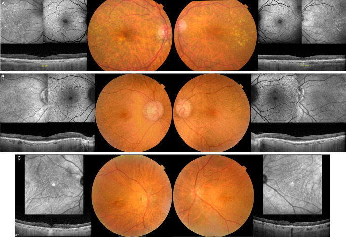
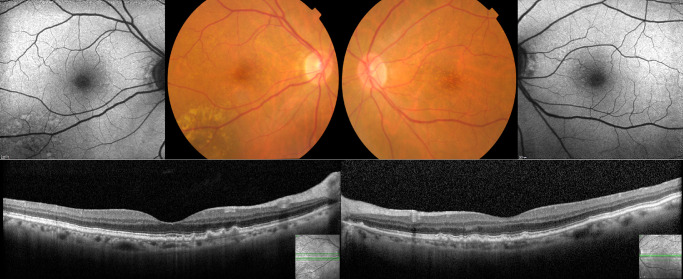
Methods: AMD patients from the Incidence CES (NCT02748824) underwent ophthalmologic examination and color fundus photography, spectral-domain optical coherence tomography (SD-OCT), fundus autofluorescence, and near-infrared imaging. Multimodal phenotypic characterization was carried out in a centralized reading center. The coding and splice-site regions of the CFH gene were sequenced through single-molecule molecular inversion probe-based next-generation sequencing in association with the EYE-RISK consortium. Variants with minor allele frequency <0.05 resulting in splice-site or protein change were selected. Differences in phenotypic features between carriers and noncarriers were analyzed using generalized estimated equations logistic regression models, considering intereye correlations.

Results: We included 39 eyes of 23 patients carrying rare CFH variants and 284 eyes of 188 noncarriers. Carrier status was associated with having higher drusen burden in the macula in the inner Early Treatment Diabetic Retinopathy Study circle (odds ratio [OR], 5.44 [95% confidence interval {CI}, 1.61-18.37]; P = 0.006), outer circle (OR, 4.37 [95% CI, 1.07-17.77]; P = 0.04), and full grid (OR, 4.82 [95% CI, 1.13-20.52]; P = 0.033). In SD-OCT, a lower total macular volume and lower inner retinal layers' volume (OR, 0.449 [95% CI, 0.226-0.894]; P = 0.023; OR, 0.496 [95% CI, 0.252-0.979]; P = 0.043) and pigment epithelial detachments (PEDs) (OR, 5.24 [95% CI, 1.08-25.44]; P = 0.04) were associated with carrying a rare CFH variant. Carriers with subretinal drusenoid deposits (SDD) had the rare variant P258L in all cases except one.

Conclusions: We identified in our cohort phenotypic differences between carriers and noncarriers of rare variants in the CFH gene. Carriers had more severe disease, namely superior drusen burden, PEDs, and thinner retinas. The rare variant P258L may be associated with SDD. Carriers are probably at increased risk of progression.


Conflict of interest statement

Disclosure: C. Farinha, Novartis (C), Bayer (C); P. Barreto, None; R. Coimbra, None; A. Iutis, None; M.L. Cachulo, Bayer (C), Novartis (C); J. Cunha-Vaz, Precision Ocular Ltd (C), Roche (C), Carl Zeiss Meditec (C), AlimeraSciences (C), Allergan (C), Bayer (C), Gene Signal (C), Novartis (C), Pfizer (C), Sanofi-Aventis (C), Vifor Pharma (C); Y.T.E. Lechanteur, Novartis (C), Bayer (C); C.B. Hoyng, Bayer (C), Novartis (C), Horus Pharma Abbvie (C), Horama (C), Astherna (F); R. Silva, Bayer (C), Alcon (C), Thea (C), Novartis (C), Alimera Sciences (C), Allergan (C)

Figures

Figure 1.
Figure 1.
Exemplificative images of fundus features of carriers. (A) Female, 68 years old (yo) (CFH rs757785149; Arg53Cys) with extensive soft drusen both inside the ETDRS grid and outside extending beyond the vascular arcades, and crystalline drusen temporal to the macula. There is a high degree of phenotypic symmetry between both eyes. (B) Male, 68yo (CFH rs371192606; Arg341His) with large, soft, confluent drusen mainly located in the outer ETDRS grid circle, and extending to the vascular arcades and nasal peripapillary area, along with hypo and hyperpigmentation in the central macula. The OCT reveals shallow and heterogeneously hyporreflective PEDs under the fovea in both eyes, but no intraretinal or subretinal fluid. The symmetry of all pathologic changes in multimodal imaging is striking. (C) Female, 80yo (CFH 1:196694418:A:G; Ile622Leu) with large soft, confluent drusen mainly located in the outer ETDRS grid circle and temporal to the macula. They extend outside the vascular arcades and to the nasal peripapillary area. There is hypopigmentation and hyperpigmentation in the central macula in both eyes. The OCT shows PED under the fovea in both eyes, in the right eye with intraretinal fluid (type 1 MNV), and the left eye without fluid (probably quiescent MNV).
Figure 2.
Figure 2.
Exemplificative multimodal images of CFH rs768526062 (Pro258Leu) carriers. (A) Female, 72yo with soft drusen mainly clustering in the temporal macula and SDD in parafoveal, nasal, and superior distribution in both eyes. The choroid is very thin (99 micra in right eye and 101 micra in left eye, subfoveal). (B) Female, 68yo with extensive SDD in both eyes affecting the posterior pole, except for the fovea, and extending to the vascular arcades. (C) Male, 81yo with SDD in both eyes affecting the posterior pole. There is foveal geographic atrophy in the right eye and soft drusen with hyperpigmentary changes in the fovea of the left eye.
Figure 3.
Figure 3.
Female, 74yo (rare variant CFH rs35274867; Asn1050Tyr) with cuticular drusen in the fovea and nasal parafovea. Besides the rare variant, this patient harbors multiple common variants, including three other CFH variants (rs10922109, rs1410996, rs3753394) and three risk-conferring variants in ARHGAP21 (rs12357257), NPLOC4_TSPAN10 (rs6565597) and SLC16A8 (rs8135665).

References

    1. Colijn JM, Buitendijk GHS, Prokofyeva E, et al. .. Prevalence of age-related macular degeneration in Europe: the past and the future. Ophthalmology. 2017; 124: 1753–1763. - PMC - PubMed
    1. Wong WL, Su X, Li X, et al. .. Global prevalence of age-related macular degeneration and disease burden projection for 2020 and 2040: a systematic review and meta-analysis. Lancet Global Health. 2014; 2(2): e106–e116. - PubMed
    1. Lambert NG, ElShelmani H, Singh MK, et al. .. Risk factors and biomarkers of age-related macular degeneration. Prog Retin Eye Res. 2016; 54: 64–102. - PMC - PubMed
    1. Fritsche LG, Fariss RN, Stambolian D, Abecasis GR, Curcio CA, Swaroop A.. Age-related macular degeneration: genetics and biology coming together. Annu Rev Genomics Hum Genet. 2014; 15: 151–171. - PMC - PubMed
    1. Fritsche LG, Igl W, Bailey JN, et al. .. A large genome-wide association study of age-related macular degeneration highlights contributions of rare and common variants. Nat Genet. 2016; 48: 134–143. - PMC - PubMed

Publication types

MeSH terms

Substances