. 2022 Sep;6(9):e2200190.
doi: 10.1002/adbi.202200190. Epub 2022 Aug 4.

Neurotrophin Pathway Receptors NGFR and TrkA Control Perineural Invasion, Metastasis, and Pain in Oral Cancer

Affiliations

Neurotrophin Pathway Receptors NGFR and TrkA Control Perineural Invasion, Metastasis, and Pain in Oral Cancer

Coleen Doan et al. Adv Biol (Weinh). 2022 Sep.

Abstract

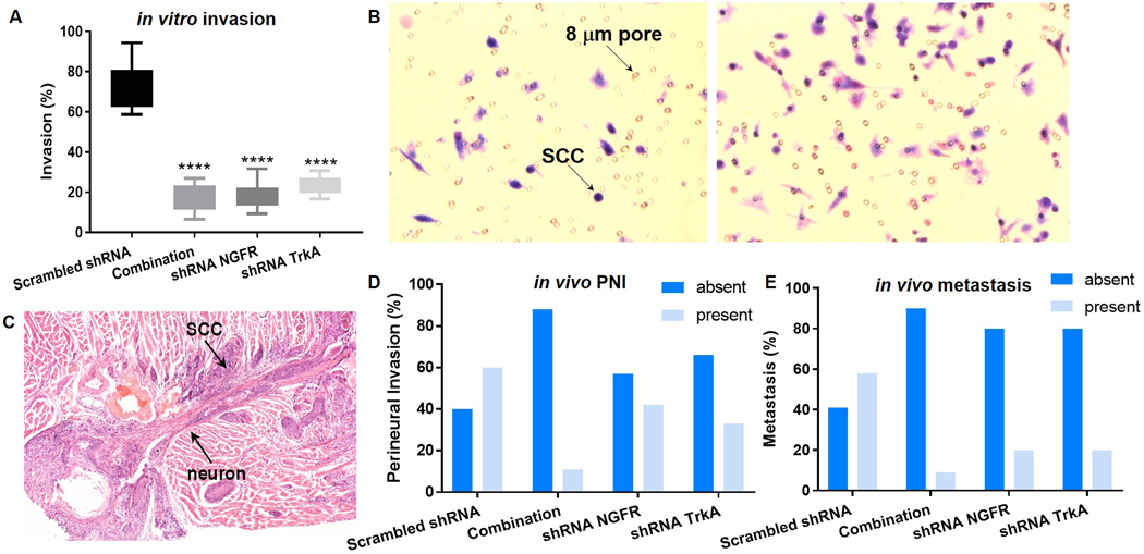
Oral squamous cell carcinoma (OSCC) patients suffer from poor survival due to metastasis or locoregional recurrence, processes that are both facilitated by perineural invasion (PNI). OSCC has higher rates of PNI than other cancer subtypes, with PNI present in 80% of tumors. Despite the impact of PNI on oral cancer prognosis and pain, little is known about the genes that drive PNI, which in turn drive pain, invasion, and metastasis. In this study, clinical data, preclinical, and in vitro models are leveraged to elucidate the role of neurotrophins in OSCC metastasis, PNI, and pain. The expression data in OSCC patients with metastasis, PNI, or pain demonstrate dysregulation of neurotrophin genes. TrkA and nerve growth factor receptor (NGFR) are focused, two receptors that are activated by NGF, a neurotrophin expressed at high levels in OSCC. It is demonstrated that targeted knockdown of these two receptors inhibits proliferation and invasion in an in vitro and preclinical model of OSCC, and metastasis, PNI, and pain. It is further determined that TrkA knockdown alone inhibits thermal hyperalgesia, whereas NGFR knockdown alone inhibits mechanical allodynia. Collectively the results highlight the ability of OSCC to co-opt different components of the neurotrophin pathway in metastasis, PNI, and pain.

Keywords: cancer pain; metastasis; neurotrophin; oral cancer; perineural invasion.


Figures

Figure 1.
Figure 1.
The heat maps depict the top most differentially expressed genes between patients with and without PNI, with and without neck metastasis, or with and without pain. Neurotrophin pathway genes are differentially expressed in patients with neck metastasis, PNI and pain. Patients are dichotomized by “0” vs “1” to denote “absent” and “present” for PNI and metastasis, and “low” vs “severe” for pain.
Figure 2.
Figure 2.
A) Functional network analysis was performed on genes differentially expressed based on metastasis status. Dotplot of over-representation analysis (ORA) GO Biological Processes (BP) pathways reveals additional pathways that converge with the neurotrophin pathway to mediate metastasis in the cohort of patients. B) The differential genes when sorted by metastasis status underwent enrichment mapping with ORA GO BP pathways. These results relate neuroptrophin genes to multiple pathways that are critical in metastasis. C) Similarly, functional network analysis of differential genes by methylation status was performed with ORA KEGG pathways. D) Differentially expressed genes based on PNI status underwent gene concept network of ORA Reactome gene sets, revealing cytokine signaling and integrin signaling as inter-related pathways with the neurotrophin pathway, with both of these pathways having established roles in cancer pain and metastasis. E) Differential genes based on PNI status analyzed by KEGG also reveals additional gene pathways (i.e., ECM-receptor interaction, PI3K-Akt signaling pathway) that have established roles in metastasis of different cancers. F) Functional network analysis of differential genes based on pain levels in the cohort shows pathways like opioid signaling and NMDA receptors, which control multiple pain conditions. Additionally, cell-cell communication, cell junction organization, and extracellular matrix organization pathways are mediated by genes that control metastasis.
Figure 3.
Figure 3.
(A and B) Transduction efficiency is comparable amongst all four treatment groups. Using GFP as a proxy for gene expression, the percentage of positive cells is similar amongst all four treatment groups. (C) NGFR expression is significantly lower in oral SCC tumors treated with shRNA to NGFR (*p<.05, compared to scrambled shRNA group, One way ANOVA, Holm Sidak). (D) TrkA expression is significantly lower in oral SCC tumors treated with shRNA to TrkA (***p<.001, compared to scrambled shRNA group, One way ANOVA, Holm Sidak). (E) NGFR and TrkA silencing inhibits oral SCC proliferation in vitro. The graph shows MTS assay absorbance as an index of cell proliferation for each treatment group. Silencing of NGFR or TrkA alone or in combination has a significant antiproliferative effect on oral SCC (Hela-O3) cells in vitro (**p<.01, ***p<.001, One way ANOVA and Tukey’s test.) (F) NGFR and TrkA silencing inhibits oral SCC proliferation in vivo. Tumor volume is quantified after tissue harvest of the in vivo model for the four treatment groups: scrambled RNA (n=10), combination treatment (n=9), shRNA NGFR (n=10), and shRNA TrkA (n=7). Combination silencing of NGFR and TrkA produces the most robust decrease in OSCC (Hela-O3) tumor growth (***p<.001, One way ANOVA, Holm Sidak).
Figure 4.
Figure 4.
(A) Box and whisker plot (showing minimum-maximum) of invasion assay results demonstrate inhibition of invasion with shRNA treatment to silence NGFR and TrkA. All three treatment groups have significantly lower percent invasion than control scrambled RNA (****p<.0001, One way ANOVA, Holm Sidak). (B) The two images show our co-culture model, where transwells are seeded with cancer cell, and the bottom well is loaded with serum media. SCC cells migrate through the pores at different frequency based on the treatment group (right greater than left). (C) Representative image of a mouse tongue SCC with PNI. (D) The contingency graph demonstrates the frequency of perineural invasion (PNI) in each treatment group in the preclinical model of oral SCC. The four treatment groups are significantly different from each other (p<0.0001, Chi square test, based on percentage of each group). (E) The contingency graph demonstrates the frequency of cervical metastasis in each treatment group in the preclinical model of oral SCC. The four treatment groups are significantly different from each other (p<.0001, Chi square test, based on percentage of each group).
Figure 5.
Figure 5.
The graphs demonstrate the change in (A) mechanical and (B) thermal threshold after cancer inoculation (day 0). Cancer cells are transduced with adenovirus expressing shRNA to the indicated genes. Combination knockdown of NGFR and TrkA produces antinociception to both mechanical and thermal stimuli, compared to the control scrambled shRNA group. NGFR knockdown alone produces significant mechanical antinociception, whereas TrkA knockdown alone produces significant thermal antinociception (*p<.05, **p<.01, ***p<.001, Two way ANOVA, Holm Sidak, see Table 3 for statistical analyses).

References

    1. Bapat AA, Hostetter G, Von Hoff DD, and Han H, Perineural invasion and associated pain in pancreatic cancer. Nat Rev Cancer, 2011. 11(10): p. 695–707. - PubMed
    1. Connelly ST and Schmidt BL, Evaluation of pain in patients with oral squamous cell carcinoma. J Pain, 2004. 5(9): p. 505–10. - PubMed
    1. Hammerlid E, Bjordal K, Ahlner-Elmqvist M, Boysen M, Evensen JF, Biorklund A, Jannert M, Kaasa S, Sullivan M, and Westin T, A prospective study of quality of life in head and neck cancer patients. Part I: at diagnosis. Laryngoscope, 2001. 111(4 Pt 1): p. 669–80. - PubMed
    1. Kolokythas A, Cox DP, Dekker N, and Schmidt BL, Nerve Growth Factor and Tyrosine Kinase A Receptor in Oral Squamous Cell Carcinoma: Is There an Association With Perineural Invasion? Journal of Oral and Maxillofacial Surgery, 2010. 68(6): p. 1290–1295. - PubMed
    1. Ye Y, Dang D, Zhang J, Viet CT, Lam DK, Dolan JC, Gibbs JL, and Schmidt BL, Nerve growth factor links oral cancer progression, pain, and cachexia. Mol Cancer Ther, 2011. 10(9): p. 1667–76. - PMC - PubMed

Publication types