. 2022 Aug 4;17(8):e0269962.
doi: 10.1371/journal.pone.0269962. eCollection 2022.

Bioactive potentiality of secondary metabolites from endophytic bacteria against SARS-COV-2: An in-silico approach

Affiliations

Bioactive potentiality of secondary metabolites from endophytic bacteria against SARS-COV-2: An in-silico approach

Yasmin Akter et al. PLoS One. .

Abstract

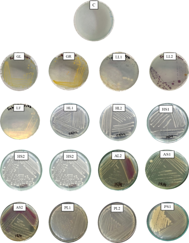
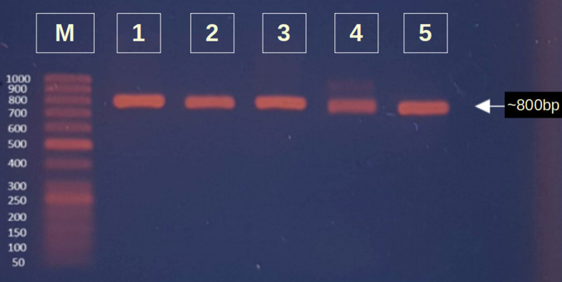
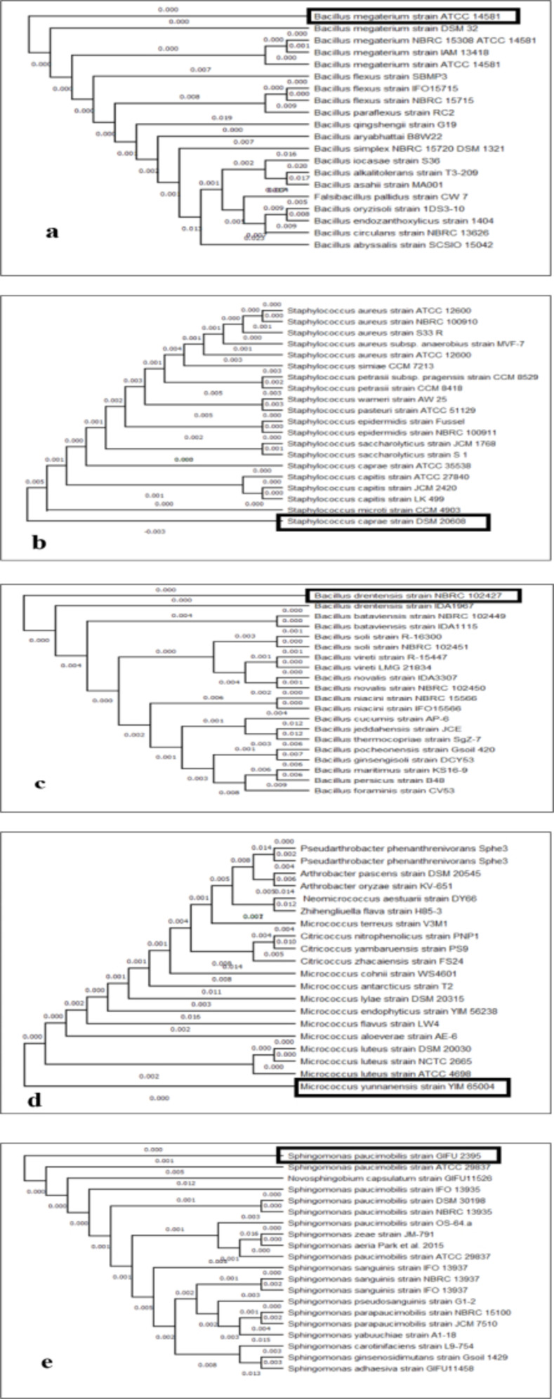
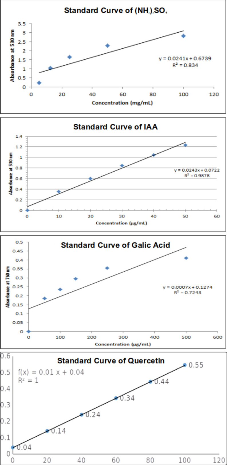
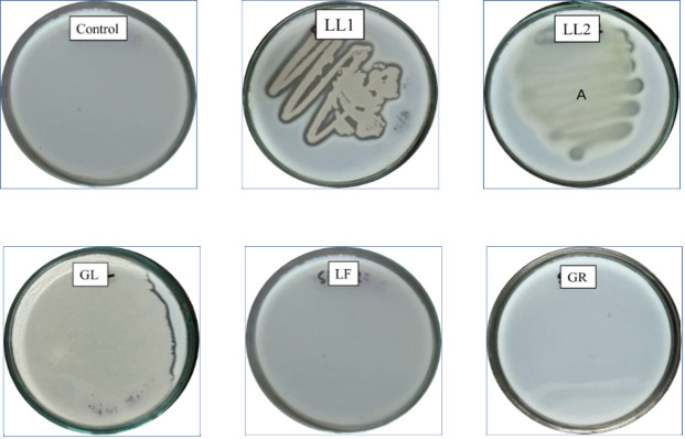
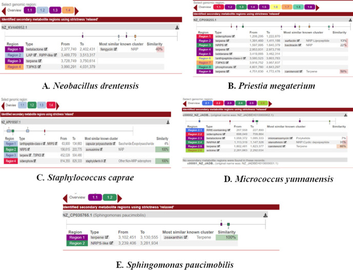
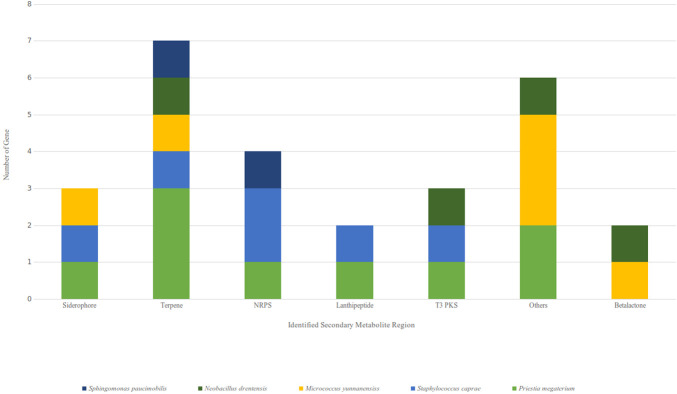
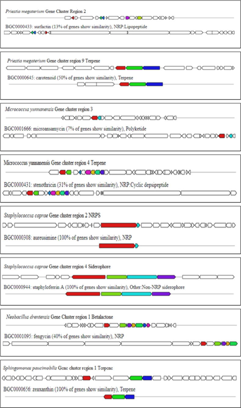
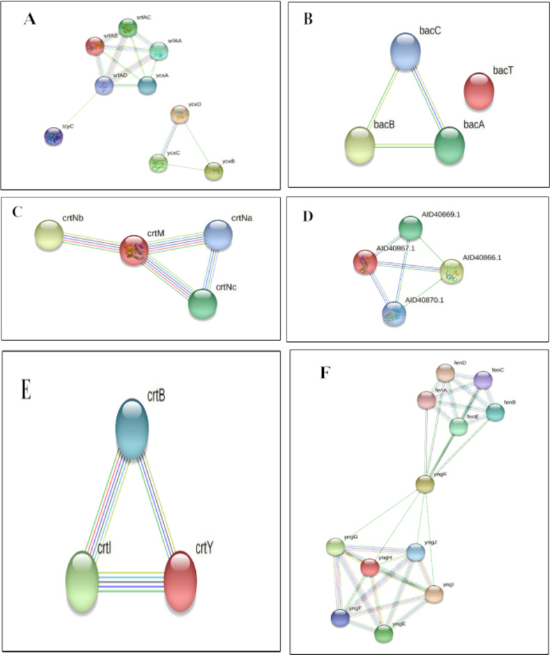
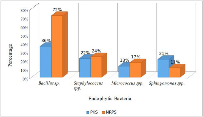
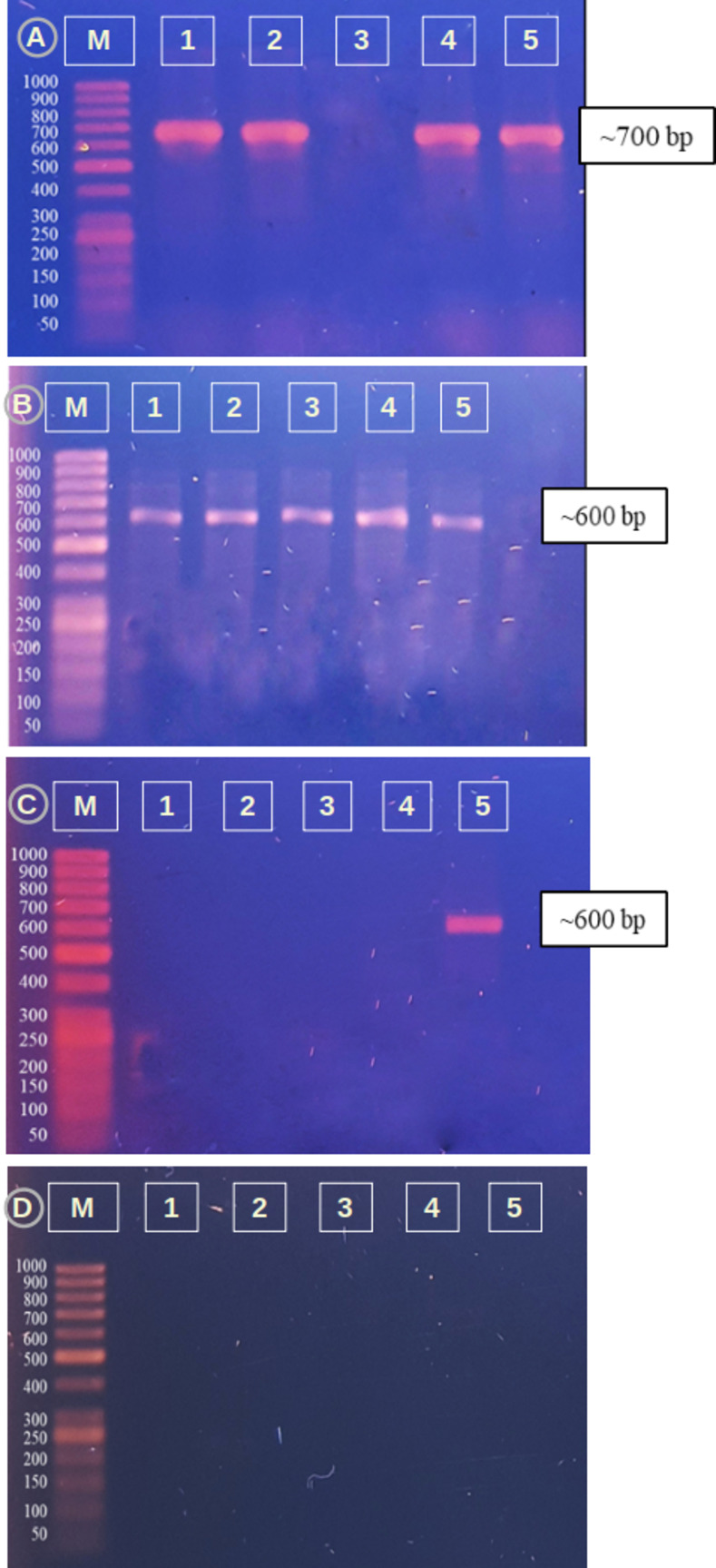
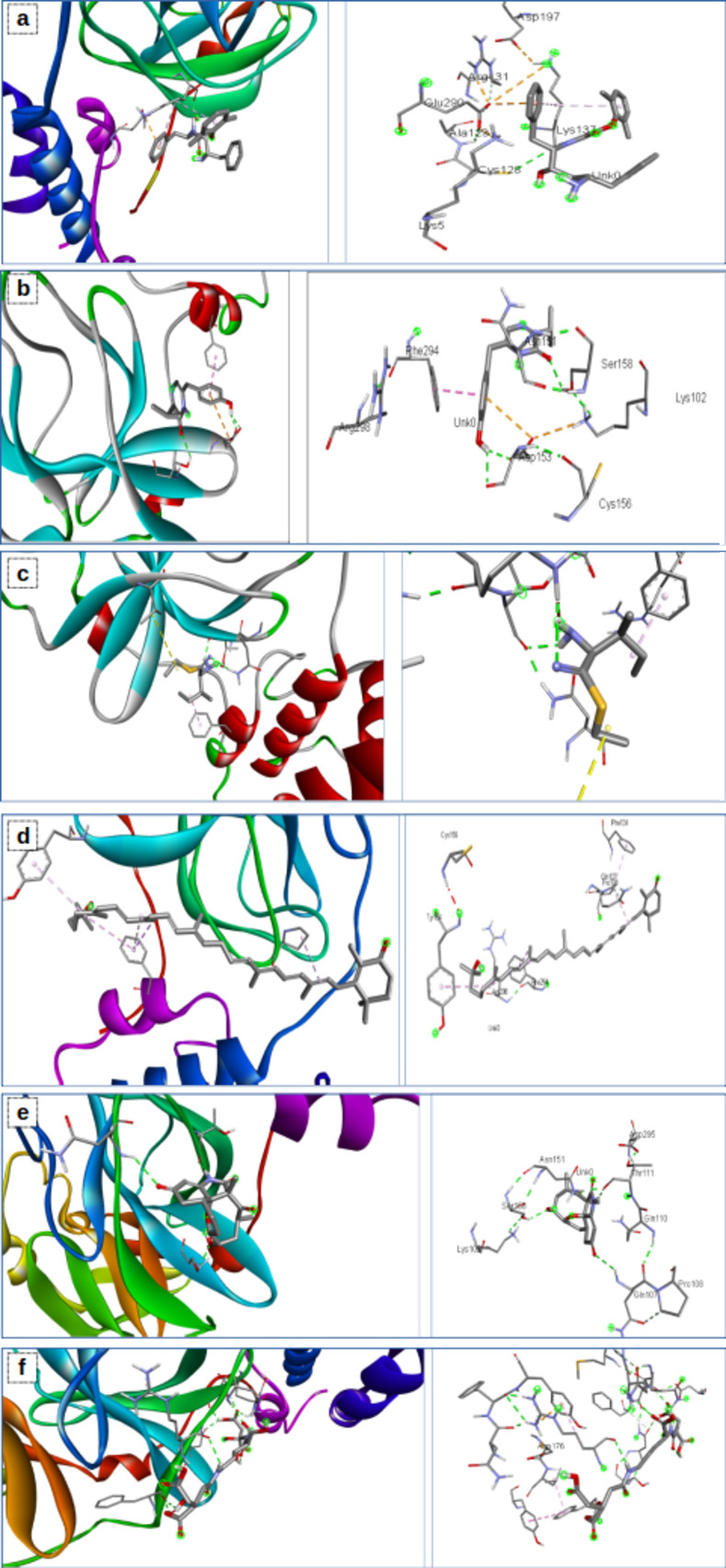
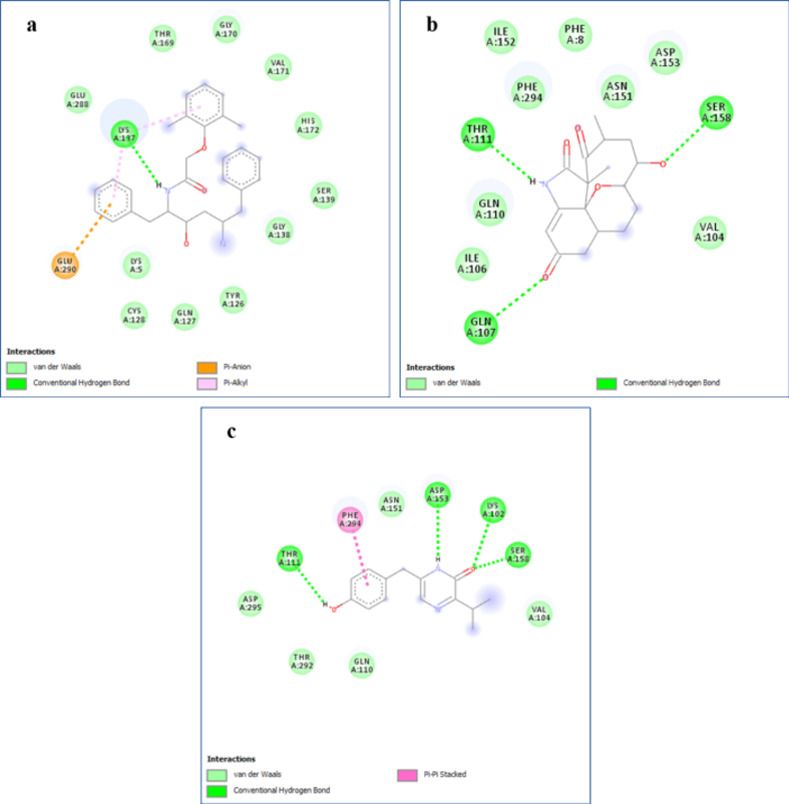
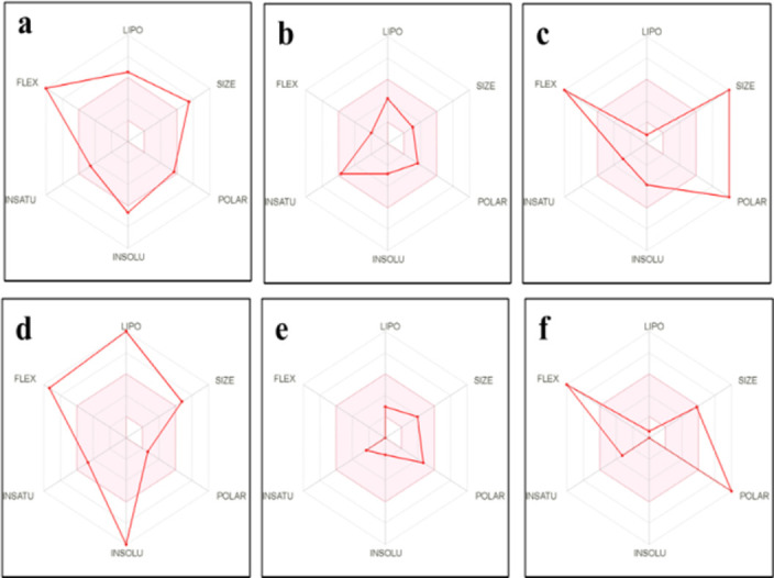
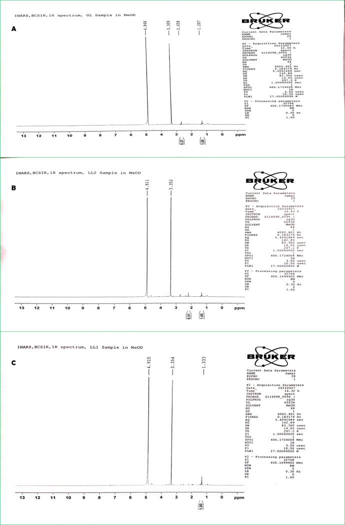
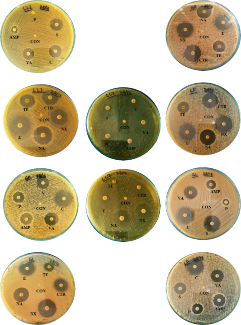
Five endophytic bacterial isolates were studied to identify morphologically and biochemically, according to established protocols and further confirmed by 16S rDNA Sanger sequencing, as Priestia megaterium, Staphylococcus caprae, Neobacillus drentensis, Micrococcus yunnanensis, and Sphingomonas paucimobiliz, which were then tested for phytohormone, ammonia, and hydrolytic enzyme production. Antioxidant compounds total phenolic content (TPC), and total flavonoid content (TFC) were assessed by using bacterial crude extracts obtained from 24-hour shake-flask culture. Phylogenetic tree analysis of those identified isolates shared sequence similarities with the members of Bacillus, Micrococcus, Staphylococcus, and Pseudomonas species, and after GenBank submission, accession numbers for the nucleotide sequences were found to be MW494406, MW494408, MW494401, MW494402, and MZ021340, respectively. In silico analysis was performed to identify their bioactive genes and compounds in the context of bioactive secondary metabolite production with medicinal value, where nine significant bioactive compounds according to six different types of bioactive secondary metabolites were identified, and their structures, gene associations, and protein-protein networks were analyzed by different computational tools and servers, which were reported earlier with their antimicrobial, anti-infective, antioxidant, and anti-cancer capabilities. These compounds were then docked to the 3-chymotrypsin-like protease (3CLpro) of the novel SARS-COV-2. Docking scores were then compared with 3CLpro reference inhibitor (lopinavir), and docked compounds were further subjected to ADMET and drug-likeness analyses. Ligand-protein interactions showed that two compounds (microansamycin and aureusimine) interacted favorably with coronavirus 3CLpro. Besides, in silico analysis, we also performed NMR for metabolite detection whereas three metabolites (microansamycin, aureusimine, and stenothricin) were confirmed from the 1H NMR profiles. As a consequence, the metabolites found from NMR data aligned with our in-silico analysis that carries a significant outcome of this research. Finally, Endophytic bacteria collected from medicinal plants can provide new leading bioactive compounds against target proteins of SARS-COV-2, which could be an effective approach to accelerate drug innovation and development.


Conflict of interest statement

The authors have declared that no competing interests exist.

Figures

Fig 1
Fig 1. Pure cultures of sixteen isolated endophytic bacteria labeled as their sample codes (GL, GR, LL1, LL2 LF, HL1, HL2, HS1, HS2, AL1, AL2, AS1, AS2, PL1, PL2, PS1); Control (C) indicates no growth of bacteria in the medium.
Fig 2
Fig 2. Electrophoretic separation (2% agarose) of the 16S rDNA gene of different isolates.
M: 50 bp DNA ladder; 1: LL1; 2: LL2; 3: LF; 4: GL; 5: GR.
Fig 3
Fig 3. Phylogenetic tree of isolates.
(a) Priestia megaterium, (b) Staphylococcus caprae, (c) Bacillus drentensis, (d) Micrococcus yunnanensis, (e) Sphingomonas paucimobiliz.
Fig 4
Fig 4. Standard curves of ammonium sulfate, indole acetic acid, gallic acid and quercetin.
Fig 5
Fig 5. Isolates in skim milk agar.
LL1 (Priestia megaterium) and LL2 (Staphylococcus caprae) represented protease enzyme activity with a clear hollow degradation of protein throughout the inoculation area.
Fig 6
Fig 6. Identification of secondary metabolite regions using the antiSMASH server.
Fig 7
Fig 7. Distribution of genes in identified biosynthetic gene clusters: NRPS Non ribosomal peptide synthetase, T3PKS = Type 3 polyketide synthase.
Fig 8
Fig 8. Graphical representations of most similar known clusters as an output of ClusterBlast specified by the MIBiG repository.
Color codes represent similar gene regions.
Fig 9
Fig 9. Predicted association of proteins of biosynthetic genes demonstrated by the STRING server.
(A) Surfactin, (B) bacitracin, (C) carotenoid, (D) staphyloferrin A, (E) zeaxanthin and (F) fengycin protein network. Line Indicator: Red Line–Presence of fusion evidence, Green Line- neighborhood evidence, Blue Line- cooccurrence evidence, Purple Line- experimental evidence, Yellow Line- textmining evidence, Light Blue Line- database evidence, Black Line- coexpression evidence.
Fig 10
Fig 10. Distribution of PKS and NRPS domains among the endophytic isolates at the genus level.
Fig 11
Fig 11. PCR amplifications of biosynthetic genes.
(A) PKS gene, (B) NRPS gene, (C) ACCD gene and (D) CHS gene. M: DNA Marker for each case; 1: LL1; 2: LL2; 3: LF; 4: GL; 5: GR.
Fig 12
Fig 12
Visualization of SARS-CoV-2 3CLpro amino acid interactions with the ligands (A) lopinavir, (B) aureusimine, (C) bacitracin, (D) carotenoid, (E) microanasamycin, and (F) staphyloferrin.
Fig 13
Fig 13. 2D representation of protein–ligand interactions.
Binding mode of 3CLpro with A) lopinavir, B) microanasamycin, and C) aureusimine.
Fig 14
Fig 14. Summary of the pharmacokinetic properties of the compounds.
(a) Lopinavir, (b) aureusimine, (c) bacitracin, (d) carotenoid, (e) microanamycin, (f) staphyloferrin The color space is a suitable physiochemical space for oral bioavailability. LIPO Lipophility: -0.7 < XLOGP3 < +5.0. SIZE: 150 g/mol: < MW < 500 g/mol. POLAR (Polarity): 20Å2 < TPSA < 130 Å 2. INSOLU (insolubility): 0 < Log S (ESOL) < 6. INSATU (insaturation): 0.25 < Fraction Csp3 < 1. FLEX (Flexibity): 0 < Num. rotatable bonds < 9.
Fig 15
Fig 15. NMR 1H spectroscopy result: In the left the metabolite structure and on the right 1H NMR spectrum.
Every peak is responsible for a respective different proton type. (A) Microansamycin, (B) Aureusimine, and (C) Surfactin.
Fig 16
Fig 16. Antibiotic sensitivity test of isolates showing inhibition zones (mm).
Control (CON) indicates discs without antibiotics in each case. Disc codes are used to show the antibiotic discs for every isolate.

Similar articles

Cited by

References

    1. Schulz B. Mutualistic interactions with fungal root endophytes. Microbial root endophytes. Springer; 2006. pp. 261–279.
    1. Wilson D. Endophyte: the evolution of a term, and clarification of its use and definition. Oikos. 1995; 274–276.
    1. Zhang HW, Song YC, Tan RX. Biology and chemistry of endophytes. Nat Prod Rep. 2006;23: 753–771. doi: 10.1039/b609472b - DOI - PubMed
    1. Santoyo G, Moreno-Hagelsieb G, del Carmen Orozco-Mosqueda M, Glick BR. Plant growth-promoting bacterial endophytes. Microbiol Res. 2016;183: 92–99. doi: 10.1016/j.micres.2015.11.008 - DOI - PubMed
    1. Wang S-S, Liu J-M, Sun J, Sun Y-F, Liu J-N, Jia N, et al. Diversity of culture-independent bacteria and antimicrobial activity of culturable endophytic bacteria isolated from different Dendrobium stems. Sci Rep. 2019;9: 1–12. - PMC - PubMed