

Single-cell genomics in AML: extending the frontiers of AML research
- PMID: 35926108
- PMCID: PMC10082362
- DOI: 10.1182/blood.2021014670
Single-cell genomics in AML: extending the frontiers of AML research
Abstract
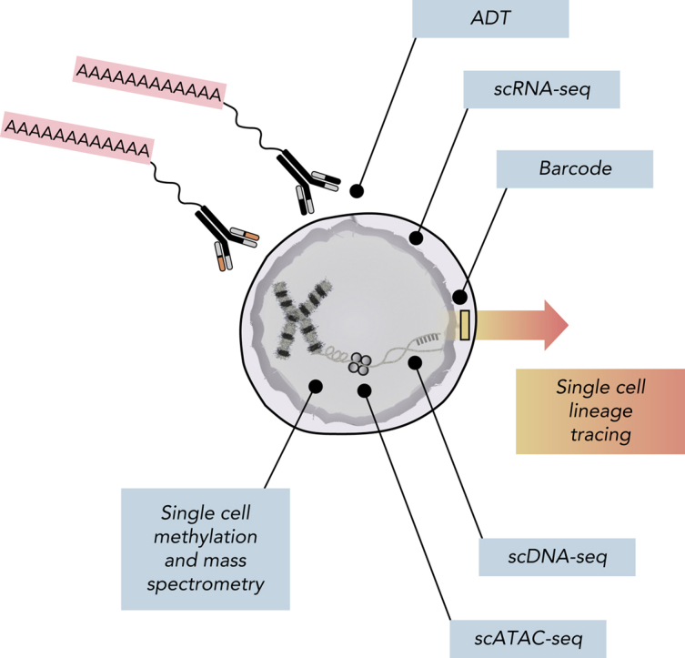
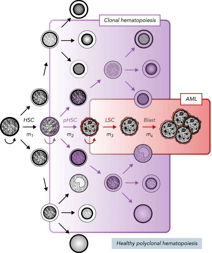
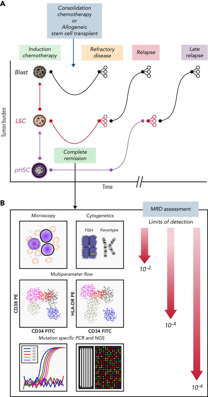
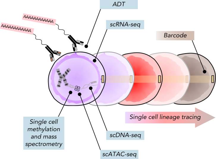
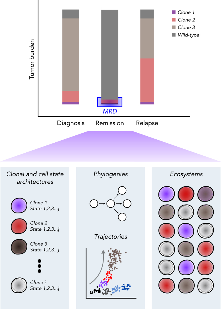
The era of genomic medicine has allowed acute myeloid leukemia (AML) researchers to improve disease characterization, optimize risk-stratification systems, and develop new treatments. Although there has been significant progress, AML remains a lethal cancer because of its remarkably complex and plastic cellular architecture. This degree of heterogeneity continues to pose a major challenge, because it limits the ability to identify and therefore eradicate the cells responsible for leukemogenesis and treatment failure. In recent years, the field of single-cell genomics has led to unprecedented strides in the ability to characterize cellular heterogeneity, and it holds promise for the study of AML. In this review, we highlight advancements in single-cell technologies, outline important shortcomings in our understanding of AML biology and clinical management, and discuss how single-cell genomics can address these shortcomings as well as provide unique opportunities in basic and translational AML research.
© 2023 by The American Society of Hematology.
Conflict of interest statement
Conflict-of-interest disclosure: R.M. is on the board of directors of CircBio, Inc., and the advisory boards of Kodikaz Therapeutic Solutions, Inc., and Syros Pharmaceuticals and is an inventor on a number of patents related to CD47 cancer immunotherapy licensed to Gilead Sciences, Inc.; receives research support from Gilead Sciences, Inc.; and is a cofounder of and equity holder in CircBio, Inc., Pheast Therapeutics, MyeloGene, Inc., and RNAC Therapeutics, Inc. The remaining authors declare no competing financial interests.
Figures
Similar articles
-
Genomics-based Approach and Prognostic Stratification Significance of Gene Mutations in Intermediate-risk Acute Myeloid Leukemia.Chin Med J (Engl). 2015 Sep 5;128(17):2395-403. doi: 10.4103/0366-6999.163400. Chin Med J (Engl). 2015. PMID: 26315090 Free PMC article. Review.
-
Application of High Throughput Technologies in the Development of Acute Myeloid Leukemia Therapy: Challenges and Progress.Int J Mol Sci. 2022 Mar 5;23(5):2863. doi: 10.3390/ijms23052863. Int J Mol Sci. 2022. PMID: 35270002 Free PMC article. Review.
-
[Precision genomic and translational medicine for acute myeloid leukemia].Yi Chuan. 2018 Nov 20;40(11):988-997. doi: 10.16288/j.yczz.18-188. Yi Chuan. 2018. PMID: 30465531 Review. Chinese.
-
Emerging therapies for acute myeloid leukemia: translating biology into the clinic.JCI Insight. 2017 Sep 21;2(18):e95679. doi: 10.1172/jci.insight.95679. eCollection 2017 Sep 21. JCI Insight. 2017. PMID: 28931762 Free PMC article. Review.
-
[Precision medicine for acute myeloid leukemia based on genomic profiling].Rinsho Ketsueki. 2019;60(7):847-853. doi: 10.11406/rinketsu.60.847. Rinsho Ketsueki. 2019. PMID: 31391376 Japanese.
Cited by
-
Cytoplasmic TP53INP2 acts as an apoptosis partner in TRAIL treatment: the synergistic effect of TRAIL with venetoclax in TP53INP2-positive acute myeloid leukemia.J Exp Clin Cancer Res. 2024 Jun 22;43(1):176. doi: 10.1186/s13046-024-03100-0. J Exp Clin Cancer Res. 2024. PMID: 38909249 Free PMC article.
-
CAR-T cell therapy for cancer: current challenges and future directions.Signal Transduct Target Ther. 2025 Jul 4;10(1):210. doi: 10.1038/s41392-025-02269-w. Signal Transduct Target Ther. 2025. PMID: 40610404 Free PMC article. Review.
-
Chimeric antigen receptor (CAR) modified T Cells in acute myeloid leukemia: limitations and expectations.Front Cell Dev Biol. 2024 Apr 17;12:1376554. doi: 10.3389/fcell.2024.1376554. eCollection 2024. Front Cell Dev Biol. 2024. PMID: 38694825 Free PMC article. Review.
-
Enhanced Antitumor Efficacy of Cytarabine and Idarubicin in Acute Myeloid Leukemia Using Liposomal Formulation: In Vitro and In Vivo Studies.Pharmaceutics. 2024 Sep 19;16(9):1220. doi: 10.3390/pharmaceutics16091220. Pharmaceutics. 2024. PMID: 39339256 Free PMC article.
-
Reduced Proteolipid Protein 2 promotes endoplasmic reticulum stress-related apoptosis and increases drug sensitivity in acute myeloid leukemia.Mol Biol Rep. 2023 Dec 12;51(1):10. doi: 10.1007/s11033-023-08994-1. Mol Biol Rep. 2023. PMID: 38085372
References
-
- Forkner CE. Clinical and pathological differentiation of acute leukemias: with special reference to acute monocytic leukemia. Arch Intern Med (Chic) 1934;53(1):1–34.
-
- Vardiman JW, Thiele J, Arber DA, et al. The 2008 revision of the World Health Organization (WHO) classification of myeloid neoplasms and acute leukemia: rationale and important changes. Blood. 2009;114(5):937–951. - PubMed
-
- Bennett JM, Catovsky D, Daniel MT, et al. Proposals for the classification of the acute leukaemias. French-American-British (FAB) co-operative group. Br J Haematol. 1976;33(4):451–458. - PubMed
-
- Griffin JD, Löwenberg B. Clonogenic cells in acute myeloblastic leukemia. Blood. 1986;68(6):1185–1195. - PubMed
-
- Fialkow PJ, Singer JW, Adamson JW, et al. Acute nonlymphocytic leukemia: heterogeneity of stem cell origin. Blood. 1981;57(6):1068–1073. - PubMed
Publication types
MeSH terms
Grants and funding
LinkOut - more resources
Full Text Sources
Medical
