. 2022 Sep 14;22(17):6866-6876.
doi: 10.1021/acs.nanolett.2c00582. Epub 2022 Aug 4.

Programmable Bispecific Nano-immunoengager That Captures T Cells and Reprograms Tumor Microenvironment

Affiliations

Programmable Bispecific Nano-immunoengager That Captures T Cells and Reprograms Tumor Microenvironment

Lu Zhang et al. Nano Lett. .

Abstract

Immune checkpoint blockade (ICB) therapy has revolutionized clinical oncology. However, the efficacy of ICB therapy is limited by the ineffective infiltration of T effector (Teff) cells to tumors and the immunosuppressive tumor microenvironment (TME). Here, we report a programmable tumor cells/Teff cells bispecific nano-immunoengager (NIE) that can circumvent these limitations to improve ICB therapy. The peptidic nanoparticles (NIE-NPs) bind tumor cell surface α3β1 integrin and undergo in situ transformation into nanofibrillar network nanofibers (NIE-NFs). The prolonged retained nanofibrillar network at the TME captures Teff cells via the activatable α4β1 integrin ligand and allows sustained release of resiquimod for immunomodulation. This bispecific NIE eliminates syngeneic 4T1 breast cancer and Lewis lung cancer models in mice, when given together with anti-PD-1 antibody. The in vivo structural transformation-based supramolecular bispecific NIE represents an innovative class of programmable receptor-mediated targeted immunotherapeutics to greatly enhance ICB therapy against cancers.

Keywords: T cells capture; fibrillar transformation; immune checkpoint blockade (ICB) therapy; nano-immuno-engager.


Conflict of interest statement

The authors declare the following competing financial interest(s): K.S.L. and L.Z. are the coinventors of a patent on fibrillar transformable nanoparticles (PCT/US2020/046495). K.S.L. is the founding scientist of LamnoTherapeutics Inc., which plans to develop the nanotherapeutics described in the manuscript. The remaining authors declare no competing interests.

Figures

Scheme 1
Scheme 1. Programmable Bispecific Nano-immunoengager (NIE) Synergizing Immune Checkpoint Blockage (ICB) Therapy via Capturing Teff Cells and Reprogramming the Tumor Microenvironment
(a and b) Schematic illustration of (a) co-self-assembly of both TPM1 and TPM2 into NIE-NPs, in situ fibrillar transformation into NIE-NFs through binding α3β1 integrin (tumor cell membrane), exposure of LLP2A ligands binding α4β1 integrin (T cell membrane), and release of TLRs 7/8 agonist (R848) from NIE-NFs through esterase hydrolysis. (b) Steps II–V show the processes for how a programmable bispecific NIE synergizes ICB therapy in tumor tissue: (I) NIE-NPs bind tumor cells and in situ transforms them into nanofibrils (NIE-NFs) on the surface of tumor cells. (II) NIE-NFs expose LLP2A and release R848 for (III) capturing T cells to tumors cells and (IV) re-educating TAMs from M2 to M1 phenotype. (V) Meanwhile, anti-PD-1 antibody greatly activates the NIE homed cytotoxic T cells for ICB therapy. (TPMs, transformable peptide monomers; NIE-NPs, nano-immuno-engager nanoparticles; NIE-NFs, nano-immuno-engager nanofibrils; R848, resiquimod, a TLRs 7/8 agonist; M1-TAM, M1-like tumor-associated macrophage; M2-TAM, M2-like tumor-associated macrophage.)
Figure 1
Figure 1
Assembly and fibrillar transformation of the programmable bispecific NIE, as well as esterase-induced exposure of LLP2A ligands and R848 release. (a) Schematic illustration of the molecular structure and function of TPM1 (LXY30-KLVFFK(Pa)) and TPM2 (proLLP2A-KLVFFK(R848)). (b) Changes in fluorescence (FL) of a DMSO solution of TPM1 and TPM2 at a 1:1 ratio following the gradual addition of water (from 0 to 99%) forming micellar NIE-NPs: excitation wavelength, 405 nm. (c) Schematic illustration and TEM images of initial NIE-NPs and subsequently transformed nanofibrils (NIE-NFs) upon interaction with soluble α3β1 integrin protein for 24 h (H2O to DMSO ratio of 99:1). The concentration of NIE-NPs used in the experiment was 20 μM. Scale bars are 100 nm. (d) Variation in the fluorescence (FL) signal of Pa in the fibrillar-transformation process of NIE-NPs to NIE-NFs over time. (e) Schematic illustration and TEM images of NIE-NPs upon interaction with soluble α4β1 integrin protein or α4β1 integrin protein plus esterase for 24 h (H2O to DMSO ratio of 99:1), respectively. The concentration of NIE-NPs used in the experiment was 20 μM. Scale bars are 100 nm. (f and g) Variation in the size distribution (f) and circular dichroism spectra (g) of NIE-NPs and NIE-NFs under different conditions. (h) In vitro release profile of R848 from NIE-NFs over time under different conditions. Data are presented as mean ± s.d., n = 3 independent experiments. The molar ratio of α3β1 or α4β1 integrin protein to peptide ligand was approximately 1:1000. a.u., arbitrary units; mdeg, millidegrees.
Figure 2
Figure 2
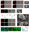
In vitro bispecific NIE binds both 4T1 breast cancer cells and CD8 T cells and re-educates tumor-associated macrophages. (a) Cellular fluorescence distribution images of interaction of NIE-NPs and CNIE-NPs for 6 h with 4T1 tumor cells to show NIE-NPs around cells and CNIE-NPs inside cells. Scale bar is 10 μm. (b) Cellular fluorescence signal retention images of 4T1 tumor cells after exposure to NIE-NPs and CNIE-NPs for 6 h followed by incubation with fresh medium without nanoparticles for 18 h to show long retention of NIE and short retention of CNIE. Scale bar is 10 μm. (c) Representative SEM images of untreated 4T1 tumor and 4T1 tumor cells treated with NIE-NPs for 6 h. Scale bar is 10 μm. The concentration of NIE-NPs was 50 μM. (d and e) (d) Cellular fluorescence distribution images and (e) representative SEM images of murine CD8 T cells (isolated from mouse spleen, CellTracker green CMFDA dye labeled, green fluorescence) after incubation with esterase-pretreated NIE-NPs to show NIE around cells. α4β1 integrins on the surface of murine CD8 T cells were preactivated by Mn2+ (1 mM). Scale bar is 10 μm. (f) Experimental scheme and cellular fluorescence distribution images of NIE-NPs (fluorescent red), after interaction with 4T1 tumor and murine CD8 T cells (fluorescent green, α4β1 integrins were preactivated by Mn2+). It shows that nanofibrillar networks (bispecific NIE) cover 4T1 tumor cells, which in turn capture CD8 T cells. More incubation time, more bound CD8 T cells. Scale bar is 10 μm. (g) Representative SEM images of 4T1 tumor and CD8 T cells after treatment with NIE-NPs (see part f). (h) Representative images of M2-like murine macrophages and subsequent re-education by NIE-NFs, NIE-NFs plus esterase, or R848 at different time points. Scale bar is 20 μm. Statistical significance was calculated using one-way ANOVA followed by Tukey’s post hoc analysis: *P < 0.05, **P < 0.01, ***P < 0.001.
Figure 3
Figure 3
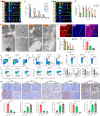
In vivo evaluation of NIE targeting tumor cells, and in situ nanofibrillar transformation to capture Teff cell and facilitate the long-term retention and activate immunity. (a and b) Time-dependent ex vivo fluorescence (FL) images (a) and quantitative analysis (b) of tumor tissues and major organs (heart (H), liver (Li), spleen (Sp), lung (Lu), kidney (K), intestine (I), muscle (M), and skin (Sk)) collected at 10, 24, 48, 72, 120, and 168 h post-injection of NIE-NPs. Data are presented as mean ± s.d., n = 3 independent experiments. (c) Time-dependent ex vivo fluorescence images of tumor tissues collected at 10, 24, 48, 72, 120, and 168 h post-injection of CNIE-NPs. (d) Fluorescence (FL) quantification of tumor tissues collected at 10, 24, 48, 72, 120, and 168 h post-injection of NIE-NPs and CNIE-NPs. Data are presented as mean ± s.d., n = 3 independent experiments. (e) Representative TEM images of distribution in tumor tissue and in situ fibrillar transformation of NIE-NPs, CNIE-NPs, and the untreated control group at 72 h post-injection. “N” depicts nucleus. (f) Fluorescence distribution images of NIE-NPs in the tumor tissue region and normal skin tissue at 72 h post-injection (red, Pa of NIE-NPs; blue, DAPI; scale bars, 50 μm). (g) R484 distribution retention in tumor tissues at different time points post-injection of NIE-NPs and CNIE-NPs. Injection dose of R848:0.94 mg kg–1; data were mean ± s.d., n = 3 for each time point. (h) Expression of CXCL10 chemokine within the tumor tissues after 5 days of NIE-NPs, CNIE-NPs, and saline treatment (n = 3; data were mean ± s.d.; single injection). (i) Representative flow cytometric analysis images and corresponding quantification of CD45+CD3+, CD8+/CD4+, CD4+Foxp3+, CD8+CD49+, and CD8+CD29+ T cells within the 4T1 tumors excised from mice treated with NIE-NPs, CNIE-NPs, or the saline control. (j) Immunohistochemistry (IHC) of tumors excised from mice after treatment with NIE-NPs or CNIE-NPs. Representative images are shown for the IHC staining of T cells (CD8+, CD4+, and Foxp3+) and macrophage markers (CD68 and CD163). Scale bar is 100 μm. (k) The expression levels (qPCR assay) of IFN-γ, TGF-β, IL12, IL10, Nos2, and Arg-1 in 4T1 tumors excised from mice 15 days after treatment with NIE-NPs or CNIE-NPs (n = 3; data were mean ± s.d.). Statistical significance was calculated using a two-sided unpaired t test compared to the NIE-NPs group: *P < 0.05, **P < 0.01, ***P < 0.001, ****P < 0.0001.
Figure 4
Figure 4
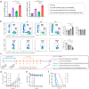
Intrinsic antitumor immune efficacy of bispecific NIE in Balb/c mice bearing 4T1 breast tumor. (a) Expression of CXCL10 and CXCL9 chemokine on day 7 within the excised tumor tissues of mice after different treatments (13 mg/kg each dose, every other day; n = 3; data were mean ± s.d.). (b) Representative flow cytometric analysis images and relative quantification of CD8+/CD4+ and CD4+Foxp3+ T cells within the 4T1 tumors excised from mice after different treatments at day 15. All treatment regimens were tail vein injected consecutively three times q.o.d. (13 mg/kg each dose, every other day, total three times; n = 5; data were mean ± s.d.). In parts a and b, (1) saline; (2) LXY30-KLVFFK(Pa)/(EK)3-KLVFFK(R848) (fibrillar-transformation but absence of LLP2A ligand); (3) LXY30-KLVFFK(Pa)/proLLP2A-KLVFFK(Pa) (fibrillar-transformation but absence of R848); and (4) LXY30-KLVFFK(Pa)/proLLP2A-KLVFFK(R848) (complete NIE-NPs). (c) Experimental design: orthotopic tumor inoculation and treatment protocol; regimen 6 is NIE-NPs with all four critical components: (1) saline; (2) (EK)3-KLVFFK(Pa)/(EK)3-KLVFFK(R848) (“R848 only” in micellar formulation); (3) proLLP2A-KLVFFK(R848) (single monomer); (4) LXY30-KAAGGK(Pa)/proLLP2A-KAAGGK(R848) (untransformable negative control CNIE-NPs); (5) LXY30-KLVFFK(Pa)/proLLP2A-KLVFFK(Pa) (fibrillar-transformation but absence of R848); (6) LXY30-KLVFFK(Pa)/proLLP2A-KLVFFK(R848). (d and e) Observation of the tumor inhibitory effect (d) and weight change (e) of mice bearing orthotopic 4T1 tumor over 21 d after initiation of treatment (n = 8 per group). Data are presented as mean ± s.d. (f) Cumulative survival of different treatment groups of mice bearing 4T1 breast tumors. Data are presented as mean ± s.d. Statistical significance was calculated using one-way ANOVA followed by Tukey’s post hoc analysis: *P < 0.05, **P < 0.01, ***P < 0.001, ****P < 0.0001.
Figure 5
Figure 5
Bispecific NIE greatly enhances ICB therapy in mice bearing 4T1 breast tumor and Lewis lung tumor. (a) Experimental design: orthotopic tumor inoculation and treatment protocol (four treatment arms; regimens 4, 5, and 6 are the same as those shown in Figure 4a). (b) Tumor response in mice bearing orthotopic 4T1 tumor over 21 d of treatment (n = 8 per group). Data are presented as mean ± s.d. (c) Cumulative survival of the four treatment groups. (d) Experimental design: Mice previously treated with regimen 6 plus anti-PD-1 Ab were rechallenged with reinoculation of 4T1 breast cancer cells on day 90. The same operation was carried out on the same age naïve mice as a negative control group. (e) No antitumor immune memory effect was observed in the same age naïve mice. (f) An antitumor immune memory effect was observed in mice previously treated with regimen 6 and anti-PD-1 Ab. (g) Cumulative survival of naïve mice and previously regimen 6 plus anti-PD-1-treated mice. (h and i) IFN-γ (h) and TNF-α (i) level in mice sera 6 days after mice were rechallenged with 4T1 tumor cells. (j and k) Observation of tumor inhibitory effect (j) and weight change (k) of mice bearing subcutaneous murine Lewis lung tumor over 21 d after initiation of treatment (n = 8 per group). The treatment protocol followed the experimental design in part a; five cycles (i.v. regimens 4–6 and i.p. anti-PD-1). Data are presented as mean ± s.d. (l) Cumulative survival of different treatment groups of mice bearing subcutaneous murine Lewis lung tumors. Statistical significance was calculated using one-way ANOVA followed by Tukey’s post hoc analysis: *P < 0.05, **P < 0.01, ***P < 0.001, ****P < 0.0001.

References

    1. Postow M. A.; Sidlow R.; Hellmann M. D. Immune-related adverse events associated with immune checkpoint blockade. New. Engl. J. Med. 2018, 378, 158–168. 10.1056/NEJMra1703481. - DOI - PubMed
    1. Topalian S. L.; Taube J. M.; Anders R. A.; Pardoll D. M. Mechanism-driven biomarkers to guide immune checkpoint blockade in cancer therapy. Nat. Rev. Cancer 2016, 16, 275–287. 10.1038/nrc.2016.36. - DOI - PMC - PubMed
    1. Sackstein R.; Schatton T.; Barthel S. R. T-lymphocyte homing: an underappreciated yet critical hurdle for successful cancer immunotherapy. Lab. Invest. 2017, 97, 669–697. 10.1038/labinvest.2017.25. - DOI - PMC - PubMed
    1. Fares C. M.; Allen E. M. V.; Drake C. G.; Allison J. P.; Hu-Lieskovan S. Mechanisms of resistance to immune checkpoint blockade: why does checkpoint inhibitor immunotherapy not work for all patients?. ASCO Educational Book 2019, 39, 147–164. 10.1200/EDBK_240837. - DOI - PubMed
    1. Riley R. S.; June C. H.; Langer R.; Mitchell M. J. Delivery technologies for cancer immunotherapy. Nat. Rev. Drug Discovery 2019, 18, 175–196. 10.1038/s41573-018-0006-z. - DOI - PMC - PubMed

Publication types