. 2022 Aug 2;40(5):111168.
doi: 10.1016/j.celrep.2022.111168.

Human UFSP1 is an active protease that regulates UFM1 maturation and UFMylation

Affiliations

Human UFSP1 is an active protease that regulates UFM1 maturation and UFMylation

David Millrine et al. Cell Rep. .

Abstract

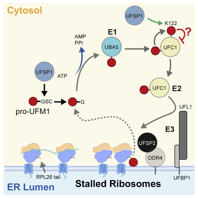
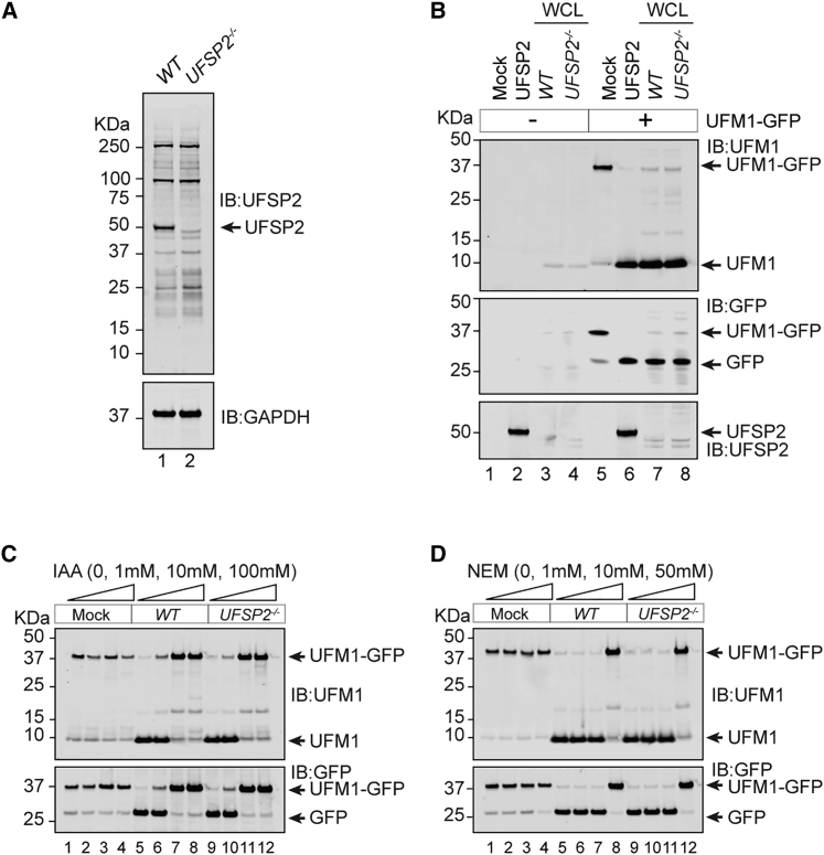
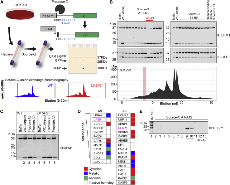
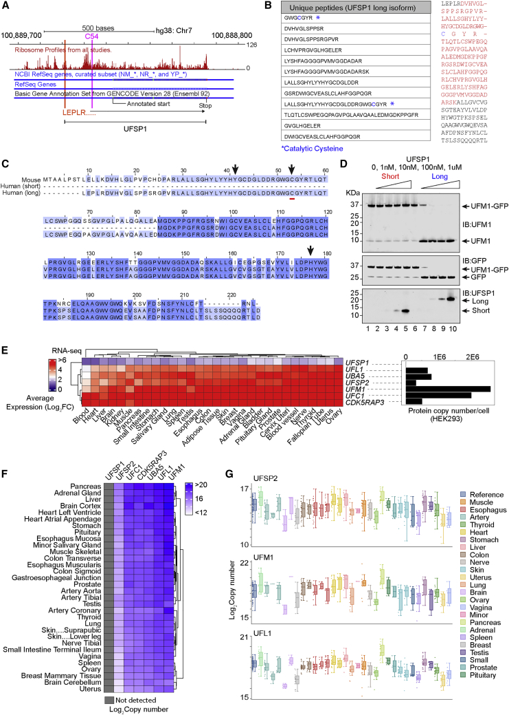
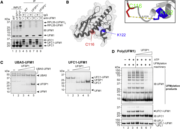
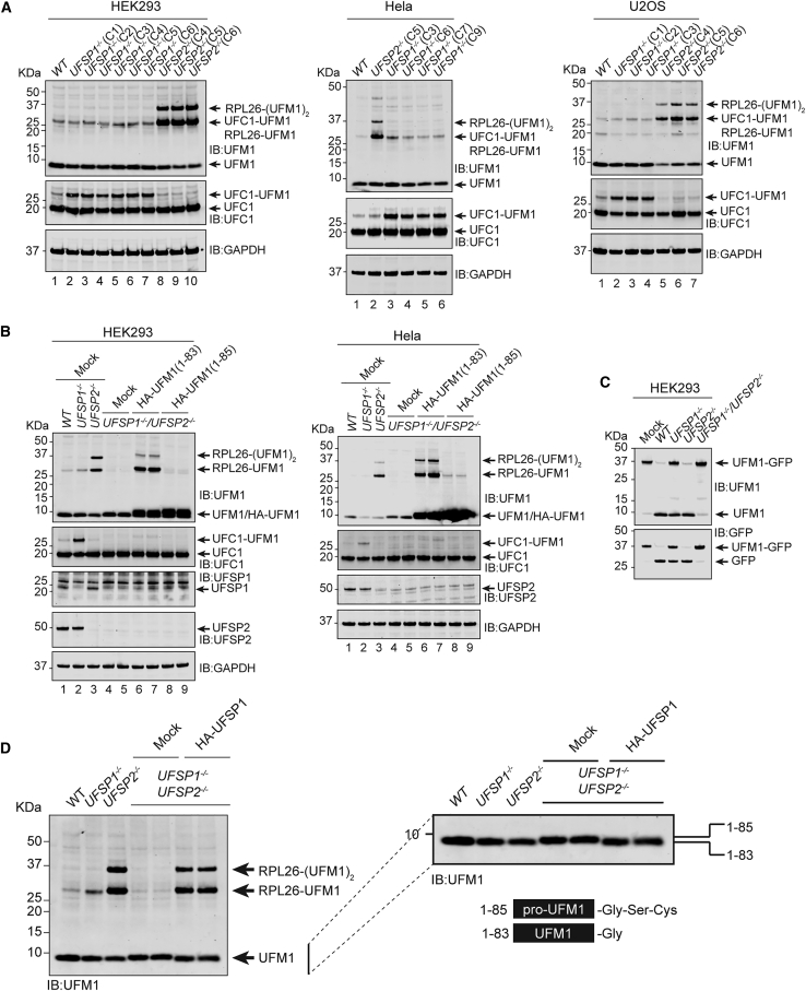
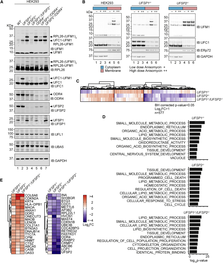
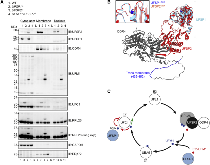
An essential first step in the post-translational modification of proteins with UFM1, UFMylation, is the proteolytic cleavage of pro-UFM1 to expose a C-terminal glycine. Of the two UFM1-specific proteases (UFSPs) identified in humans, only UFSP2 is reported to be active, since the annotated sequence of UFSP1 lacks critical catalytic residues. Nonetheless, efficient UFM1 maturation occurs in cells lacking UFSP2, suggesting the presence of another active protease. We herein identify UFSP1 translated from a non-canonical start site to be this protease. Cells lacking both UFSPs show complete loss of UFMylation resulting from an absence of mature UFM1. While UFSP2, but not UFSP1, removes UFM1 from the ribosomal subunit RPL26, UFSP1 acts earlier in the pathway to mature UFM1 and cleave a potential autoinhibitory modification on UFC1, thereby controlling activation of UFMylation. In summary, our studies reveal important distinctions in substrate specificity and localization-dependent functions for the two proteases in regulating UFMylation.

Keywords: CP: Cell biology; CP: Molecular biology; ER; UBA5; UFC1; UFM1; cysteine protease; endoplasmic reticulum; membrane protein; ribosome; ubiquitin; ubiquitin-like modifier.


Conflict of interest statement

Declaration of interests The authors declare no competing interests.

Figures

None
Graphical abstract
Figure 1
Figure 1
UFSP2 is not the sole UFM1-specific peptidase in human cells (A) Confirmation of UFSP2 knockout by western blotting. (B) In vitro assay incubating cell lysates from wild-type (WT) and UFSP2−/− HEK293 cells with a UFM1-GFP fusion protein. Cleavage of a recombinant UFM1-GFP fusion protein into its constituent parts, UFM1 and GFP, is interpreted as peptidase activity. Recombinant UFSP2 (2 μM) is included as a positive control. (C and D) Prevention of UFM1-GFP cleavage by the cysteine peptidase inhibitors iodoacetamide (IAA, C) and N-ethylmaleimide (NEM, D). Cell lysates were pretreated for 1 h in darkness at room temperature prior to mixing with the recombinant UFM1-GFP probe. Probe-lysate incubations were performed at 37°C for 2 h. Data are representative of more than three independent experiments.
Figure 2
Figure 2
Screening for alternative UFM1 protease identifies UFSP1 as a candidate peptidase (A) (Top) Schematic overview of the screening process. HEK293 cells are lysed by mechanical stress and fractionated sequentially over heparin and Source-Q columns. Eluted fractions are screened for activity by incubation with the UFM1-GFP fusion protein. (Bottom) Chromatograms showing protein eluted from Source-Q columns on a salt gradient. Shown are experiments performed in parallel using unmodified HEK293 wild-type (WT) (blue) and UFSP2−/− (red) HEK293 cells. (B) Representative screening results. Heparin-binding proteins have been eluted in a single fraction (“Heparin binding”) while Source-Q-binding proteins have been eluted in fractions A1–B9. Eluted fractions were incubated with UFM1-GFP fusion protein for 2 h at 37°C and analyzed by immunoblot (IB). Heparin and Q-column flow-through (FT) are shown on the far right. Protein cleavage activity is detected in Q-column fractions A8 and A9 (red lines). (C) In vitro assay incubating active fractions (A8 and A9) purified from WT and UFSP2−/− HEK293 cell lysates with the UFM1-GFP fusion protein. Experiment is the same as depicted by chromatograms in (A). (D) Identity of proteases identified in the active fractions. The output of mass spectrometry analysis of the active fractions was aligned with MEROPS database annotations to identify proteases. (E) Immunoblot analysis of endogenous UFSP1 in active fractions.
Figure 3
Figure 3
UFSP1 translated from non-canonical start site is an active protease (A) Ribosome profiling data downloaded from GWIPS-viz (https://gwips.ucc.ir/). Global aggregate protected reads from multiple studies are shown in red (UCSC-track) aligned to Refseq and GENCODE (v28) gene annotation. (B) (Left) LC-MS data showing peptides mapping to long-isoform UFSP1 identified in active fractions. (Right) Amino acid sequence of proposed full-length UFSP1. Sequences in red are identified in LC-MS analysis of active fractions. The catalytic cysteine is highlighted blue. (C) Cross-species multiple sequence alignment of UFSP isoforms. Human short (Q6NVU6), human long (A0A5F9ZGY7), and mouse (Q9CZP0) UFSP1. (D) In vitro assay incubating recombinant long- and short-isoform UFSP1 variants with the UFM1-GFP fusion protein. (E) (Left) RNA-sequencing analysis of human tissues by the Genotype Tissue (GTEx) consortium (https://gtexportal.org/home/). Shown are the log2 transformed transcripts per million. (Right) Data independent acquisition quantitative proteomics analysis of UFM1 pathway components in HEK293 cells. (F and G) Copy number estimation of UFM1 pathway components in human tissues derived from published proteomics data (PXD016999). Heatmaps are clustered using the Euclidean method.
Figure 4
Figure 4
UFSP1 is active against diverse substrates in vitro (A) Immunoprecipitation (IP) of UFM1 from the indicated cell lysates. (B) Crystal structure of UFC1 (PDB: 2Z60) with K122 (blue) and C116 (red) highlighted. (C) Activity of recombinant UFSP1 against the indicated substrates. (D) Activity of UFSP1 against UFMylation products. UFM1 pathway components were reconstituted in vitro (UBA5, UFC1, UFBP1-UFL1) in the presence of ATP. After 1 h the reaction was quenched with apyrase and incubated with increasing molar concentrations of recombinant UFSP1.
Figure 5
Figure 5
UFSP1 is the UFM1-activating peptidase in vivo (A) Immunoblot analysis of UFSP1−/− and UFSP2−/− cell lines as indicated. Labels include abbreviated clone IDs (e.g., C1 is clone 1). (B) Rescue of RPL26 UFMylation by expression of mature UFM1. Constructs expressing HA-tagged mature (UFM11−83) or precursor (UFM11−85) UFM1 were transiently transfected into UFSP1−/−/UFSP2−/− double knockout cell lines. Twenty-four hours later cells were lysed and analyzed by immunoblot with the indicated antibodies. (C) In vitro assay incubating HEK293 cell lysates from the indicated knockout cell lines with the UFM1-GFP probe. (D) Immunoblot analysis of HEK293 cells transiently transfected with HA-tagged UFSP1. (Right) zoomed-in section of the blot shown on the left to highlight changes in electrophoretic mobility of UFM1. This western blot is reproduced complete with loading controls in Figure S5B.
Figure 6
Figure 6
Distinct substrates and functions for the UFSPs (A) Immunoblot analysis of the indicated knockout cell lines (HEK293 Flp-in TREx). (B) Cell lines were treated with 200 nM (+) or 50 μM (++) anisomycin for 20 min prior to cell lysis. Immunoblot analysis of cytoplasmic and membrane fractions is shown. (C–E) Comparison of total proteomes of indicated cell lines. (C) Heatmap showing Log2 fold change (FC) in abundance of proteins passing statistical thresholds in at least one experimental condition (Benjamini-Hochberg adjusted p < 0.05; log2 FC > 1). Heatmap is clustered using the k-means method (n = 3). Data for all significant proteins (padj < 0.05) are shown in Figures S6D and S6E. (D) Gene ontology (GO) enrichments calculated using a hypergeometric distribution test (Broad Institute; http://www.gsea-msigdb.org/gsea/msigdb/compute_overlaps.jsp). Top ten GO enrichments are shown for each dataset. (E) Proteins subject to the most extreme changes (top/bottom ten). Proteomics data show LIMMA differential analyses (UFSP1−/− versus WT, UFSP2−/− versus WT, UFSP1/UFSP2−/− versus WT). Volcano plots and principal component analyses are included in supplemental information. Heatmaps in (C) and (E) are shown at the same scale.
Figure 7
Figure 7
Distinct intracellular localization of UFSPs determines function (A) Immunoblot analysis of membrane, cytosol, and nuclear fractions derived from UFSP1−/−, UFSP2−/−, and UFSP1−/−/UFSP2−/− cell lines. (B) AlphaFold prediction of UFSP2-ODR4 complex aligned to the predicted structure of human UFSP1. The catalytic cysteine is highlighted by the box. (C) Schematic showing suggested model of the UFM1 pathway. In brief, precursor UFM1 is proteolytically activated through the removal of a C-terminal serine-cysteine peptide prior to sequential loading onto the E1, E2, and E3 conjugating enzymes. This culminates in modification of the ribosomal subunit RPL26. UFSPs act at several points in this pathway; (1) both UFSP1 and UFSP2 contribute to pro-UFM1 processing; (2) UFSP1 catalyzes the removal of UFM1 from UFC1, releasing UFC1 from a potentially autoinhibitory state; (3) UFSP2 catalyzes the removal of UFM1 from RPL26, preventing excess ribosome modification. ODR4 is essential for stabilizing UFSP2 and anchoring it at the ER membrane in proximity to the ribosome.

Similar articles

Cited by

References

    1. Banerjee S., Kumar M., Wiener R. Decrypting UFMylation: how proteins are modified with UFM1. Biomolecules. 2020;10:E1442. doi: 10.3390/biom10101442. - DOI - PMC - PubMed
    1. Chen C., Itakura E., Weber K.P., Hegde R.S., de Bono M. An ER complex of ODR-4 and ODR-8/Ufm1 specific protease 2 promotes GPCR maturation by a Ufm1-independent mechanism. PLoS Genet. 2014;10:e1004082. doi: 10.1371/journal.pgen.1004082. - DOI - PMC - PubMed
    1. Colin E., Daniel J., Ziegler A., Wakim J., Scrivo A., Haack T.B., Khiati S., Denommé A.S., Amati-Bonneau P., Charif M., et al. Biallelic variants in UBA5 reveal that disruption of the UFM1 cascade can result in early-onset encephalopathy. Am. J. Hum. Genet. 2016;99:695–703. doi: 10.1016/j.ajhg.2016.06.030. - DOI - PMC - PubMed
    1. Demichev V., Messner C.B., Vernardis S.I., Lilley K.S., Ralser M. DIA-NN: neural networks and interference correction enable deep proteome coverage in high throughput. Nat. Methods. 2020;17:41–44. doi: 10.1038/s41592-019-0638-x. - DOI - PMC - PubMed
    1. Duan R., Shi Y., Yu L., Zhang G., Li J., Lin Y., Guo J., Wang J., Shen L., Jiang H., et al. UBA5 mutations cause a new form of autosomal recessive cerebellar ataxia. PLoS One. 2016;11:e0149039. doi: 10.1371/journal.pone.0149039. - DOI - PMC - PubMed

Publication types

MeSH terms