. 2022 Sep 26;32(18):4071-4078.e4.
doi: 10.1016/j.cub.2022.07.033. Epub 2022 Aug 3.

Conversion of anterograde into retrograde trains is an intrinsic property of intraflagellar transport

Affiliations

Conversion of anterograde into retrograde trains is an intrinsic property of intraflagellar transport

Adrian Pascal Nievergelt et al. Curr Biol. .

Abstract

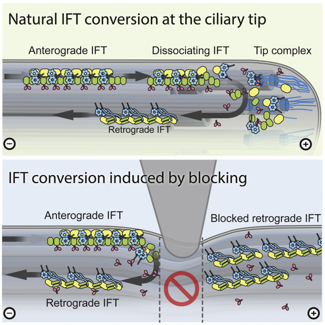
Cilia or eukaryotic flagella are microtubule-based organelles found across the eukaryotic tree of life. Their very high aspect ratio and crowded interior are unfavorable to diffusive transport of most components required for their assembly and maintenance. Instead, a system of intraflagellar transport (IFT) trains moves cargo rapidly up and down the cilium (Figure 1A).1-3 Anterograde IFT, from the cell body to the ciliary tip, is driven by kinesin-II motors, whereas retrograde IFT is powered by cytoplasmic dynein-1b motors.4 Both motors are associated with long chains of IFT protein complexes, known as IFT trains, and their cargoes.5-8 The conversion from anterograde to retrograde motility at the ciliary tip involves (1) the dissociation of kinesin motors from trains,9 (2) a fundamental restructuring of the train from the anterograde to the retrograde architecture,8,10,11 (3) the unloading and reloading of cargo,2 and (4) the activation of the dynein motors.8,12 A prominent hypothesis is that there is dedicated calcium-dependent protein-based machinery at the ciliary tip to mediate these processes.4,13 However, the mechanisms of IFT turnaround have remained elusive. In this study, we use mechanical and chemical methods to block IFT at intermediate positions along the cilia of the green algae Chlamydomonas reinhardtii, in normal and calcium-depleted conditions. We show that IFT turnaround, kinesin dissociation, and dynein-1b activation can consistently be induced at arbitrary distances from the ciliary tip, with no stationary tip machinery being required. Instead, we demonstrate that the anterograde-to-retrograde conversion is a calcium-independent intrinsic ability of IFT.

Keywords: TIRF microscopy; cilia and flagella; ciliary tip; intraflagellar transport; micromanipulator; total-internal reflection microscopy.


Conflict of interest statement

Declaration of interests The authors declare no competing interests.

Figures

None
Graphical abstract
Figure 1
Figure 1
The intraflagellar transport (IFT) machinery of gliding Chlamydomonas can be observed and manipulated in total internal reflection microscopy (TIRFM) (A) ① Anterograde trains, driven by the heterotrimeric motor kinesin-II, move towards the ciliary tip. ② At the ciliary tip, kinesin-II dissociates from the train, and the trains convert into retrograde form by an unknown mechanism. ③ Reassembled retrograde trains move away from the tip towards the cell body by the dynein-1b motor specific for IFT. (B) A sharp but soft wedge is made by drop casting silicone onto a supporting capillary, followed by trimming off excess after curing. (C) Illustration of mechanical blockage by lowering a silicone wedge onto the cilium of a Chlamydomonas reinhardtii cell by micro-manipulator on a TIRF microscope. (D) Individual frames from a TIRFM movie show how IFT is locally and reversibly blocked inside a cilium by the force applied using a silicone wedge (dashed line, non-blocking in blue, blocking in red). See also Video S1.
Figure 2
Figure 2
Kymograms visualizing the time-dependent response of the IFT machinery to external manipulation (A) Compressing the cilium with a soft wedge creates a small band where IFT cannot pass (horizontal white lines in the kymogram, created by the accumulation of stopped fluorescent trains). After the blockage is applied, trains build up and shortly after start moving in retrograde direction. The approximate location of the ciliary tips is outlined for clarity in purple. See also Video S1. (B) Detail view of the region between the cell body and the block (white square) in (A). About 2.5 s after IFT is locally blocked, retrograde trains can be seen moving towards the cell body at normal speed (white arrows). (C) Kymogram of wedge blocking experiment in severely calcium-depleted cilia (see also Figure S1 and Videos S2 and S3). Transport and conversion show no discernable difference to (A) where calcium is available. (D) In lithium-treated cells, the motors are chemically inhibited and pile up at variable distances from the ciliary tip. Gliding of the cell dislodges the stuck trains, which immediately move in retrograde direction at high speed (≈10 μm s−1) (insert). Anterograde trains are colored in green, while retrograde trains are colored in magenta.
Figure 3
Figure 3
Kinetics of anterograde to retrograde transport conversion of IFT trains (A and B) Tomographic slice through ciliary tip showing an anterograde train in the process of disassembly (scale bar, 200 nm). Highlighted rectangular region is magnified in (B), showing loss of structural cohesion of the train localized at the end of the B-tubule (scale bar, 100 nm). The structured part of the train is colored in blue, the disordered part of the train in violet, the microtubule in green, and the ciliary coat in yellow. See also Video S4. (C) Retrograde train appearance frequency at physical blockage. Transport recovers to normal steady-state level with a half-time of 2.6 ± 1.7 s (first-order response fit in blue, uncertainty bounds in light gray). (D) Retrograde train appearance frequency at the ciliary tip after releasing the physical blockage. Steady-state retrograde transport recovers with a half-time of 0.77 ± 0.74 s (fit same as C; error bars in C and D represent SEM). (E) Box plot of train recovery half-time at the wedge and at the ciliary tip after release. Data at the wedge (C and E) from 895 trains in 23 experiments on 12 individual cells. Data at the tip (D and E) from 580 individual trains in 18 experiments on 10 individual cells. See also Figure S2.
Figure 4
Figure 4
Dynamics of the IFT motors kinesin-II and dynein during anterograde to retrograde conversion (A) Denoised kymograms of dual-labeled cilia show distinct behavior between dynein (labeled with D1bLIC-mCherry) and kinesin-II (labeled with KAP-GFP), both in the undisturbed flagellum and when blocked by a wedge. Inserts illustrate regions highlighted in (B) and (C). (B) Background-subtracted detail at the ciliary tip. Positions of some prominent retrograde trains are marked with white arrows, with the same positions shown in the KAP-GFP detail for reference. (C) Background-subtracted detail at the wedge. White arrows denote the location of visible retrograde trains, the positions of which are duplicated in the KAP-GFP detail for reference. See also Figure S3.

Comment in

Similar articles

Cited by

References

    1. Kozminski K.G., Johnson K.A., Forscher P., Rosenbaum J.L. A motility in the eukaryotic flagellum unrelated to flagellar beating. Proc. Natl. Acad. Sci. USA. 1993;90:5519–5523. doi: 10.1073/pnas.90.12.5519. - DOI - PMC - PubMed
    1. Lechtreck K.F. IFT–Cargo Interactions and Protein Transport in Cilia. Trends Biochem. Sci. 2015;40:765–778. doi: 10.1016/j.tibs.2015.09.003. - DOI - PMC - PubMed
    1. Picariello T., Brown J.M., Hou Y., Swank G., Cochran D.A., King O.D., Lechtreck K., Pazour G.J., Witman G.B. A global analysis of IFT-A function reveals specialization for transport of membrane-associated proteins into cilia. J. Cell Sci. 2019;132:jcs220749. doi: 10.1242/jcs.220749. - DOI - PMC - PubMed
    1. Rosenbaum J.L., Witman G.B. Intraflagellar transport. Nat. Rev. Mol. Cell Biol. 2002;3:813–825. doi: 10.1038/nrm952. - DOI - PubMed
    1. Cole D.G., Diener D.R., Himelblau A.L., Beech P.L., Fuster J.C., Rosenbaum J.L. Chlamydomonas Kinesin-II-dependent Intraflagellar Transport (IFT): IFT Particles Contain Proteins Required for Ciliary Assembly in Caenorhabditis elegans Sensory Neurons. J. Cell Biol. 1998;141:993–1008. doi: 10.1083/jcb.141.4.993. - DOI - PMC - PubMed

Publication types