. 2022 Sep;23(9):901-910.
doi: 10.3348/kjr.2021.0914. Epub 2022 Jul 25.

Efficacy and Safety of the Safe Triangular Working Zone Approach in Percutaneous Vertebroplasty for Spinal Metastasis

Affiliations

Efficacy and Safety of the Safe Triangular Working Zone Approach in Percutaneous Vertebroplasty for Spinal Metastasis

Bi Cong Yan et al. Korean J Radiol. 2022 Sep.

Abstract

Objective: This study aimed to assess the technical feasibility, efficacy, and safety of the safe triangular working zone (STWZ) approach applied in percutaneous vertebroplasty (PV) for spinal metastases involving the posterior part of the vertebral body.

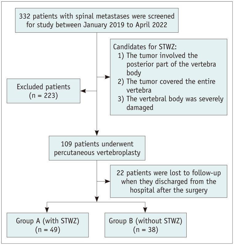
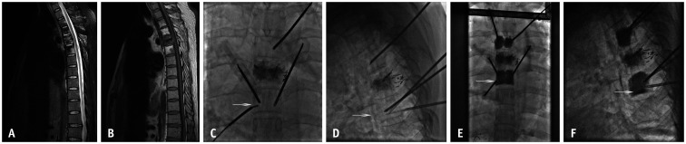
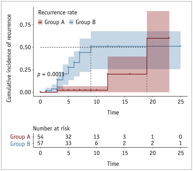
Materials and methods: We prospectively enrolled 87 patients who underwent PV for spinal metastasis involving the posterior part of the vertebral body, with or without the STWZ approach, from January 2019 to April 2022. Forty-nine patients (27 females and 22 males; mean age ± standard deviation [SD], 57.2 ± 11.6 years; age range, 31-76 years) were included in group A (with STWZ approach), accounting for 54 vertebrae. Thirty-eight patients (18 females and 20 males; 59.1 ± 10.9 years; 29-81 years) were included in group B (without STWZ approach), accounting for 57 vertebrae. Patient demographics, procedure-related variables, and pain relief as assessed using the visual analog scale (VAS) were collected at different time points. Tumor recurrence in the vertebrae after PV was analyzed using Kaplan-Meier curves.

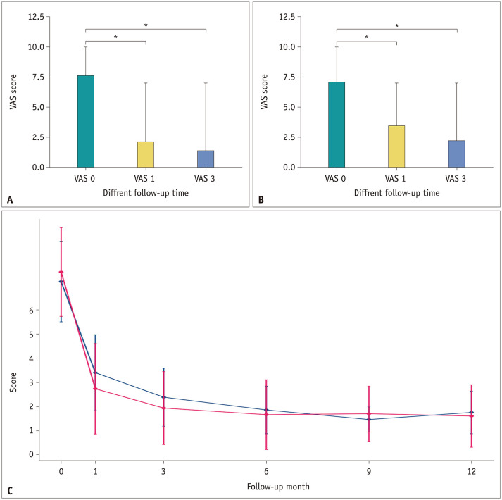
Results: The STWZ approach was successful from T1 to L5 without severe complications. Cement filling was satisfactory in 47/54 (87.0%) and 25/57 (43.9%) vertebrae in groups A and B, respectively (p < 0.001). Cement leakage was not significantly different between groups A and B (p = 1.000). Mean VAS score ± SD before and 1 week and 1, 3, 6, 9, and 12 months after PV were 7.6 ± 1.8, 4.2 ± 2.0, 2.7 ± 1.9, 1.9 ± 1.5, 1.7 ± 1.4, 1.7 ± 1.1, and 1.6 ± 1.3, respectively, in group A and 7.2 ± 1.7, 4.0 ± 1.3, 3.4 ± 1.6, 2.4 ± 1.2, 1.8 ± 1.0, 1.4 ± 0.5, and 1.7 ± 0.9, respectively, in group B. Kaplan-Meier analysis showed a lower tumor recurrence rate in group A than in group B (p = 0.001).

Conclusion: The STWZ approach may represent a new, safe, alternative/auxiliary approach to target the posterior part of the vertebral body in the PV for spinal metastases.

Keywords: Percutaneous vertebroplasty; Puncture approach; Spinal metastases.


Conflict of interest statement

The authors have no potential conflicts of interest to disclose.

Figures

Fig. 1
Fig. 1. Patient flow diagram.
STWZ = safe triangular working zone
Fig. 2
Fig. 2. Diagrams (A to F) showing the steps involved in the technique to perform PV via the STWZ approach.
A. Local anesthesia administered via the STWZ approach targeted the posterior point of the periosteum. B. In the LP view, the disposable coaxial biopsy needle with a blunt core needle was carefully advanced until it approached the vertebral body posteriorly, and the patient’s response was continuously monitored. C. The EV catheter needle replaced the blunt core needle coaxially. D, E. The bevel needle sheath was punctured through the EV, then the EV was removed with the inner needle inserted. F. With the help of a hammer, the bevel needle was guided toward the target zone or adjusted to many angles within the vertebral body. LP = lateral projection, PV = percutaneous vertebroplasty, STWZ = safe triangular working zone approach
Fig. 3
Fig. 3. Primary origin of vertebral tumors in all included patients.
The number on top of the bar indicates the number of certain tumor types, the different colors represent the different tumor types.
Fig. 4
Fig. 4. Example patient.
A-D. Sagittal CT view (A1, A2) and axial T2-weighted MR image (B1, B2) show the lumbar spine in a 70-year-old male who suffered spinal pain after screw fixation and fracture of the L4 vertebra following metastasis from lung cancer with rupture of the posterior wall (A1 to B1 for preoperative imaging; A2 to B2 for postoperative imaging). C1, C2 and D1, D2 show the digital subtraction angiography scan, A2 to B2 show the cement covering the vertebra and tumor, B2 image shows that the tumor is smaller after vertebroplasty than before, with relief of the spinal pain. The arrows indicate the safe triangular working zone approach.
Fig. 5
Fig. 5. Example patient.
A-F. MRI scans of tumors occurring in the posterior vertebral body in a 46-year-old male with adenocarcinoma of the liver with bone metastases (A, B). STWZ was performed to target the bone metastasis tumor (C, D) with radiofrequency ablation and achieved satisfactory cement fulfillment (E, F). The arrows indicate the STWZ approach. STWZ = safe triangular working zone
Fig. 6
Fig. 6. VAS score in groups A and B.
A. VAS at baseline (VAS 0) and after 1 and 3 months (VAS 1 and VAS 3) in group A. B. VAS at baseline (VAS 0) and after 1 and 3 months (VAS 1 and VAS 3) in group B. C. The improvement in VAS scores are shown for both groups. For the included patients, a new baseline was established before the procedure, and follow-up was performed at 1, 3, 6, 9 and 12 months after percutaneous vertebroplasty in groups A (red) and B (blue). *p < 0.001 in comparison with baseline (preoperation). VAS = visual analog scale
Fig. 7
Fig. 7. Kaplan–Meier curve shows the cumulative proportion of vertebral recurrence after vertebroplasty.
Group A (red) had lower tumor recurrence rate in the vertebrae than group B (blue).

Similar articles

Cited by

References

    1. Health Quality Ontario. Vertebral augmentation involving vertebroplasty or kyphoplasty for cancer-related vertebral compression fractures: a systematic review. Ont Health Technol Assess Ser. 2016;16:1–202. - PMC - PubMed
    1. Tsoumakidou G, Too CW, Koch G, Caudrelier J, Cazzato RL, Garnon J, et al. CIRSE guidelines on percutaneous vertebral augmentation. Cardiovasc Intervent Radiol. 2017;40:331–342. - PubMed
    1. Kallmes DF, Schweickert PA, Marx WF, Jensen ME. Vertebroplasty in the mid- and upper thoracic spine. AJNR Am J Neuroradiol. 2002;23:1117–1120. - PMC - PubMed
    1. Robinson Y, Heyde CE, Försth P, Olerud C. Kyphoplasty in osteoporotic vertebral compression fractures--guidelines and technical considerations. J Orthop Surg Res. 2011;6:43. - PMC - PubMed
    1. Ringer AJ, Bhamidipaty SV. Percutaneous access to the vertebral bodies: a video and fluoroscopic overview of access techniques for trans-, extra-, and infrapedicular approaches. World Neurosurg. 2013;80:428–435. - PubMed