

Diagnostic Errors in Cerebrovascular Pathology: Retrospective Analysis of a Neuroradiology Database at a Large Tertiary Academic Medical Center
- PMID: 35926887
- PMCID: PMC9451623
- DOI: 10.3174/ajnr.A7596
Diagnostic Errors in Cerebrovascular Pathology: Retrospective Analysis of a Neuroradiology Database at a Large Tertiary Academic Medical Center
Abstract
Background and purpose: Diagnostic errors affect 2%-8% of neuroradiology studies, resulting in significant potential morbidity and mortality. This retrospective analysis of a large database at a single tertiary academic institution focuses on diagnostic misses in cerebrovascular pathology and suggests error-reduction strategies.
Materials and methods: CT and MR imaging reports from a consecutive database spanning 2015-2020 were searched for errors of attending physicians in cerebrovascular pathology. Data were collected on missed findings, study types, and interpretation settings. Errors were categorized as ischemic, arterial, venous, hemorrhagic, and "other."
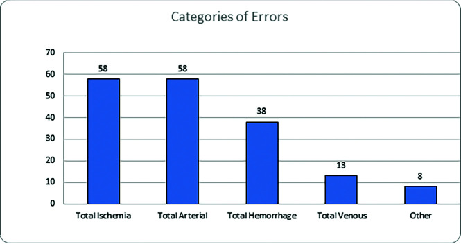
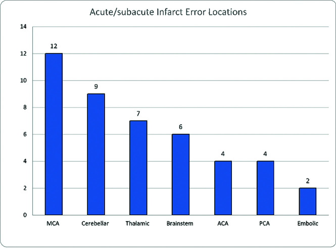
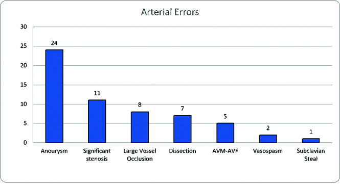
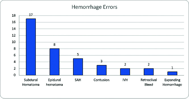
Results: A total of 245,762 CT and MR imaging neuroradiology examinations were interpreted during the study period. Vascular diagnostic errors were present in 165 reports, with a mean of 49.6 (SD, 23.3) studies on the shifts when an error was made, compared with 34.9 (SD, 19.2) on shifts without detected errors (P < .0001). Seventy percent of examinations occurred in the hospital setting; 93.3% of errors were perceptual; 6.7% were interpretive; and 93.9% (n = 155) were clinically significant (RADPEER 2B or 3B). The distribution of errors was arterial and ischemic each with 33.3%, hemorrhagic with 21.8%, and venous with 7.5%. Most errors involved brain MR imaging (30.3%) followed by head CTA (27.9%) and noncontrast head CT (26.1%). The most common misses were acute/subacute infarcts (25.1%), followed by aneurysms (13.7%) and subdural hematomas (9.7%).
Conclusions: Most cerebrovascular diagnostic errors were perceptual and clinically significant, occurred in the emergency/inpatient setting, and were associated with higher-volume shifts. Diagnostic errors could be minimized by adjusting search patterns to ensure vigilance on the sites of the frequently missed pathologies.
© 2022 by American Journal of Neuroradiology.
Figures
Similar articles
-
Impact of Shift Volume on Neuroradiology Diagnostic Errors at a Large Tertiary Academic Center.Acad Radiol. 2023 Aug;30(8):1584-1588. doi: 10.1016/j.acra.2022.08.035. Epub 2022 Sep 27. Acad Radiol. 2023. PMID: 36180325
-
Analysis of misses in imaging of head and neck pathology by attending neuroradiologists at a single tertiary academic medical centre.Clin Radiol. 2021 Oct;76(10):786.e9-786.e13. doi: 10.1016/j.crad.2021.06.011. Epub 2021 Jul 23. Clin Radiol. 2021. PMID: 34304864
-
Factors Associated With Neuroradiologic Diagnostic Errors at a Large Tertiary-Care Academic Medical Center: A Case-Control Study.AJR Am J Roentgenol. 2023 Sep;221(3):355-362. doi: 10.2214/AJR.22.28925. Epub 2023 Mar 29. AJR Am J Roentgenol. 2023. PMID: 36988269
-
Role of noncontrast head CT in the assessment of vascular abnormalities in the emergency room.Emerg Radiol. 2013 Dec;20(6):529-41. doi: 10.1007/s10140-013-1136-6. Epub 2013 Jun 7. Emerg Radiol. 2013. PMID: 23739799 Review.
-
Magnetic resonance applications in cerebral injury.Radiol Clin North Am. 1992 Mar;30(2):353-66. Radiol Clin North Am. 1992. PMID: 1535861 Review.
Cited by
-
Shift Volume Directly Impacts Neuroradiology Error Rate at a Large Academic Medical Center: The Case for Volume Limits.AJNR Am J Neuroradiol. 2024 Apr 8;45(4):374-378. doi: 10.3174/ajnr.A8119. AJNR Am J Neuroradiol. 2024. PMID: 38238099 Free PMC article.
References
-
- Balogh EP, Miller BT, Ball JR, eds; Committee on Diagnostic Error in Health Care, Board on Health Care Services, Institute of Medicine, and National Academies of Sciences, Engineering, and Medicine. Improving Diagnosis in Health Care. National Academies Press; (US: ) 2015 - PubMed
-
- Kohn LT, Corrigan JM, Donaldson MS, eds; Institute of Medicine Committee on Quality of Health Care in America. To Err is Human: Building a Safer Health System. National Academies Press; (US: ) 2000 - PubMed