Review
. 2022 Dec;71(12):2587-2597.
doi: 10.1136/gutjnl-2021-326562. Epub 2022 Aug 4.

Recent advances in clinical practice: advances in cross-sectional imaging in inflammatory bowel disease

Affiliations
Review

Recent advances in clinical practice: advances in cross-sectional imaging in inflammatory bowel disease

Jordi Rimola et al. Gut. 2022 Dec.

Abstract

Endoscopy remains the reference standard for the diagnosis and assessment of patients with inflammatory bowel disease (IBD), but it has several important limitations. Cross-sectional imaging techniques such as magnetic resonance enterography (MRE) and intestinal ultrasound (IUS) are better tolerated and safer. Moreover, they can examine the entire bowel, even in patients with stenoses and/or severe inflammation. A variety of cross-sectional imaging activity scores strongly correlate with endoscopic measures of mucosal inflammation in the colon and terminal ileum. Unlike endoscopy, cross-sectional techniques allow complete visualisation of the small-bowel and assess for extraintestinal disease, which occurs in nearly half of patients with IBD. Extramural findings may predict outcomes better than endoscopic mucosal assessment, so cross-sectional techniques might help identify more relevant therapeutic targets. Coupled with their high sensitivity, these advantages have made MRE and IUS the primary non-invasive options for diagnosing and monitoring Crohn's disease; they are appropriate first-line investigations, and have become viable alternatives to colonoscopy. This review discusses cross-sectional imaging in IBD in current clinical practice as well as research lines that will define the future role of these techniques.

Keywords: CROHN'S DISEASE; GASTROINTESTINAL ULTRASOUND; INFLAMMATORY BOWEL DISEASE; MAGNETIC RESONANCE IMAGING.


Conflict of interest statement

Competing interests: JR has received grants from AbbVie and consulting fees from Boehringer Ingelheim, Janssen, Origo, Lument, Takeda and Alimentiv. JT has received grants from AbbVie and Janssen, advisory board fees and/or speaker fees from Janssen, Galapagos and Pfizer. SK has no declarations. SAT is a previous research consultant to Alimentiv. Shareholder in Motilent. TK has received grants from AbbVie, Janssen and Takeda, advisory board fees and/or speaker fees from AbbVie, Amgen, Arena Pharmaceuticals, Biogen, Boehringer Ingelheim, Bristol Myers Squibb, Celgene, Celltrion, Dr Falk Pharma, Ferring Arzneimittel, Galapagos, Gilead, Janssen, MSD Sharp & amp; Dome GmbH, Pfizer, Roche, Takeda Pharma and Vifor Pharma.

Figures
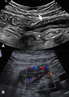
Figure 1
Figure 1
Small-bowel inflammation in Crohn’s disease detected using intestinal ultrasound. Diffusely increased bowel wall thickness (measured using the line that appears in the image, resulting in a distance of 5.3 mm) in the terminal ileum (arrow) is associated with partial loss of echostratification. The surrounding mesenteric fat appears (asterisk) is hyperechogenic due to perienteric inflammatory changes (A). Intestinal ultrasound in a different patient shows hypervascularisation of the small-bowel measured by colour Doppler (B). The wall of the terminal ileum is thickened and has a high colour Doppler signal that extends outside the bowel wall. These findings are consistent with active inflammatory small-bowel Crohn’s disease.
Figure 2
Figure 2
Small-bowel inflammation in Crohn’s disease detected using magnetic resonance enterography. Axial post-contrast fat-saturated T1-weighted images (A) show moderate bowel-wall thickening and hyperenhancement (arrow); compare with the normal thickness in the uninvolved segments (arrowhead). The same segment seen on axial fat-saturated T2-weighted images (B). Note the increased signal intensity within the wall due to oedema (arrows) and the thin perienteric rim, due to the presence of fluid around the ileum. In the proximal sections of the same segment (C), ulcerations are seen as small disruptions of the inner surface of the thickened bowel wall (arrowhead).
Figure 3
Figure 3
Evolving role of cross-sectional imaging in the field of inflammatory bowel disease. DWI, diffusion-weighted imaging; US, ultrasound.
Figure 4
Figure 4
Coronal (A, C) and axial (B, D) T2 non-fat saturated weighted images from an MRE enterography examination before (A, B) and after (C, D) anti TNF-α therapy. Before treatment there is active small-bowel Crohn’s disease with mural thickening and increased mural T2 signal (arrows). Following 6 months of anti-TNF-α therapy the bowel has returned to near normal with subtle low T2 signal thickening only (arrows). MRE, magnetic resonance enterography; TNF, tumour necrosis factor.
Figure 5
Figure 5
Monitoring disease activity using intestinal ultrasound. Composite image of small-bowel inflammation due to Crohn’s disease in the terminal ileum showing a mural wall thickness of 7 mm and partially abrogated echostratification at week 0 (A). The same part of terminal ileum at week 48 after treatment with ustekinumab showing normalisation of mural thickness, normal echostratification (B). Overall, findings are consistent with achievement of transmural remission.
Figure 6
Figure 6
A single image from a dynamic set with a region of interest (white circle) placed on a diseased terminal Ileum (A). Motion is assessed by assigning each pixel in the image with an associated displacement value, and expressed as quantitative value (green circle) (B).

Similar articles

Cited by

References

    1. Torres J, Mehandru S, Colombel J-F, et al. . Crohn’s disease. The Lancet 2017;389:1741–55. 10.1016/S0140-6736(16)31711-1 - DOI - PubMed
    1. Ungaro R, Mehandru S, Allen PB, et al. . Ulcerative colitis. Lancet 2017;389:1756–70. 10.1016/S0140-6736(16)32126-2 - DOI - PMC - PubMed
    1. Turner D, Ricciuto A, Lewis A, et al. . STRIDE-II: an update on the selecting therapeutic targets in inflammatory bowel disease (STRIDE) initiative of the International organization for the study of IBD (IOIBD): determining therapeutic goals for Treat-to-Target strategies in IBD. Gastroenterology 2021;160:1570–83. 10.1053/j.gastro.2020.12.031 - DOI - PubMed
    1. Goodsall TM, Noy R, Nguyen TM, et al. . Systematic review: patient perceptions of monitoring tools in inflammatory bowel disease. J Can Assoc Gastroenterol 2021;4:e31–41. 10.1093/jcag/gwaa001 - DOI - PMC - PubMed
    1. Wu Y-W, Tang Y-H, Hao N-X, et al. . Crohn's disease: CT enterography manifestations before and after treatment. Eur J Radiol 2012;81:52–9. 10.1016/j.ejrad.2010.11.010 - DOI - PubMed