. 2022 Aug 4;13(1):4543.
doi: 10.1038/s41467-022-32174-7.

The molecular mechanism of snake short-chain α-neurotoxin binding to muscle-type nicotinic acetylcholine receptors

Affiliations

The molecular mechanism of snake short-chain α-neurotoxin binding to muscle-type nicotinic acetylcholine receptors

Mieke Nys et al. Nat Commun. .

Erratum in

Abstract

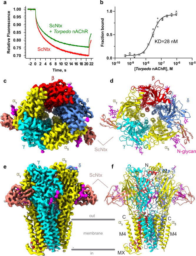
Bites by elapid snakes (e.g. cobras) can result in life-threatening paralysis caused by venom neurotoxins blocking neuromuscular nicotinic acetylcholine receptors. Here, we determine the cryo-EM structure of the muscle-type Torpedo receptor in complex with ScNtx, a recombinant short-chain α-neurotoxin. ScNtx is pinched between loop C on the principal subunit and a unique hairpin in loop F on the complementary subunit, thereby blocking access to the neurotransmitter binding site. ScNtx adopts a binding mode that is tilted toward the complementary subunit, forming a wider network of interactions than those seen in the long-chain α-Bungarotoxin complex. Certain mutations in ScNtx at the toxin-receptor interface eliminate inhibition of neuronal α7 nAChRs, but not of human muscle-type receptors. These observations explain why ScNtx binds more tightly to muscle-type receptors than neuronal receptors. Together, these data offer a framework for understanding subtype-specific actions of short-chain α-neurotoxins and inspire strategies for design of new snake antivenoms.


Conflict of interest statement

The authors declare no competing interests.

Figures

Fig. 1
Fig. 1. Binding of ScNtx to the muscle-type Torpedo nAChR: biophysical and structural characterization.
a MST traces of fluorescently labeled unbound (red) and bound (green) ScNtx to purified Torpedo muscle-type nAChR reconstituted in asolectin-MSP1E3D1 lipidic nanodiscs. b MST concentration-response curve: change in normalized fluorescence as a function of Torpedo nAChR concentration. Data are presented as single data points in addition to the mean values ± standard deviations. c, e Top and side view of the cryo-EM map. d, f Top and side view of the 3D reconstruction in cartoon representation. N-glycans are shown as sticks. Source data are provided as a Source Data file.
Fig. 2
Fig. 2. Comparison of the ScNtx and α-Bgtx binding mode to the Torpedo nAChR.
a Sequence alignment of ScNtx and α-Bgtx generated in ESPRIPT. Residue numbering and secondary structure information at the top is for ScNtx. TT indicates a β-turn. Yellow digits indicate disulfide bonds. Blue frames indicate regions of similarity. Red boxes indicate strict conservation, red characters indicate similarity. b, c Differences in binding mode of ScNtx (salmon) and α-Bgtx (cyan) at the α-δ subunit interface after superposition of the ScNtx-Torpedo nAChR complex onto the α-Bgtx-Torpedo nAChR complex (PDB ID: 6UWZ). The principal subunit (αδ) is shown in yellow; the complementary subunit (δ) is shown in blue. The orientations are chosen so that the tilt at the base of the three-fingered hand (b) and the conformational difference of finger I (c) are obvious. Roman numbers indicate the three fingers of ScNtx and α-Bgtx. d, e Cartoon representation of ScNtx and α-Bgtx. The orientation and color code are the same as in b. The disulfide bonds are shown in yellow. The tilt is indicated by dashed lines connecting the tip of finger II, which is in a fixed position, to disulfide bridges 1 and 3. The inset shows an enlarged view of the base of the three-fingered hand which is formed by loops connecting the different β-strands. These loops adopt markedly different positions in ScNtx (salmon) and α-Bgtx (cyan) as indicated by dashed lines.
Fig. 3
Fig. 3. Molecular contacts at the ScNtx and α-Bgtx-receptor interface.
Receptor and toxins are in cartoon representation. Interacting residues and glycans are shown as sticks, colored by subunit. Dashed lines indicate hydrogen bonds or salt bridges. Color code as in Fig. 2. The orientations are chosen so that the described interactions are clear (Supplementary Movie 1). The PDB ID for the α-Bgtx-Torpedo nAChR complex is 6UWZ. a, b Interactions between the principal subunit (αδ) and finger I of ScNtx and α-Bgtx. For α-Bgtx, additional interactions with the extended C-terminus are included. c, d Interactions between the principal subunit (αδ) and finger II of ScNtx and α-Bgtx. e, f Interactions between the complementary subunit (δ) and finger II of ScNtx and α-Bgtx. g, h Interactions between the complementary subunit (δ) and finger III of ScNtx and α-Bgtx.
Fig. 4
Fig. 4. Pharmacological characterization of ScNtx.
ag Electrophysiological recordings on Xenopus laevis oocytes expressing different subtypes of nAChRs: the adult muscle-type nAChR, the neuronal α7 nAChR and the neuronal α4β2 nAChR. Horizontal bars indicate duration of acetylcholine application. Increasing concentrations of ScNtx or mutant ScNtx (R28A or R31A) were applied as indicated in salmon. Traces in salmon indicate co-application of acetylcholine (at a concentration near the EC50) and (mutant) ScNtx. Blue traces indicate application of acetylcholine alone. The inset depicts ScNtx in a cartoon representation. Residues subjected to mutagenesis are shown in spheres. Roman numbers indicate the three fingers. h, i Concentration-Inhibition curves: normalized currents in oocytes expressing the muscle-type nAChR or the neuronal α7 nAChR as a function of ScNtx concentration. Data are presented as mean values ± standard deviations. Green curves are for wild-type ScNtx, red curves for mutant ScNtx (R28A or R31A). Source data are provided as a Source Data file.

References

    1. Gutiérrez JM, et al. Snakebite envenoming. Nat. Rev. Dis. Prim. 2017;3:17063. doi: 10.1038/nrdp.2017.63. - DOI - PubMed
    1. Miwa JM, et al. Lynx1, an endogenous toxin-like modulator of nicotinic acetylcholine receptors in the mammalian CNS. Neuron. 1999;23:105–114. doi: 10.1016/S0896-6273(00)80757-6. - DOI - PubMed
    1. Arredondo J, Chernyavsky AI, Webber RJ, Grando SA. Biological effects of SLURP-1 on human keratinocytes. J. Invest. Dermatol. 2005;125:1236–1241. doi: 10.1111/j.0022-202X.2005.23973.x. - DOI - PubMed
    1. Arredondo J, Chernyavsky AI, Jolkovsky DL, Webber RJ, Grando SA. SLURP-2: A novel cholinergic signaling peptide in human mucocutaneous epithelium. J. Cell. Physiol. 2006;208:238–245. doi: 10.1002/jcp.20661. - DOI - PubMed
    1. Nirthanan, S., Awal, W. & Niranjan, N. R. Snake α-Neurotoxins and the Nicotinic Acetylcholine Receptor. 10.1007/978-94-007-6648-8 (2016).

Publication types